L2 Resorption 1 - Introduction, General Internal Resorption
Internal Root Resorption - Aetiology and Treatment1
Resorption – Introduction, General Principles, Classification; Internal Resorption — W/Prof. Paul V. Abbott AO
Internal Root Resorption - Aetiology and Treatment
Prof. Paul V. Abbott AO BDSc, MDS, FRACDS(Endo), FPFA, FADI, FICD, FACD, FIADT
- Specialist Endodontist
- Winthrop Professor of Clinical Dentistry
- UWA Dental School, The University of Western Australia
Oral Health Centre of Western Australia
UWA Dental School – DMD 3rd Year
Introduction and Principles of Root Resorption
Related resorption topics
Definition and Requirements for Resorption
Outline2
- Definition and Requirements for Resorption
- Classification of Resorption
- Prevalence of Internal Resorption
- For Each Type of Internal Resorption:
- Pathogenesis
- Features
- Diagnosis
- Management / Treatment
Root Resorption — Definition
Resorption is either a physiological or a pathological process which results in the loss of substance from a tissue.
- Root resorption specifically refers to loss of substance from the roots, namely cementum and dentine.
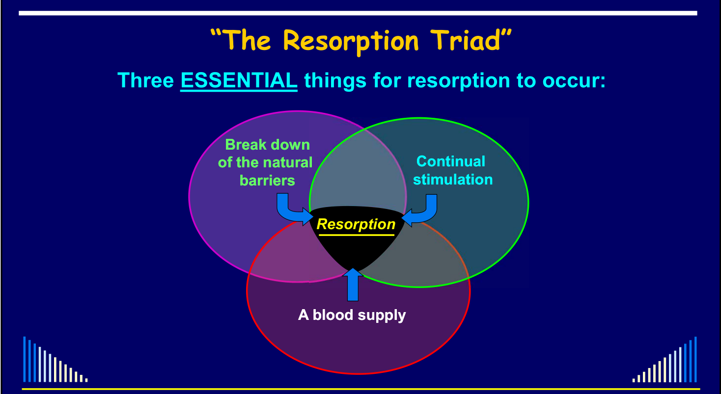
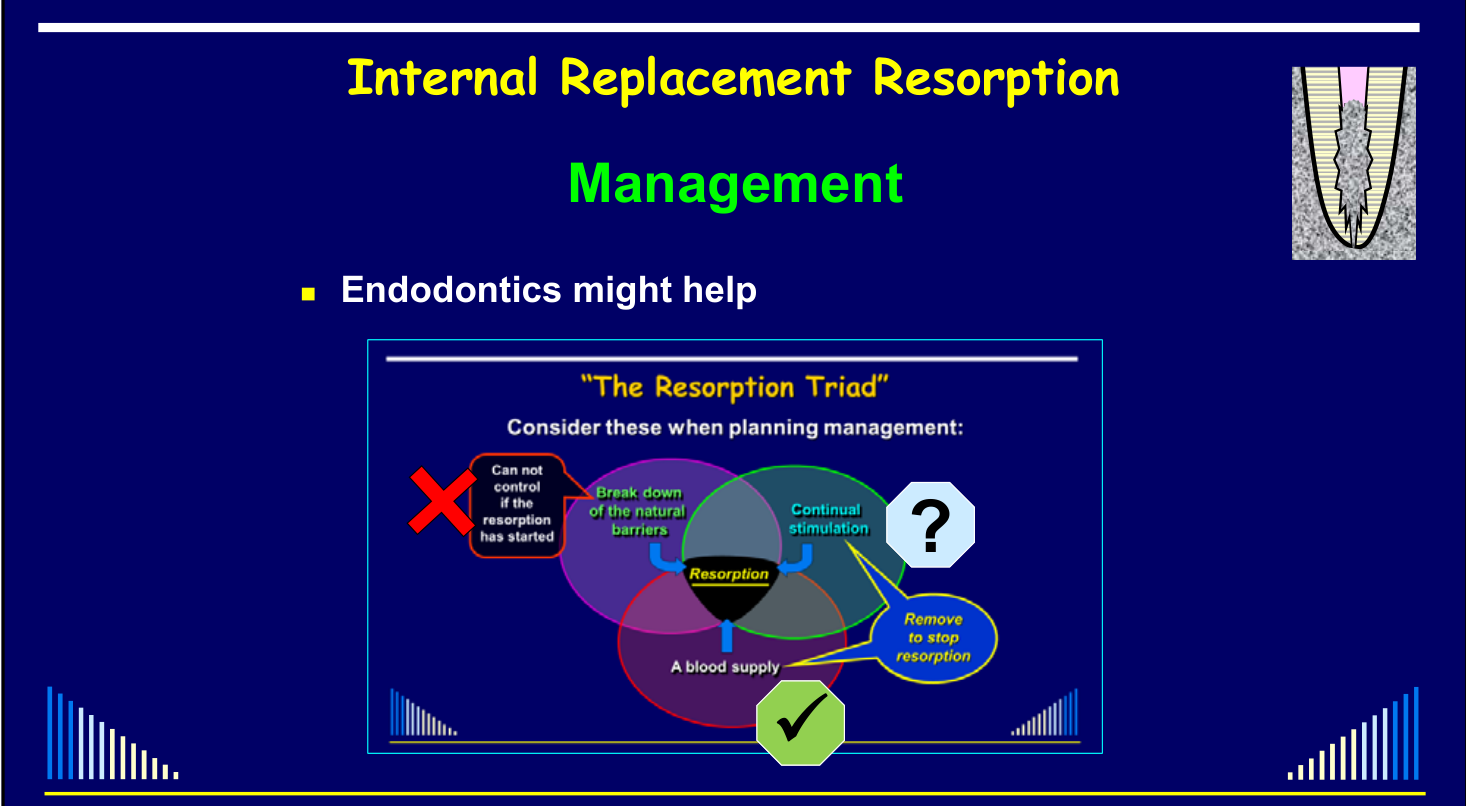
Three Essential Requirements for Resorption to Occur3
- Break down of the natural barriers
- Continual stimulation
- A blood supply
- The lecturer referred to these three requirements collectively as the resorption triad.
- A blood supply is needed to keep clastic cells alive and functioning.
- When all three requirements are present, resorption of tissue is likely to occur.
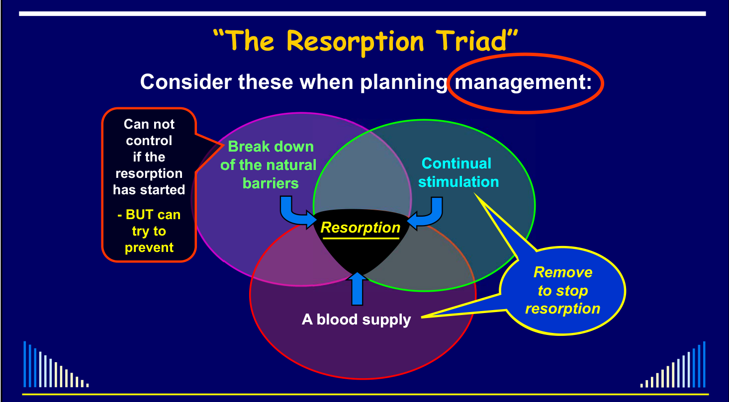
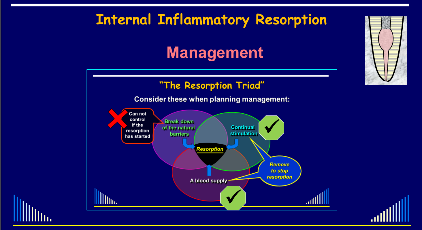
- In internal resorption, breakdown of the natural barriers has already occurred and generally cannot be reversed.
- Management therefore focuses on removing or reducing the continual stimulation and removing the blood supply that maintains the clastic cells. These three factors lead to resorption.
 |  |
Related topic
Considerations When Planning Management
-
If the resorption has started, it cannot be controlled — BUT it may be possible to try to prevent it by addressing the contributing factors.
-
Remove the continual stimulation to stop resorption.
-
Not every case requires direct treatment, but every case requires appropriate management.
-
Understanding the situation and the requirements for resorption helps determine whether treatment is necessary, the condition can be monitored, or bacteria, tissue, or blood supply can be removed to stop the resorptive process.
Classification of Resorption4
- The traditional classification consists of surface resorption, inflammatory resorption, and replacement resorption.
- This classification applies to both internal and external resorption.
- Some alternative terminology describes inflammatory resorption as infection-related resorption and replacement resorption as ankylosis-related resorption.
- The lecturer preferred classifying resorption according to what is occurring in the tissues rather than what the condition is related to.
- External invasive resorption is often called cervical resorption, but the lecturer considered that term unsuitable because the condition is subgingival and does not always occur in the cervical region, particularly when recession has occurred.
Related topic
Internal Root Resorption Overview
General Definition of Internal Resorption
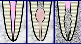
Internal Root Resorption — Types5
- Surface
- Inflammatory
- Replacement
- Internal inflammatory resorption was described as the most common of the three types.
General Definition — Internal Resorption
- The removal of dentine by clastic cells
- Begins at the root canal wall
- Progresses through dentine towards the cementum
- May also resorb cementum and perforate the external root surface
Lecturer — Perforation Risk
Perforation through to the external root surface becomes more likely in particular circumstances.
- The risk increases when the condition has been present for a long time.
- Rapidly developing internal inflammatory resorption also increases the likelihood of perforation.
Outline
See - Lecture outline
Prevalence of Internal Root Resorption6
-
A very rare condition
- Bell (1830) — the first person to describe a tooth with internal resorption
- Thoma (1935) — 1 case in 1,000 teeth
- Andreasen (1970) — approx. 2% of luxated teeth
-
Other studies have reported internal resorption — but all were following various treatments, e.g.
- Cabrini et al (1957) — 28% after pulpotomy
- Ahlberg et al (1983) — 55% after autotransplantation
Lecturer — Treatment-Related Figures
These higher figures may reflect particular treatment-related situations rather than the prevalence of internal resorption in teeth generally.
-
Haapasalo & Endal (2006) stated:
- Epidemiological data is scarce
- Estimated prevalence as: 0.01 – 1% of patients
- “Based on limited studies and clinical experience of the authors”
- “This estimate is quite rough and may be wrong”
- Typically only 1 tooth per patient
- Very occasionally 2 adjacent teeth — with trauma a likely factor
- Many cases referred as “internal resorption” are actually “external invasive resorption”
-
None of these studies / estimates mentioned which type of internal resorption
- But they probably only meant Internal Inflammatory Resorption
-
Since:
- Internal Surface Resorption is probably very rare
- → AND it cannot be detected clinically or radiographically — any histological
- Internal Replacement Resorption is also very rare
- → It is not well known or understood
- → Easily confused with External Invasive Resorption
- Internal Surface Resorption is probably very rare
Analysis of a Referral-based Endodontic Practice. Part 1. Demographic Data and Reasons for Referral7
Abbott PV.
J Endod 1994; 20: 93-96.
Analysis of a Referral-based Endodontic Practice. Part 1. Demographic Data and Reasons for Referral Abbott PV. J Endod 1994; 20: 93-96.
Types and Incidence of Root Resorption Among the 2000 Patients Referred for Treatment (Table 5)8
In Abbott’s referral-based cohort, 36% of patients with external invasive resorption had originally been referred as internal resorption; this is a subgroup denominator, not 36% of all 2,000 referrals. Full Table 5: Abbott 1994 - Referral Practice Table 5.
Outline
See - Lecture outline
Lecturer — Surface Resorption
Internal surface resorption is very rare and does not normally produce signs or symptoms. The significance and general relevance of whitening-related resorption remain uncertain, although the experimental findings suggest that it may be transient.
Internal Surface Resorption
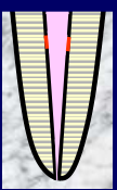
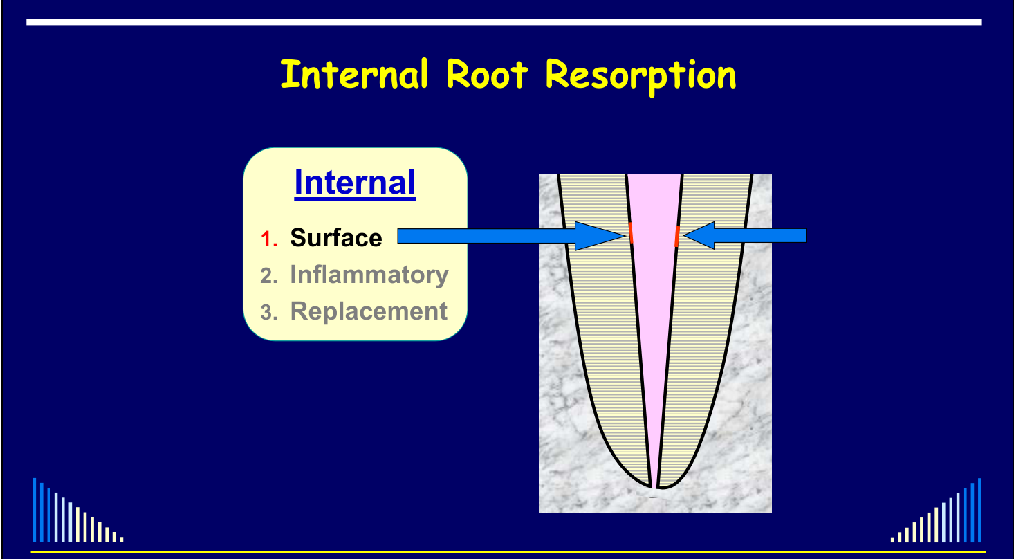
Features and Possible Causes9
 |  |
 |
Features10
- Minor areas of resorption of the root canal wall
- Not visible radiographically
- Only noted histologically
- Believed to be self-limiting — if the irritant is removed
- May be a precursor to internal inflammatory resorption
Possible Causes11
- Trauma to the tooth
- Necrosis + infection of the coronal pulp
- Due to caries, cracks, fractures, restoration breakdown, etc.
- External whitening / bleaching of the tooth
 |  |
External Whitening
Seale et al - J Dent Res 1981
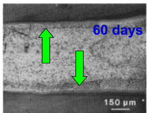
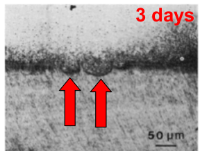
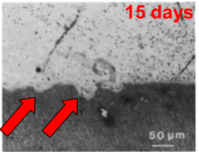
In a dog model, 35% hydrogen peroxide was applied to enamel for 30 minutes weekly over four weeks. Internal surface resorption with active dentinoclasts was reported at 3 days; by 60 days resorption had stopped and healing was evident. This does not estimate human incidence or risk. Full study details: Seale et al 1981 - Dog Bleaching Experiment.
3 days; 15 days
60 days
See - Possible Causes
The shared causes are listed under Internal Surface Resorption.
BUT — the aetiology is unknown in most cases. Mainly because this type of resorption cannot be diagnosed clinically or radiographically. Hence, we do not know when it is present !!!
Pathogenesis
- Largely unknown
- Needs a stimulus (as discussed)
- Transient in nature
- Demonstrated by Wedenberg & Lindskog (1985)
Experimental Internal Resorption in Monkey Teeth
Wedenberg & Lindskog - Endod Dent Traumatol 1985; 1: 221-227.
In this induced monkey model, non-infected sealed teeth showed transient surface resorption, while infected unsealed teeth had more extensive and prolonged resorption; spread of resorbing cells occurred only where dentine was denuded. This model-specific association does not establish that infection is required in every human case. Full methods and results: Wedenberg and Lindskog 1985 - Internal Resorption Model.
Diagnosis12
- Can not diagnose clinically
- Can not see it radiographically
Lecturer — Diagnostic Appearance
Internal inflammatory resorption is mainly diagnosed radiographically as a radiolucency within the tooth with enlargement of the root canal space. Shape and size are variable, and a periapical radiolucency may or may not be present; when present, it likely indicates that the lesion is no longer active.
Lecturer — External Differentiation
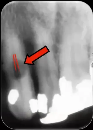
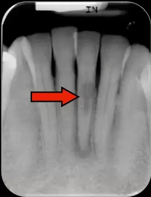
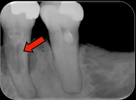
To distinguish internal from external resorption, trace the outline of the root canal. In internal resorption the canal outline joins the defect, whereas in external resorption the canal may appear to pass through or be superimposed over it; tube-shift radiographs can also help.
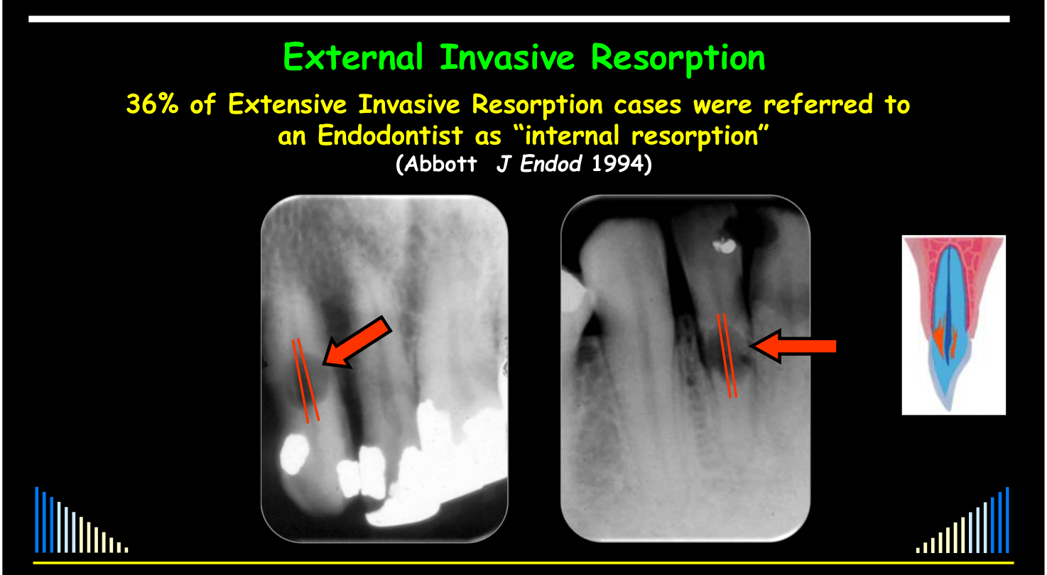 | 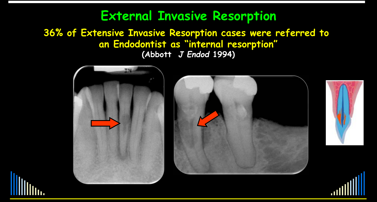 |
Management
Management (if ever suspected / diagnosed)13
- Monitor radiographically to see if resorption continues
- If it does continue:
- It is likely to be Internal Inflammatory Resorption
- Therefore do root canal treatment
- → Will discuss later
Lecturer — Treatment Sequence
Management involves removing the pulp if it is still present, thoroughly cleaning and preparing the canal, and using copious sodium hypochlorite irrigation. Ultrasonics can improve irrigant flow into the irregular defect, and intracanal medicaments may be repeated before the canal and resorptive defect are filled.
Lecturer — Inflammatory Resorption
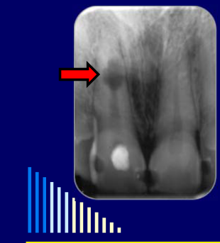
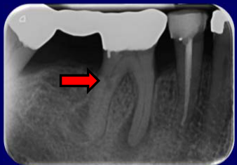
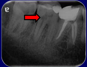
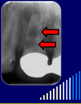
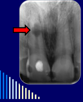
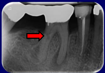
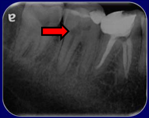
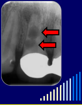
Internal inflammatory resorption is rare; in the lecturer’s review of 2,000 referred patients, it occurred in 0.5%. It is traditionally described as an oval radiolucent enlargement of the pulp space, but its shape and size can vary, and it may occur in any tooth or anywhere along the root canal.
Lecturer — Inflammatory Causes
Caries- and restoration-related infection may be at least as prominent as trauma in reported internal inflammatory resorption cases. The small selected series (28 teeth with subtype unspecified and 13 extracted human teeth) do not establish causation or population proportions; full study data are in Caliskan and Turkun 1997 - Internal Resorption Case Series and Wedenberg and Zetterqvist 1987 - Human Internal Resorption Histology.
Lecturer — Replacement Resorption
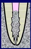
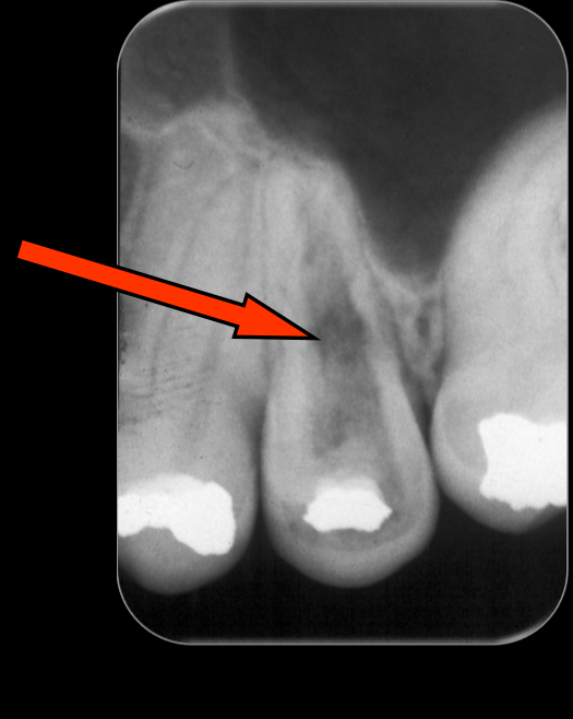
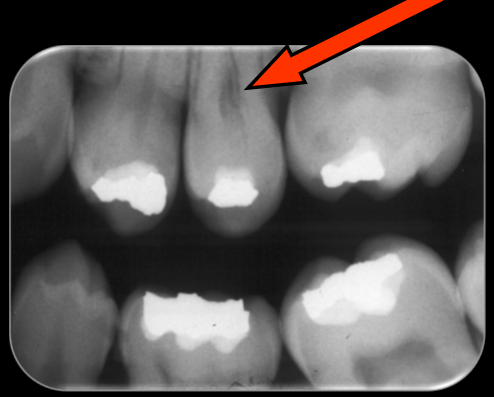
Internal replacement resorption is even rarer than internal inflammatory resorption; in the lecturer’s review of 2,000 patients, only one patient had it, representing 0.05%. It involves irregular enlargement of the pulp space and replacement of pulp and dentine with bone-like tissue, potentially replacing the entire root if it remains undetected.
Lecturer — Resorption Requirements
Internal inflammatory resorption requires bacteria in the pulp and damage to the odontoblast layer and predentine, exposing mineralized dentine to clastic cells. The precise injurious event is unknown, so any source of pulp irritation may be involved.
Lecturer — Resorption Phases
Internal inflammatory resorption has active and inactive phases. In the inactive phase, the bacterial front has moved through the defect, the entire root canal is pulpless and infected, and chronic apical periodontitis or a periapical radiolucency may develop.
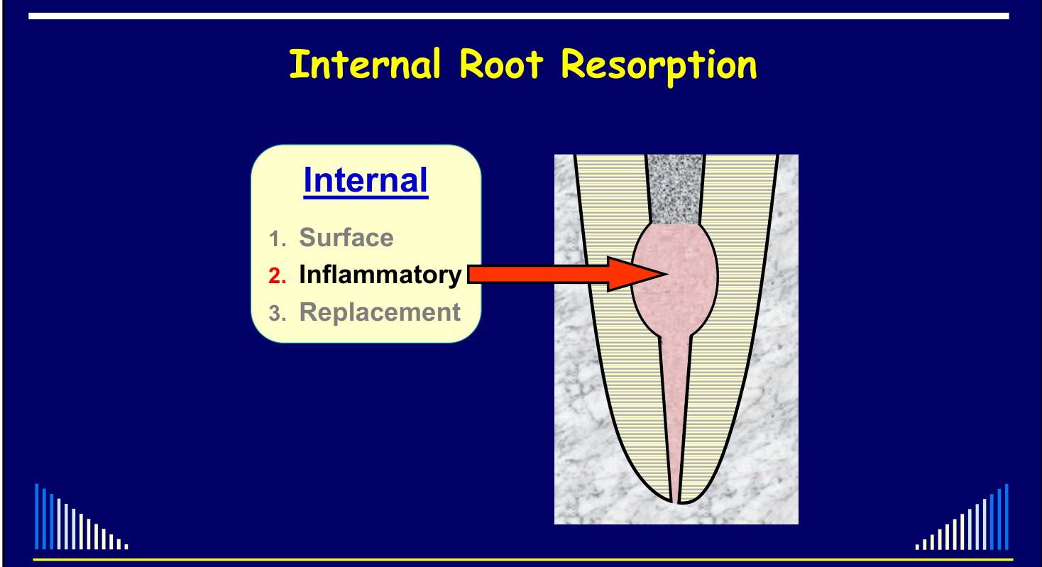
Internal Inflammatory Resorption
Features and Possible Causes14
 |
Features15
- A rare condition
- 0.5% of 2000 patients (Abbott - J Endod 1994)
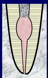
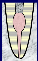
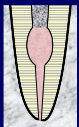
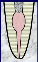
- Usually described as a radiolucent “oval-shaped” increase of the pulp space
- But it can be any shape and in any tooth
- Can occur anywhere along the length of the canal
 |  |  |
 |  |  |
 |  |  |
 |
Possible Causes
- ? Trauma to the tooth
- ? Necrosis + infection of the coronal pulp
- Due to caries, cracks, fractures, restoration breakdown, etc.
BUT - aetiology is not always known or obvious
Çalışkan & Türkün - Endod Dent Traumatol 1997
In this selected 28-tooth series (resorption type unspecified), histories of trauma, caries, periodontal disease and no identified cause were recorded; these histories do not establish causal proportions. Full figures and limitations: Caliskan and Turkun 1997 - Internal Resorption Case Series.
Wedenberg and Zetterqvist - J Endod 1987
In 13 selected extracted human teeth with internal inflammatory resorption, caries history was reported more often than trauma; overlapping histories and extraction-based selection do not establish causal proportions or population frequency. Full study data: Wedenberg and Zetterqvist 1987 - Human Internal Resorption Histology.
Pathogenesis
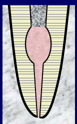
Requirements for Internal Inflammatory Resorption16
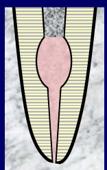
For internal inflammatory resorption - need:
- Bacteria in the pulp (Wedenberg & Lindskog 1985)
- The odontoblast layer and the predentine of the canal wall must be damaged
- This exposes the mineralised dentine to clastic cells
The exact injurious events needed to cause this damage is not known / understood:
- Could be anything that irritates the pulp
- e.g. trauma, caries, heat, restorative procedures, orthodontics, cracks, idiopathic dystrophic changes
 |  |
Experimental Evidence
See - Experimental Monkey Model
See the local model summary at Experimental Internal Resorption in Monkey Teeth and the full study record at Wedenberg and Lindskog 1985 - Internal Resorption Model.
Internal Resorption in Human Teeth17
A selected human histology series examined 13 extracted teeth with internal inflammatory resorption; all had infected root canal systems and defects in the middle or apical third. The selected sample limits generalization. Full study context and findings: Wedenberg and Zetterqvist 1987 - Human Internal Resorption Histology.
Histological Findings18
See - Histological Findings
Full microscopy findings and the original PDF page mapping are preserved in Wedenberg and Zetterqvist 1987 - Human Internal Resorption Histology.
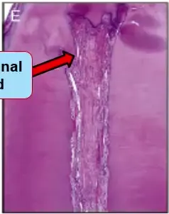
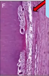
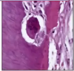
Histological Slides
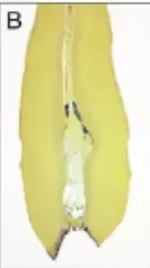
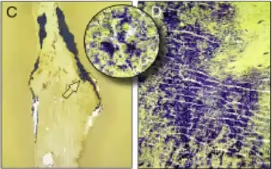
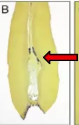
From: Internal Root Resorption: A Review. Patel S, Ricucci D, Durak C, Tay F. J Endod 2010; 36: 1107-21.
- Bacteria in coronal third of root canal
- No odontoblasts, only some predentine, resorption lacunae
- Apical third of canal - Pulp inflamed
- Odontoclast with multiple nuclei
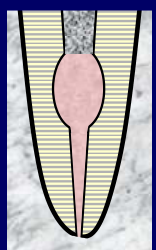
Summary Pathogenesis19
- Coronal to the resorption defect
- Necrotic and infected pulp tissue
- Within the resorptive defect
- Granulation tissue with clastic cells
- Apical to the resorption area
- The pulp will be irreversibly inflamed or replaced with “periodontal-like connective tissue”
- Unless the resorption is no longer active:
- Then the entire canal will be pulpless and infected
 | |
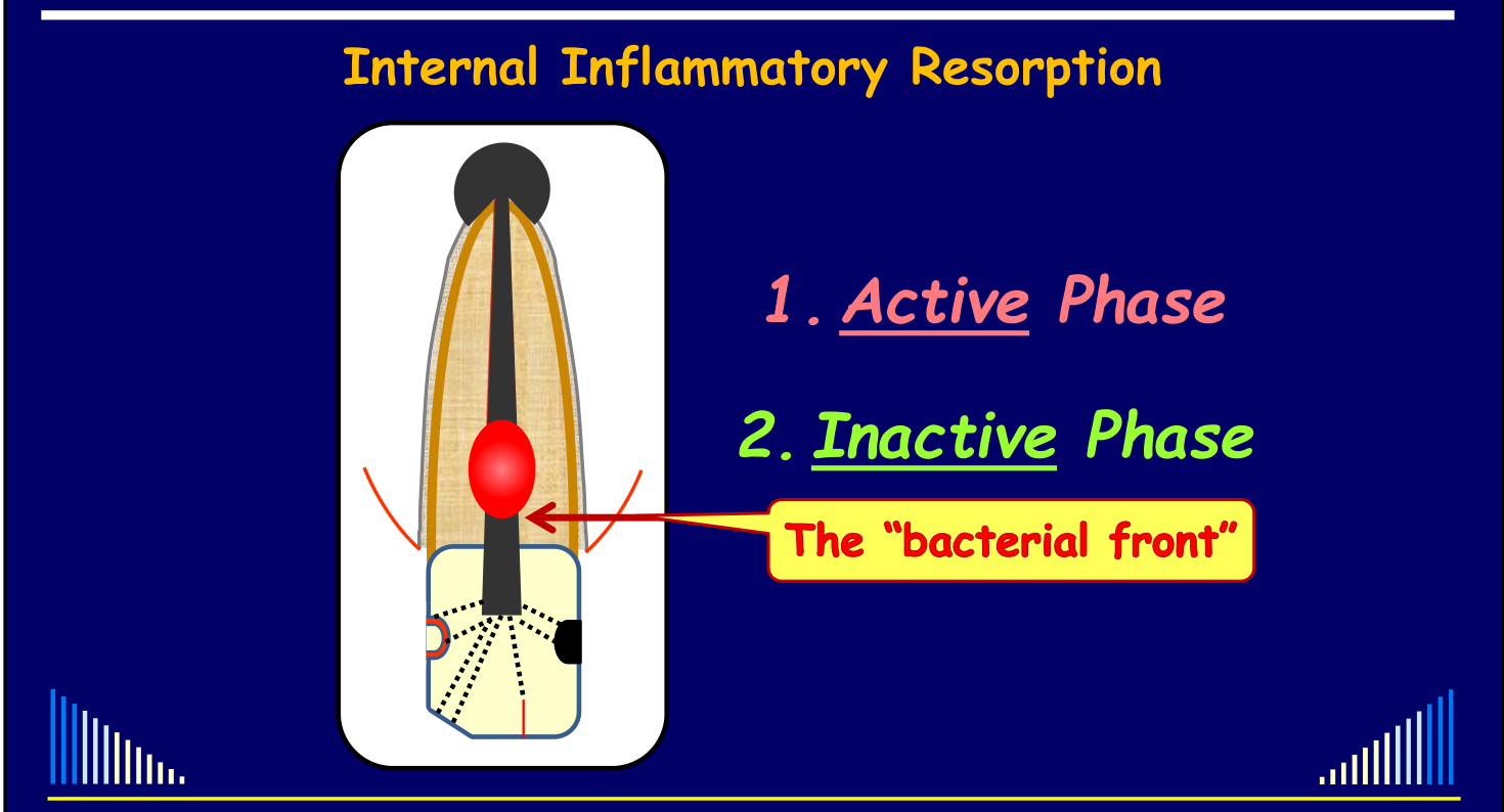
Progression and Phases
- Progression due to interaction between the bacteria and the tissue remaining apically - until it necroses
- The “bacterial front” gradually moves apically through the root canal until all the pulp necroses
- As occurs in all teeth with pulp disease
- Hence, this type of resorption has two phases:
- Active phase
- Inactive phase
Diagnosis20
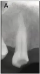
Radiographic Appearance21
Mainly based on the radiographic appearance:
- Radiolucent area within the tooth showing enlargement of the root canal space
- The shape and size are variable
- May or may not have a periapical radiolucency
 | |
Clinical Features
- Usually the same as for teeth with pulpless, infected root canal systems (e.g. no response to pulp tests, etc.)
- Periapical conditions can vary
- Chronic apical periodontitis, chronic apical abscess, acute apical periodontitis, etc, etc.
Differentiating from External Invasive Resorption22
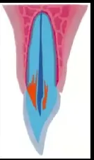
Need to differentiate from External Invasive Resorption (cervical resorption):
- INTERNAL INFLAMMATORY Resorption
- EXTERNAL INVASIVE Resorption
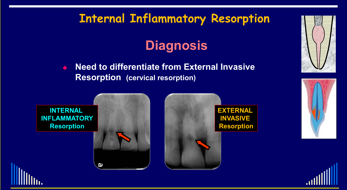 |  |
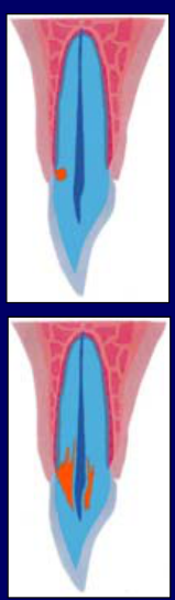 |
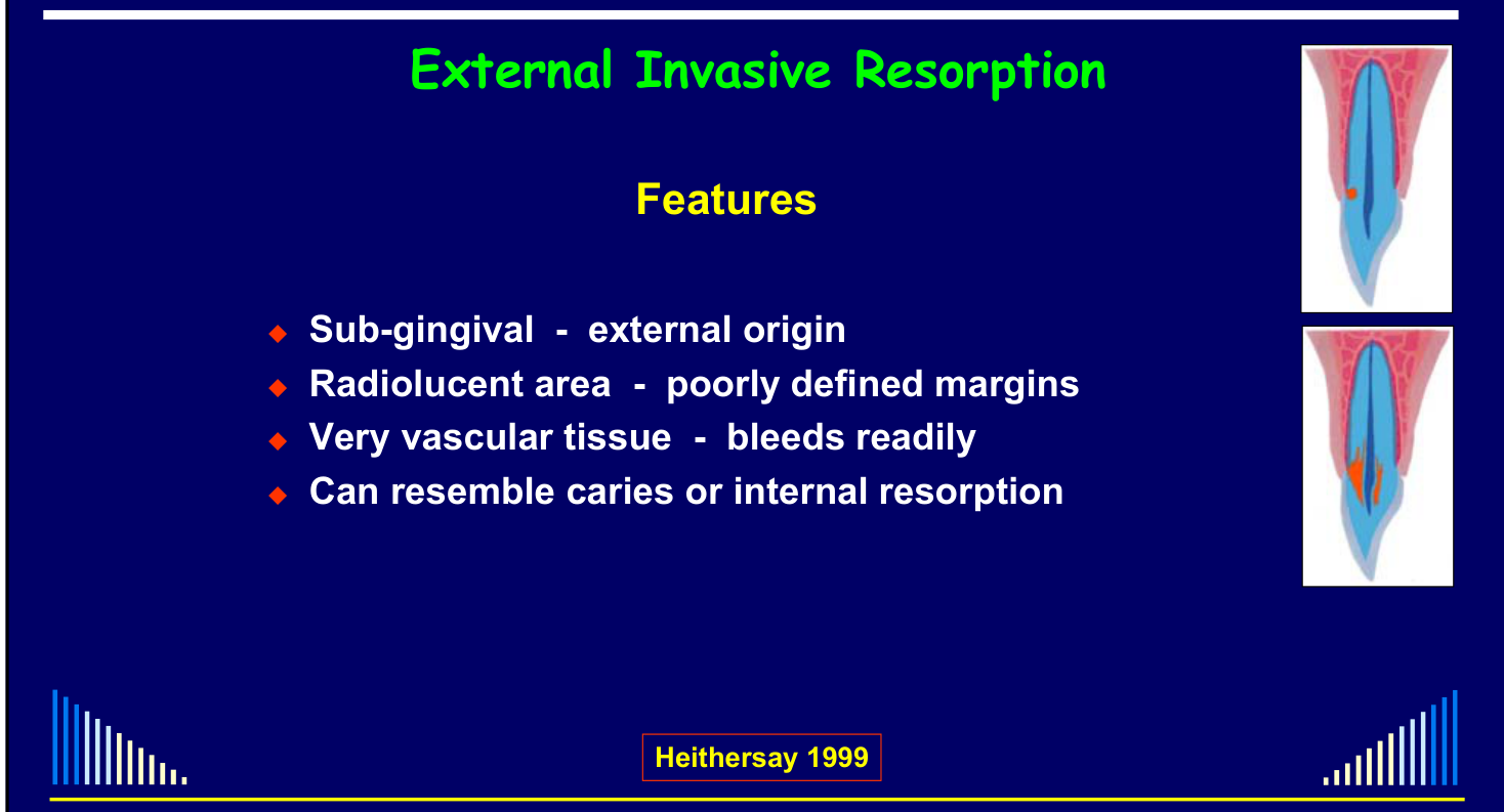
External Invasive Resorption - Features
(Heithersay 1999)
- Sub-gingival - external origin
- Radiolucent area - poorly defined margins
- Very vascular tissue - bleeds readily
- Can resemble caries or internal resorption
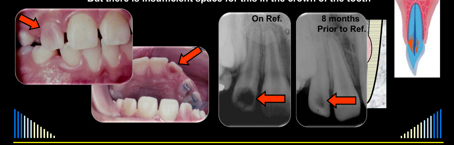
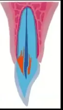
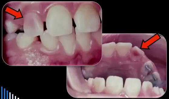
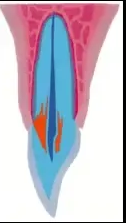
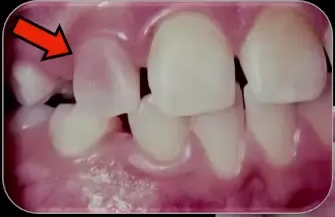
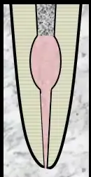
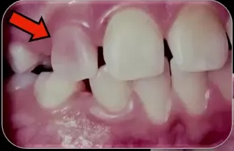
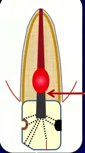
”Pink Teeth”23
“Pink teeth” are most likely due to external invasive resorption — i.e. NOT internal inflammatory resorption:
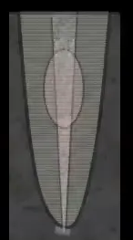
- Since internal inflammatory resorption needs necrotic & infected pulp tissue coronal to the resorbing area to stimulate the resorption
- But there is insufficient space for this in the crown of the tooth
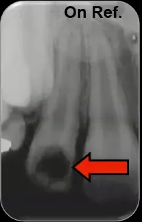
(On Ref. / 8 months Prior to Ref. — radiograph labels)
 |  |
Related diagnosis
L9 retains the fuller external-invasive features L9 Resorption 5 - External Invasive > Clinical Features and canal-outline comparison L9 Resorption 5 - External Invasive > Referred as “Caries”. Topic links: Internal and External Resorption Diagnosis.
Management - “The Resorption Triad”
Consider these when planning management:
- Resorption — can not control if the resorption has started
- Continual stimulation — break down of the natural barriers
- A blood supply — remove to stop resorption
**Management of Internal Inflammatory resorption **
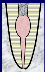
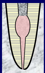
Management Steps24
- Remove the pulp (if any present)
- Active phase - necrotic coronally, inflamed / conn. tissue apically
- Inactive phase - canal will be pulpless
- Thoroughly clean and prepare the root canal
- Aim to remove resorbing cells and blood supply
- Copious irrigation - esp. with 1% NaOCl
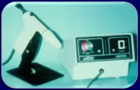
- Ultrasonics may help irrigant to flow into defect
- Medicate the root canal system
- To inhibit & destroy the clastic cells and remaining bacteria
 |  |
Options for Intra-canal Medicaments
- Corticosteroid - Antibiotic (CS-AB) compounds
- Calcium hydroxide
- Combination ⇒ CS-AB paste + Ca(OH)₂
Use of CS-AB Paste and Calcium Hydroxide
CS-AB Paste - Ledermix Paste Components
See - Ledermix formulation
The canonical formulation and concentrations are in L7 Ledermix Paste. Topic links: Ledermix Paste in Resorption.
Properties of the Components
Corticosteroids:
- Anti-inflammatory
- Anti-resorptive effects
- Direct inhibition of clastic cells
- Prevent attachment of clastic cells
- Promote healing
- Biocompatible
Tetracyclines:
- Anti-microbial
- Anti-resorptive effects
- Direct inhibition: clastic cells, collagenase
- Reduce replacement resorption & ankylosis
- Promotes PDL healing
Management Sequence
- Remove the pulp (if any present)
- Thoroughly clean and prepare the root canal
- Initially use CS-AB paste for 4 weeks
- Reduce inflammation
- Inhibit clastic cells
- Anti-bacterial
Calcium Hydroxide Phase
- Then use Ca(OH)₂
- Use 2 or 3 times - for 4 weeks each time
- Anti-bacterial
- Dissolves necrotic tissue
- Kills resorbing cells
- Inactivates LPS
- Encourages hard tissue repair
- Esp. if the resorptive lesion has perforated the root
- Will need extended treatment time
- Or may be able to repair with MTA
- Esp. if the resorptive lesion has perforated the root
Root Canal Filling and Follow-Up
(Andreasen & Andreasen 1994)
Root Canal Filling
- Root canal filling - GP and sealer
- Use a thermoplastic technique for GP
- e.g. Obtura, vertical compaction, etc
- Or: lateral condensation apically and then warm GP in resorption defect
Follow-Up - All Cases
- Clinical and radiographic reviews are essential
- Initially: 6 months after the RCF is completed
- Then: every 3-4 years
- For as long as possible !!!
- Review timing may be adjusted to the healing response and the patient’s circumstances (e.g. a second review after another 12 months)
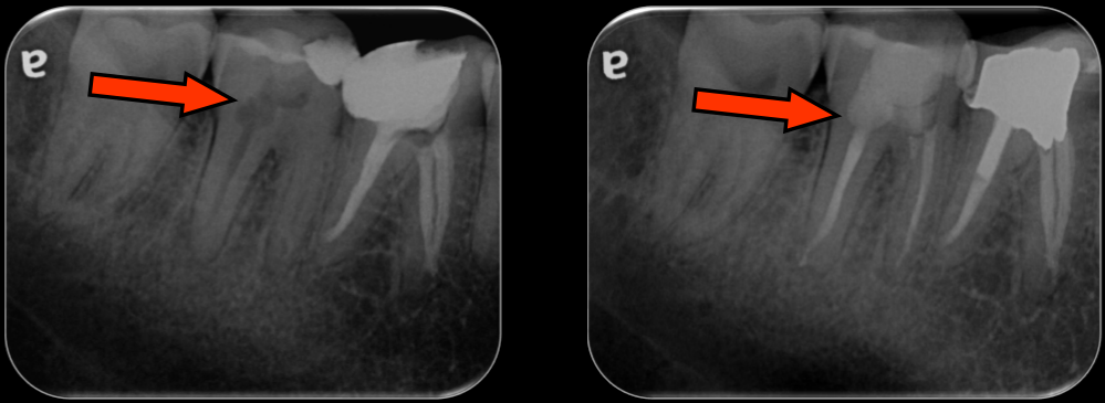
Clinical Case Examples2526
-
Pre-op
-
Post-op
-
1 Year Review
-
Pre-op
-
Post-op
-
1 Year Review
 | 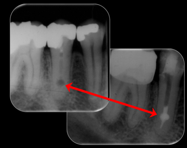 | 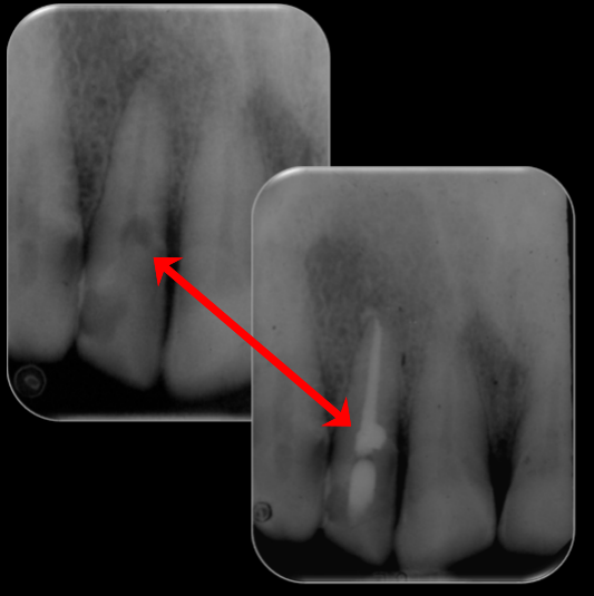 |
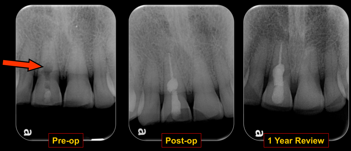 | 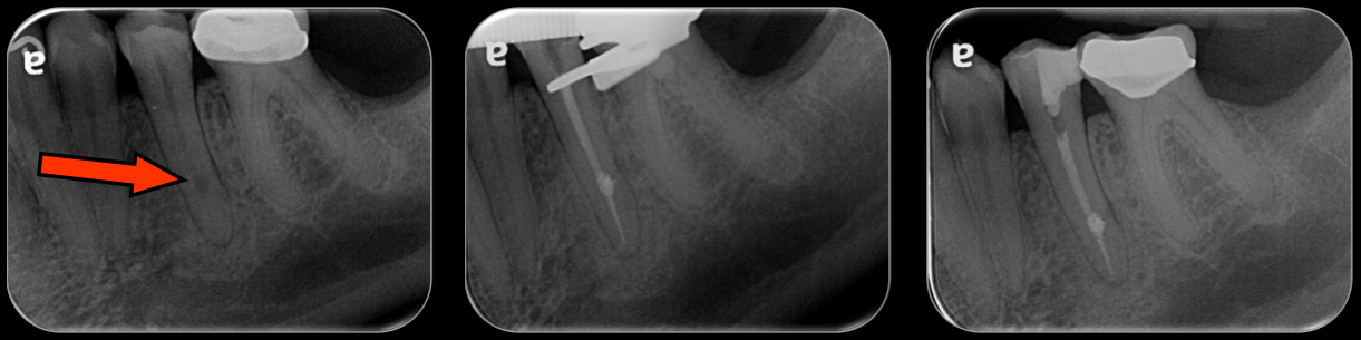 |
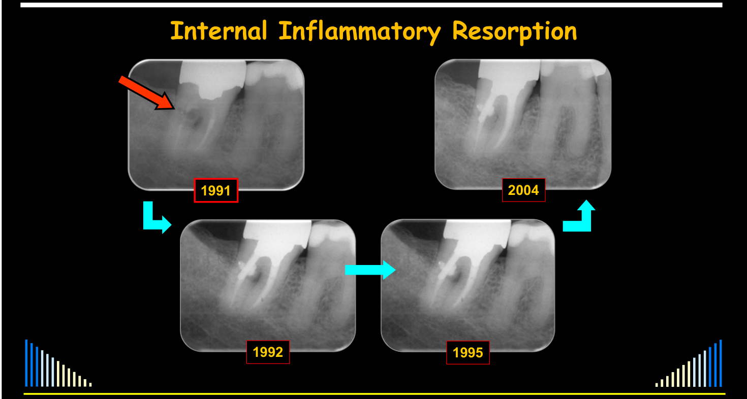
Case Timeline27
-
1991
-
1992
-
1995
-
2004
-
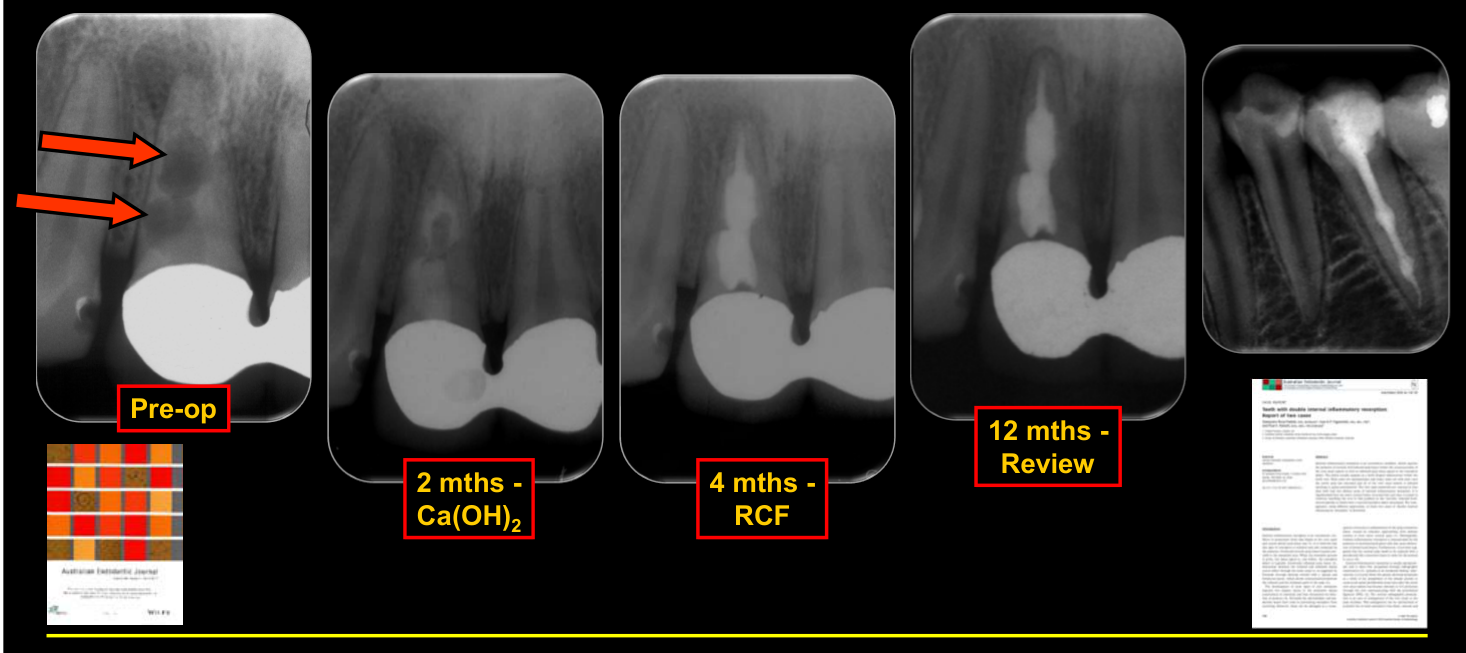
Pre-op
-
2 mths - Ca(OH)₂
-
4 mths - RCF
-
12 mths - Review
Rossi-Fedele G, Figueiredo JAP, Abbott PV. Aust Endo J 2010; 36: 122-129.
Case — Distribution and Follow-Up
Several examples showed that internal inflammatory resorption can occur in the apical, middle, or coronal third, including in posterior teeth. One case had incomplete root canal treatment with calcium hydroxide left in the tooth; treatment was later continued, the resorptive defect was filled, periapical repair was achieved, and the case had a nine-year follow-up.
 |  |
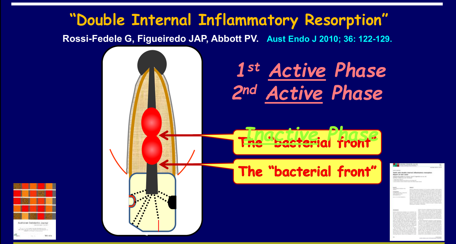
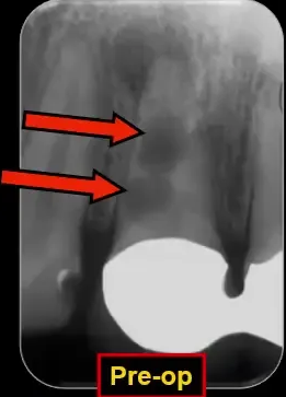
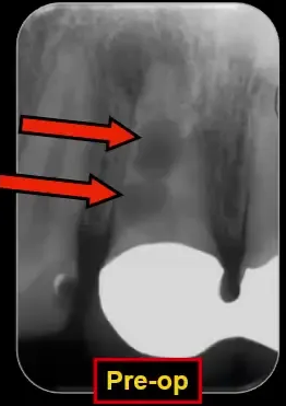
”Double Internal Inflammatory Resorption”28
Rossi-Fedele G, Figueiredo JAP, Abbott PV. Aust Endo J 2010; 36: 122-129.
- 1st Active Phase — the “bacterial front”
- Inactive Phase
- 2nd Active Phase — the “bacterial front”
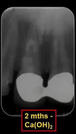
Case — Double Resorption Defects
A published case showed two separate internal inflammatory resorption defects in one root. Calcium hydroxide did not completely fill either defect after the first dressing, reinforcing the need for more than one intracanal calcium hydroxide dressing.
- Following root filling, the total treatment time was approximately four months.
- At the 12-month review, the large periapical radiolucency had reduced and progressive healing with good periapical repair was present.
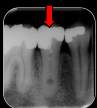
- A second lower premolar treated by the lecturer’s colleagues also had two areas of internal inflammatory resorption.
- The proposed sequence was a first active phase producing the coronal defect, progression of the bacterial front producing a second more apical active phase, and then an inactive phase in which the entire canal became pulpless and infected.
- The resulting periapical radiolucency indicated chronic apical periodontitis.
 |  |
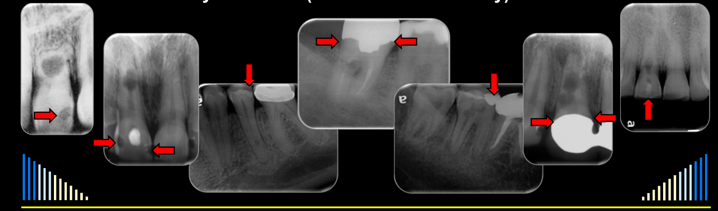 |
Interesting Observation
- All of these cases had caries or restorations breaking down
- No history of trauma (and trauma is unlikely) in these cases
Lecturer — Aetiology in Cases
The lecturer suggested that internal inflammatory resorption may more commonly be associated with infected root canal systems caused by caries, breakdown of restorations, cracks, or fractures. Trauma may be overemphasized as a cause, particularly in posterior teeth where trauma was considered unlikely.
Prognosis and Treatment Outcomes
Prognosis29
- Very few studies - due to the rare occurrence
- Generally considered to have good or excellent prognosis
- After root canal treatment
- If no perforation, and
- If tooth not weakened too much - through loss of tooth structure
| |
Treatment Outcomes
Çalışkan & Türkün - Endod Dent Traumatol 1997
Among the root-canal-treated teeth in this selected 28-tooth series, non-perforated cases had favourable outcomes, while perforated cases often required prolonged calcium hydroxide and sometimes surgery. The small series does not establish general prognosis; full outcome denominators: Caliskan and Turkun 1997 - Internal Resorption Case Series.
Lecturer — Tissue Progression
Histological findings can vary along the same root.
- In one area, a thin layer of bone-like tissue lined the canal wall while a relatively normal pulp remained.
- More apically, a thicker layer lined the canal wall and only a narrow pulp space remained.
- The condition may progress until the root is extensively or entirely replaced by bone.
Lecturer — Diagnostic Findings
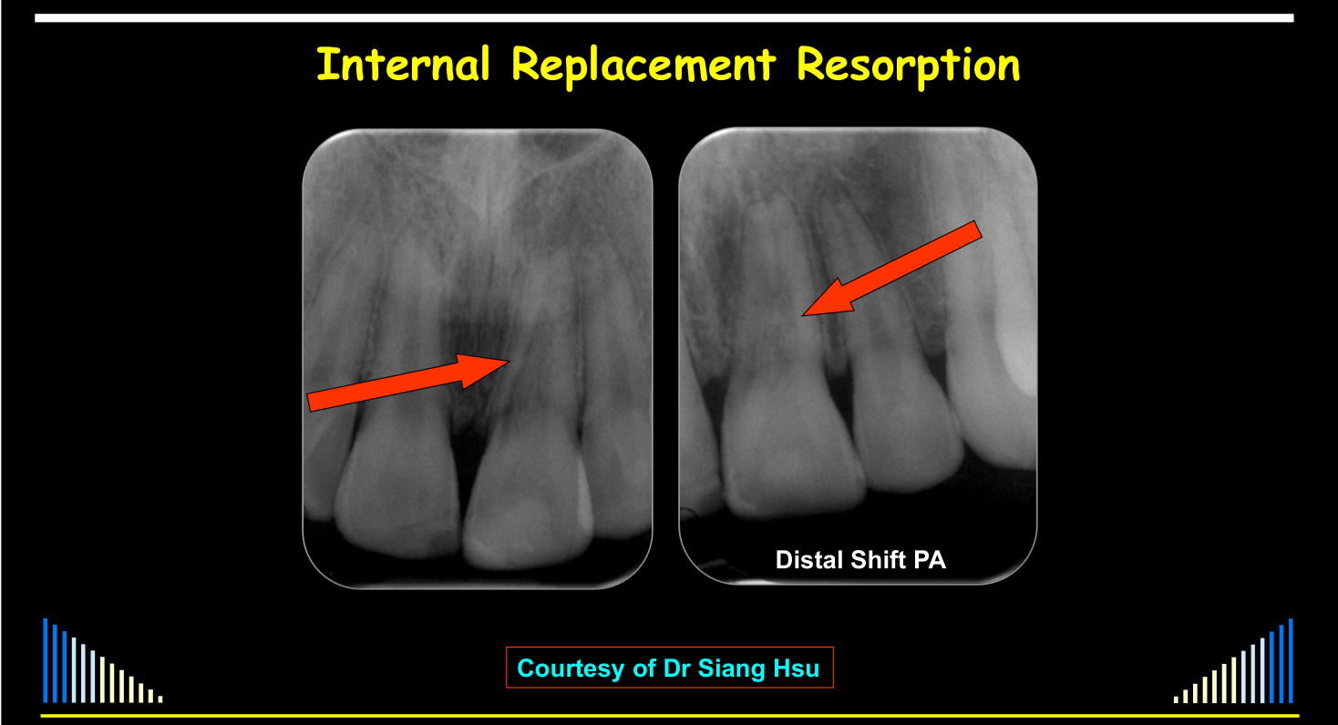
Teeth may be symptomatic as well as asymptomatic, and diagnosis is often incidental after radiographs taken for another reason.
- A distal tube-shift radiograph showed that the resorptive defect did not shift relative to the root canal, indicating that it was internal.
- Its bone-like appearance supported a diagnosis of internal replacement resorption.
Internal Replacement Resorption
Features and Possible Causes
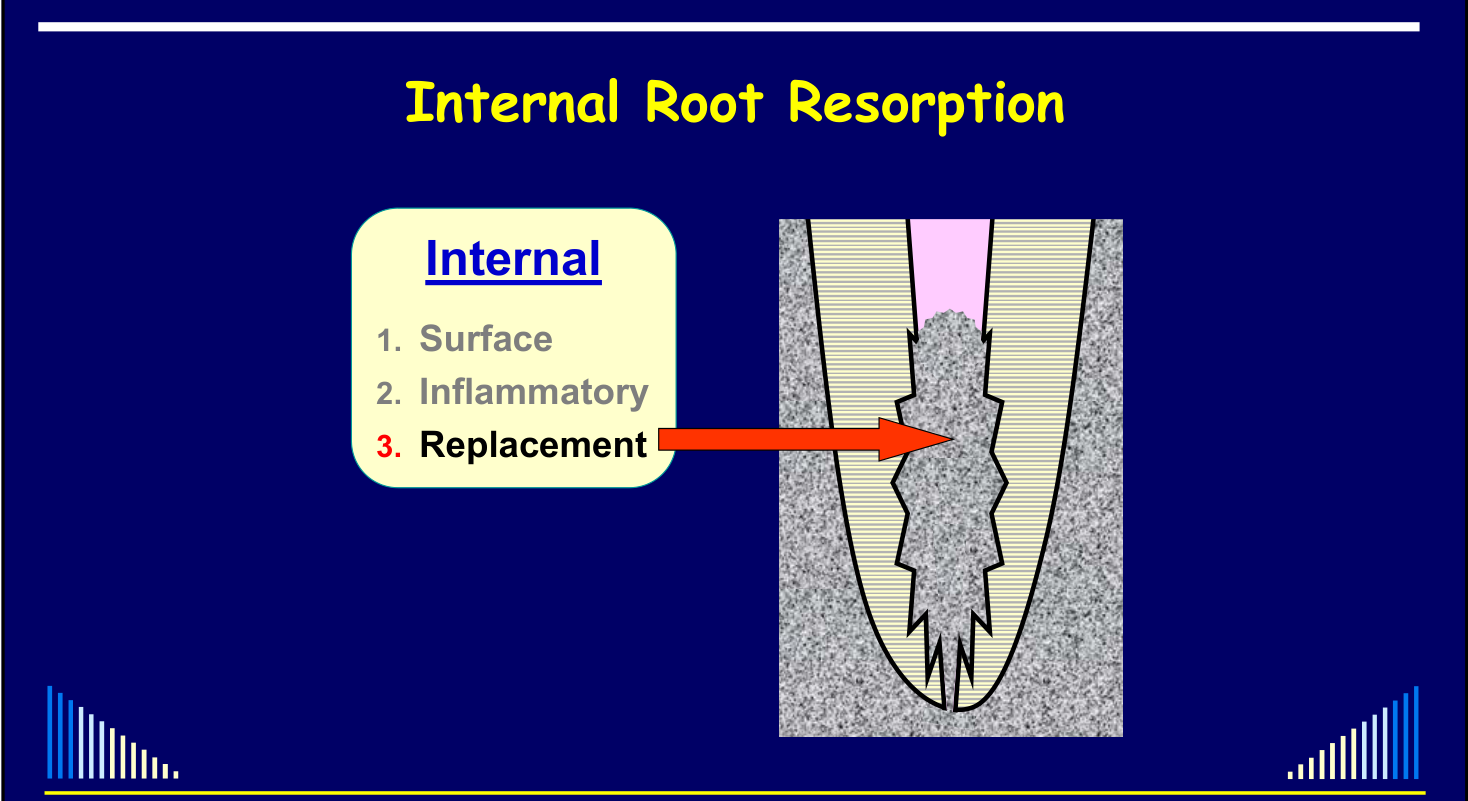
Internal Root Resorption
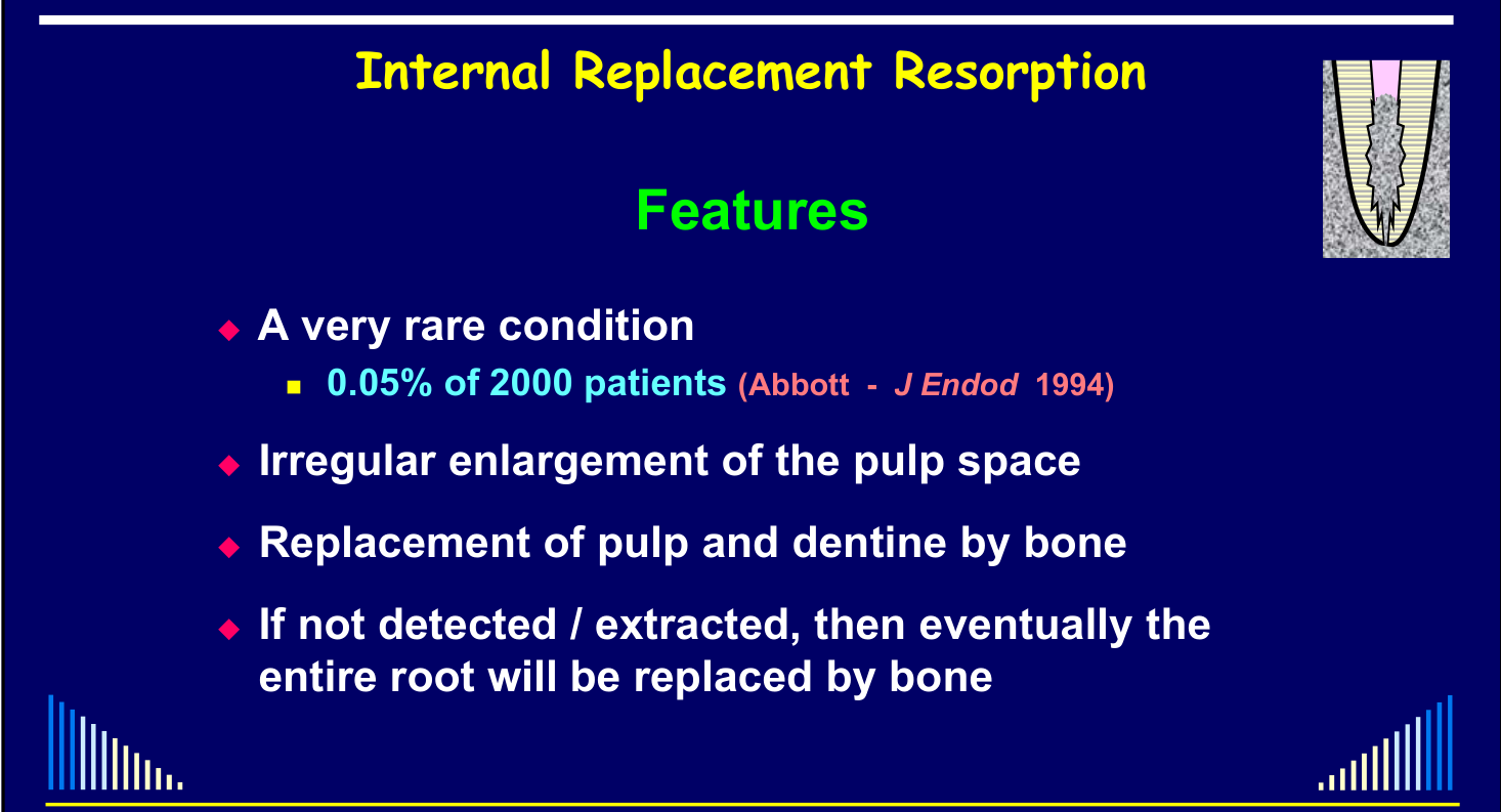
Internal Replacement Resorption — Features30
- A very rare condition
- 0.05% of 2000 patients (Abbott - J Endod 1994)
- Irregular enlargement of the pulp space
- Replacement of pulp and dentine by bone
- If not detected / extracted, then eventually the entire root will be replaced by bone
 |  |
Pathogenesis and Diagnosis
Possible Causes
- Largely unknown
- May be related to trauma
- But not all cases have a history of trauma
- A very rare condition
- Therefore, little data and no research available
Internal Replacement Resorption — Pathogenesis
- Largely unknown
- Suggested that there needs to be long-term low-grade pulpitis causing damage to, or loss of, the odontoblast layer and predentine (Patel et al 2010)
- Similar to internal surface and inflammatory resorption (Wedenderg & Lindskog 1987)
- Histologically - resorption of dentine is followed by deposition of a metaplastic hard tissue that resembles bone or cementum (Patel et al 2020)
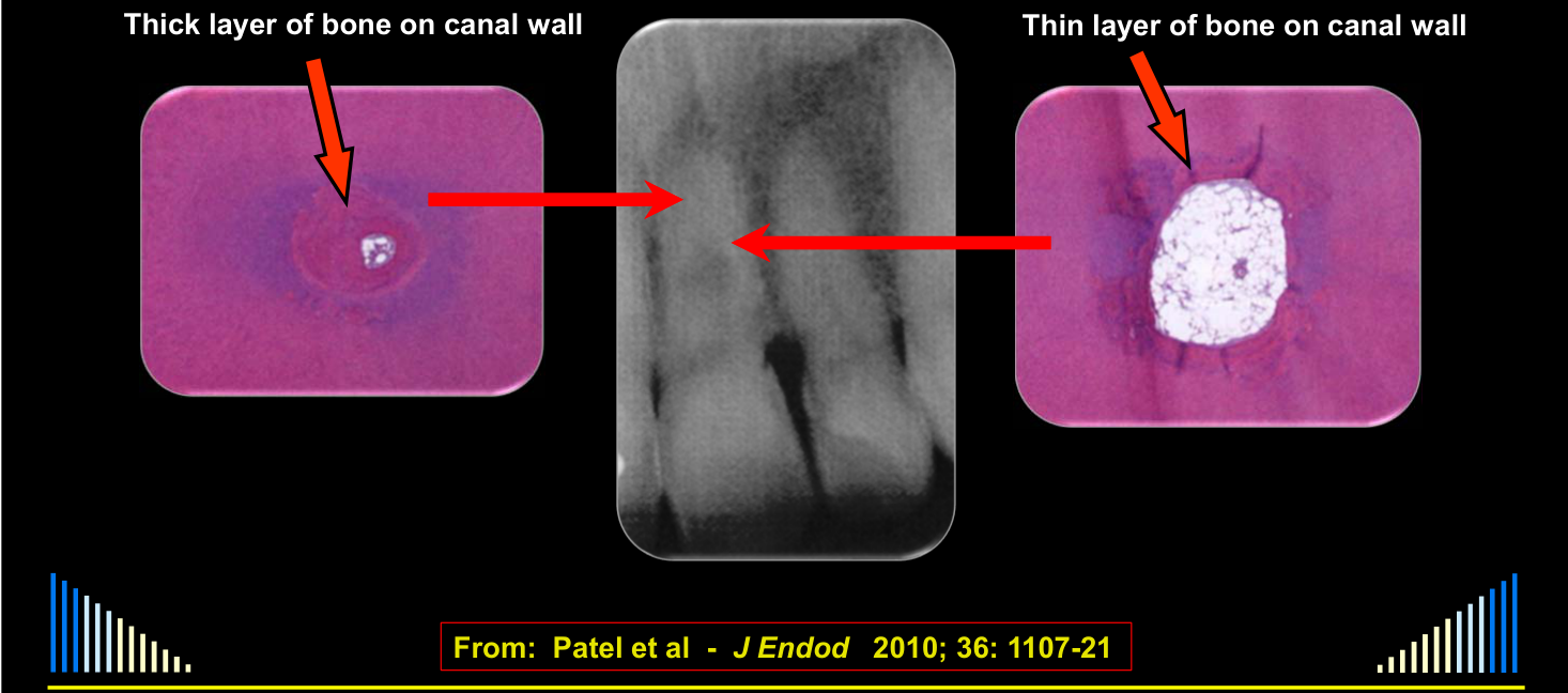
From: Patel et al - J Endod 2010; 36: 1107-21
- Thick layer of bone on canal wall
- Thin layer of bone on canal wall
Diagnosis31
- Asymptomatic
- Tooth may or may not respond to pulp sensibility tests
- Usually discovered incidentally on a radiograph
- Diagnosis is based on the radiographic appearance
- Irregular enlargement of the pulp space
- And the presence of bone replacing the pulp and dentine
- Diagnosis is based on the radiographic appearance
- Need to differentiate from External Invasive Resorption
- Probing, tube-shift PA radiographs, CBCT, etc.
Management32
 |  |
 |
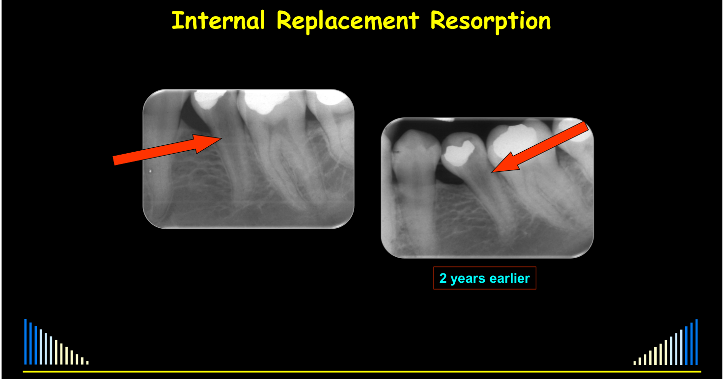
Radiographic Appearance33
- Distal Shift PA
- Internal Replacement Resorption
 |  |
Management
- Endodontics might help
- But only if diagnosed at a very early stage
- Which is rare
- But only if diagnosed at a very early stage
- Usually just monitor - especially radiographically
- Eventually may need extraction
- Potential sequelae:
- Ankylosis
- Crown or Root fracture
- Periodontal problems
- Potential sequelae:
Summary
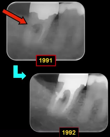
Resorption – Introduction, General Principles, Classification; Internal Resorption — W/Prof. Paul V. Abbott AO
2 years earlier
Internal Resorption is very rare: 0.05 – 0.5% of an Endodontist’s referrals.
Three Types of Internal Resorption34
- Surface
- Inflammatory — the most common type
- Replacement
Lecturer — Internal Types
The lecturer further characterized each internal resorption type.
- Internal surface resorption is usually detectable only histologically and may be transient and self-limiting.
- Internal inflammatory resorption is associated with exposed dentine, clastic cells, and bacteria, and may occur anywhere along the root canal.
- Internal replacement resorption is extremely rare and replaces pulp and dentine with bone-like tissue; it may eventually replace the entire root.
Management
- Surface
- NIL required
- Inflammatory
- Root canal treatment
- Use CS-AB + Ca(OH)₂ medicaments
- Root canal treatment
- Replacement
- Monitor
- ? RCT (?? early cases only)
- Eventual extraction
Lecturer — Management Details
Management depends on the behavior and extent of the resorption.
- Suspected surface resorption should be monitored radiographically; continued progression suggests internal inflammatory resorption.
- Inflammatory resorption requires removal of pulp and infected tissue through thorough instrumentation, sodium hypochlorite irrigation, and ultrasonics.
- Corticosteroid-antibiotic paste is followed by repeated calcium hydroxide dressings, and the irregular defect is commonly filled using a thermoplastic gutta-percha technique before clinical and radiographic review.
- Root canal treatment for replacement resorption may be considered only when detected very early.
Prognosis
- Surface
- Excellent / Transient
- Inflammatory
- Usually good if no perforation
- Uncertain if perforation
- Replacement
- Most will eventually need extraction
Lecturer — Prognostic Details
The prognosis reflects the progressive behavior of each resorption type.
- Surface resorption is considered to have an excellent prognosis because it is believed to be transient.
- Perforation makes the outcome of inflammatory resorption uncertain and less predictable.
- With replacement resorption, most teeth eventually require extraction as the resorption progresses through the root.
Clinical Cases
Case: Histology of internal surface resorption in dog teeth
Question
Scenario: A 1981 histological study on dog teeth investigating the effects of hydrogen peroxide applied to the enamel surface to induce internal surface resorption.
What’s shown: Histological images of the teeth at different time points (3 days, 15 days, and up to 60 days) after the bleaching procedure.
Consider: What are the histological findings at each time point, and what do they indicate about the nature of the resorption?
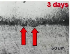
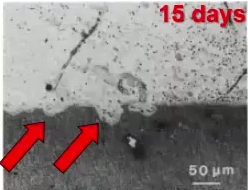
Answer
Observations:
- At 3 days, internal surface resorption is occurring with active dentinoclasts (transcribed as “dentin class”) present.
- At 15 days, the resorption is more advanced.
- At 60 days, the resorption has stopped and healing is evident.
Reasoning: The progression and subsequent cessation of resorption with evident healing indicate that internal surface resorption is likely a transient condition that can self-limit if the irritant is removed.
Outcome: The study demonstrates that the resorption is transient and heals over time when the irritant is no longer present.
Takeaway: Internal surface resorption can be a transient, self-limiting condition when the irritant is removed, though it is rarely diagnosed clinically or radiographically.
Case: Clinical photo of hard tissue forming in an anterior tooth
Question
Scenario: A clinical photo from a study showing an anterior tooth, illustrating the healing phase of a resorptive process in a sealed, non-infected group.
What’s shown: A clinical image of an anterior tooth.
Consider: What is visible in the image and what does it indicate about the resorptive process in this group?
Answer
Observations:
- Hard tissue formation is visible in the image.
Reasoning: The presence of hard tissue forming indicates that the resorptive process was transient and that healing is occurring, which contrasts with the unsealed, infected group that experienced extensive and prolonged colonization of resorbing cells.
Outcome: The image demonstrates the transient nature of the resorption and the subsequent healing and hard tissue formation when bacteria are excluded.
Takeaway: When the irritant (such as bacteria) is absent, the resorptive process can be transient, leading to hard tissue formation and healing, whereas the presence of bacteria leads to internal inflammatory resorption.
Case: Internal inflammatory resorption, histological sections
Question
Scenario: A tooth with internal inflammatory resorption from a published paper (Patel and colleagues, histology by Ricucci) is presented to illustrate the pathogenesis of the condition.
What’s shown: Histological sections (section B and higher magnification) of the tooth root.
Consider: Identify the histological features in the coronal, defect, and apical areas, and determine what they indicate about the disease process.
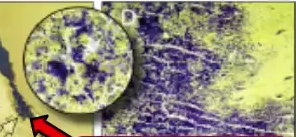
Answer
Observations:
- Coronal third: Bacteria present (dark stained areas) within necrotic and infected pulp tissue.
- Resorptive defect: Granulation tissue containing clastic cells (odontoclasts with multiple nuclei) and resorption lacunae.
- Apical third: Inflamed pulp tissue, absence of odontoblasts, and only some predentine present.
Reasoning: The histological findings demonstrate the active process of internal inflammatory resorption. The bacterial front is located at the junction of the necrotic coronal pulp and the granulation tissue in the defect. Apically, the pulp is either irreversibly inflamed or replaced by periodontal-like connective tissue. As the bacterial front progressively moves apically, it interacts with the remaining tissue until it necroses, leading to an inactive phase where the entire canal becomes pulpless and infected.
Outcome: None stated for this specific case; it is used to illustrate the pathogenesis and phases of internal inflammatory resorption.
Takeaway: Internal inflammatory resorption progresses as a “bacterial front” moves apically through the root canal, transitioning from an active phase (with vital/inflamed tissue apically) to an inactive phase (an entirely pulpless and infected canal).
Case: Radiographs of teeth with resorptive defects, differentiating internal from external
Question
Scenario: Radiographs of teeth with resorptive defects are presented to differentiate between internal inflammatory resorption and external invasive (cervical) resorption.
What’s shown: Periapical radiographs showing resorptive radiolucencies, with one image featuring red lines tracing the root canal outline.
Consider: Identify the radiographic features that differentiate internal inflammatory resorption from external invasive resorption.
Answer
Observations:
- External invasive resorption typically presents as a radiolucency with very poorly defined margins, whereas internal inflammatory resorption generally has more defined margins.
- In the specific case shown, the outline of the root canal (indicated by red lines) can be seen going through, or superimposed over, the resorptive defect.
- Two additional examples also show the root canal clearly superimposed over the defect.
Reasoning: The poorly defined margins suggest an external origin. More definitively, if the root canal outline is visible passing through the resorptive defect, it proves the defect is external to the canal space. Therefore, the condition is external invasive resorption, not internal inflammatory resorption. The lecturer notes that about a third of external invasive resorption cases are initially misdiagnosed as internal resorption due to these similarities.
Outcome: Diagnosed as external resorption.
Takeaway: Differentiating internal from external resorption relies on assessing the margins (poorly defined for external, more defined for internal) and tracing the root canal outline; if the canal outline is superimposed over the defect, it confirms external invasive resorption. Tube shift radiographs can also be utilized to determine if the defect is internal or external.
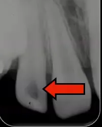
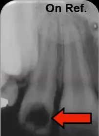
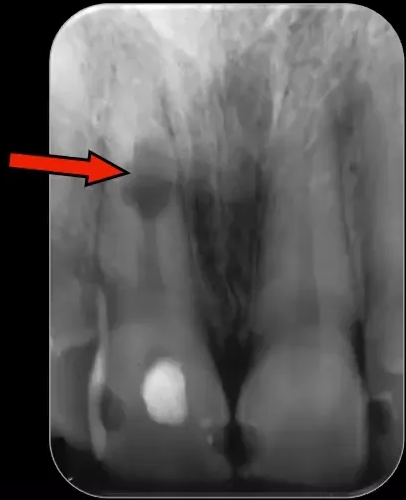
Case: Pink tooth with large coronal radiolucency
Question
Scenario: A patient is referred with a pink tooth and a large radiolucency within the crown of the tooth, which has progressed over an eight-month period.
What’s shown: Clinical photographs of a pink tooth and serial radiographs showing a large radiolucency in the crown.
Consider: What is the most likely diagnosis for a pink tooth, and how can it be radiographically distinguished from internal inflammatory resorption?
Answer
Observations:
- The tooth appears pink clinically.
- Radiographs show a very large radiolucency within the crown of the tooth.
- The radiolucency progressed over a relatively short period of eight months.
- The outline of the pulp space can be traced through the resorptive defect (superimposed).
Reasoning:
- A pink tooth is often mistakenly thought to be a sign of internal inflammatory resorption. However, internal inflammatory resorption requires necrotic and infected pulp tissue coronal to the resorbing area, which cannot occur in the crown due to a lack of space.
- Therefore, a pink tooth is most likely due to external invasive resorption.
- To distinguish between the two, one should look at the outline of the pulp space. In external resorption, the outline of the canal is superimposed over the defect and can be traced through it. In internal resorption, the outline of the pulp joins the radiolucency.
Outcome: None stated for this specific case.
Takeaway: A “pink tooth” is most likely due to external invasive resorption, not internal inflammatory resorption, and can be distinguished by tracing the outline of the root canal through the resorptive defect.
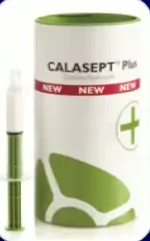
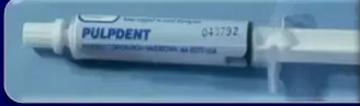
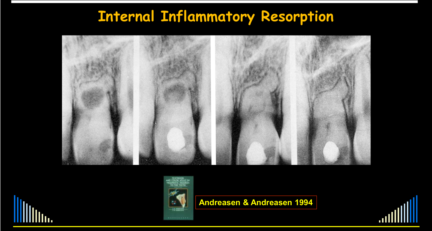
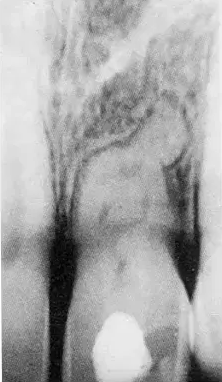
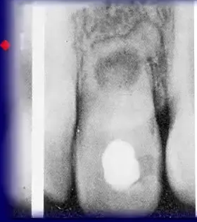
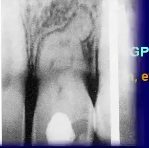
Case: Internal inflammatory resorption, progression of calcium hydroxide treatment
Question
Scenario: A case from Andreasen’s textbook demonstrating the progression of treatment for internal inflammatory resorption using calcium hydroxide.
What’s shown: A series of periapical radiographs showing a resorptive defect prior to treatment, and subsequent radiographs after the successive placement of calcium hydroxide.
Consider: How does the radiographic appearance of the resorptive defect change with successive applications of calcium hydroxide over multiple appointments?
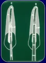
Answer
Observations:
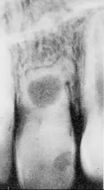
- First radiograph: Shows the resorptive defect prior to any treatment.
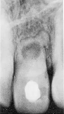
- Second radiograph: Taken after calcium hydroxide was placed in the canal for the first time; the calcium hydroxide does not entirely fill the resorptive defect, leaving a radiolucency around it.
- Third radiograph: After calcium hydroxide is replaced again after another four weeks, the defect is becoming more full of calcium hydroxide, but some areas are not completely filled.
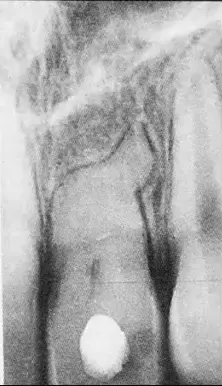
- Fourth radiograph: The resorptive defect is fully filled with calcium hydroxide.
Reasoning:
- Calcium hydroxide helps to remove the last remaining bits of necrotic or other pulp tissue in the resorptive defect.
- This process is progressive and does not happen all at once, requiring multiple dressings over several appointments to have its full effect and completely fill the defect.
Outcome: Following the complete filling of the defect with calcium hydroxide, the root canal is typically filled with gutta percha and cement. A thermoplastic technique with gutta percha is preferred to adequately fill the irregular defect, sometimes using a combination of lateral condensation apically and warm gutta percha in the resorption defect.
Takeaway: Calcium hydroxide requires several appointments and multiple dressings to progressively dissolve remaining tissue and completely fill a resorptive defect in internal inflammatory resorption.
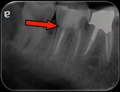
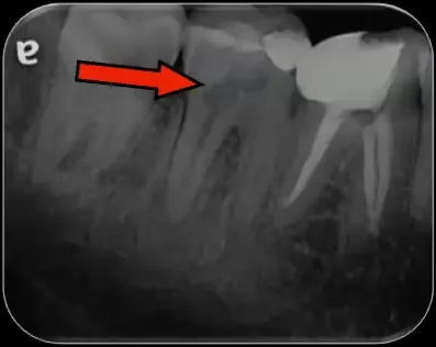
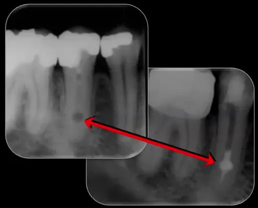
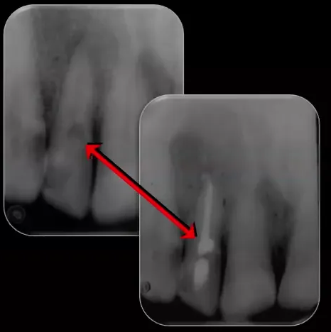
Case: Tooth with internal inflammatory resorption, preoperative and postoperative radiographs
Question
Scenario: A tooth with internal inflammatory resorption, demonstrating that the condition can occur in any tooth, including posterior teeth, and is not always associated with trauma.
What’s shown: Preoperative and postoperative radiographs showing an irregularly shaped radiolucency within the canal.
Consider: How to identify the shape of the resorptive defect and the appropriate obturation technique to fill it.
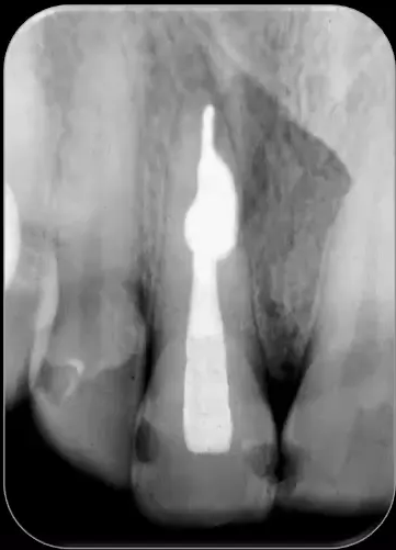
Answer
Observations:
- The radiolucency within the canal is irregular in shape, not just oval.
- The postoperative radiograph shows the canal and defect filled.
Reasoning: Because the defect is irregular, a thermoplastic technique is required to adequately fill it. The lecturer used lateral compaction in the apical part of the canal and an obtura/vertical compaction technique in the resorption defect and remaining canal.
Outcome: The canal and resorption defect were very well filled.
Takeaway: Internal inflammatory resorption can occur in any tooth (including posterior teeth) and is not always associated with trauma; irregular defects require thermoplastic obturation techniques to be adequately filled.
Case: Tooth with internal inflammatory resorption and incomplete prior treatment, 9-year follow-up
Question
Scenario: A patient presented with a tooth containing calcium hydroxide from an incomplete root canal treatment started by another dentist.
What’s shown: Radiographs showing a resorptive defect that was subsequently filled, with a 9-year follow-up showing periapical repair.
Consider: How to manage a tooth with internal inflammatory resorption where treatment was previously abandoned, and what the long-term outcome might be.

Answer
Observations:
- The tooth initially contained only calcium hydroxide from a previously started, but unfinished, root canal treatment.
- The resorptive defect was filled upon completion of the treatment.
- A 9-year follow-up radiograph shows good periapical repair.
Reasoning: Continuing the root canal treatment and adequately filling the defect leads to long-term healing.
Outcome: Treatment was completed, and a 9-year follow-up demonstrated good periapical repair.
Takeaway: Even if root canal treatment for internal inflammatory resorption is started and abandoned by another provider, completing the treatment can result in excellent long-term periapical healing.
Case: Tooth with double internal inflammatory resorption, published case
Question
Scenario: A published case of a tooth with double internal inflammatory resorption, treated and reviewed over a 12-month period.
What’s shown: A series of radiographs showing two areas of inflammatory resorption in the tooth root, the placement of calcium hydroxide, the final root filling, and a 12-month review.
Consider: The challenges of filling multiple resorptive defects with calcium hydroxide and the expected healing timeline.
Answer
Observations:
- Two distinct areas of internal inflammatory resorption are present in the tooth root.
- Calcium hydroxide was placed in the canal, but it was difficult to completely fill the defects initially.
- The tooth was eventually root filled after about four months of treatment.
- A 12-month review shows a fairly large initial radiolucency getting smaller and nicely healed.
Reasoning: The difficulty in completely filling the defects with calcium hydroxide on the first attempt emphasizes the need for multiple intracanal dressings. The progressive reduction in the periapical radiolucency indicates successful healing.
Outcome: Total treatment time was about four months. A 12-month review showed good periapical repair and healing of the initial radiolucency.
Takeaway: Multiple intracanal dressings with calcium hydroxide are often necessary to adequately treat complex internal inflammatory resorption defects, leading to good periapical healing.
Case: Lower premolar with double internal inflammatory resorption, published case
Question
Scenario: A lower premolar from the same published study as the previous case, also presenting with two areas of internal inflammatory resorption.
What’s shown: Radiographs of a lower premolar with two resorptive defects and a periapical radiolucency.
Consider: The pathogenesis and progression of multiple resorptive defects in a single tooth.
Answer
Observations:
- The lower premolar has two areas of internal inflammatory resorption.
- There is a periapical radiolucency indicating chronic apical periodontitis.
Reasoning: The lecturer theorizes that the two defects resulted from two active phases of bacterial progression. The first phase caused the more coronal defect, and as the bacterial front progressed, a second area developed. Eventually, the entire root canal system became pulpless and infected, leading to the inactive phase and the periapical radiolucency.
Takeaway: Multiple internal inflammatory resorption defects in a single tooth can result from sequential active phases of bacterial progression through the root canal system.
Case: Histology of internal replacement resorption
Question
Scenario: Histological images from a study by Patel and Rekucci are presented to illustrate tissue changes within the root canal.
What’s shown: Micrographs showing the canal wall at the mid-root level and more apically.
Consider: What histological features are visible in the pulp space and canal wall at these different levels?
Answer
Observations:
- At the mid-root level, there is a thin layer of bone on the canal wall with a relatively normal-looking pulp inside.
- More apically, there is a thicker layer of bone on the canal wall with a very narrow pulp space remaining.
Reasoning: These images demonstrate the metaplastic type of hard tissue that resembles bone or cementum, which replaces the pulp and dentine in internal replacement resorption.
Outcome: None stated for this exhibit.
Takeaway: Histologically, internal replacement resorption is characterized by metaplastic hard tissue resembling bone or cementum replacing the pulp and dentine, with the pulp space becoming progressively narrower.
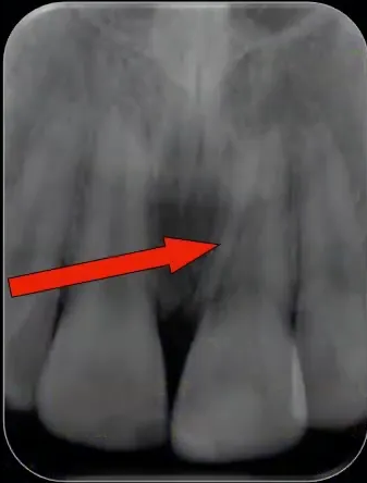
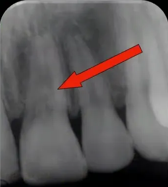
Case: Resorptive defect on tube shift radiograph
Question
Scenario: A radiograph is presented to demonstrate the differentiation between internal and external resorption.
What’s shown: Periapical and distal tube shift radiographs showing a resorptive defect in a tooth.
Consider: How to determine the origin of the resorption and the specific type of internal resorption based on the radiographic findings.
Answer
Observations:
- The distal tube shift radiograph shows no shifting of the resorptive defect with respect to the root canal.
- The tissue filling the enlarged pulp space has a bone-like appearance.
Reasoning: The lack of shifting on the tube shift radiograph indicates that the defect is centered on the root canal, confirming it is of internal origin rather than external. The bone-like appearance of the tissue replacing the pulp and dentine within the enlarged pulp space is characteristic of internal replacement resorption.
Outcome: The condition was diagnosed as internal replacement resorption.
Takeaway: Simple tube shift radiography can differentiate internal from external resorption; a defect that does not shift relative to the root canal on a tube shift radiograph is internal in origin.
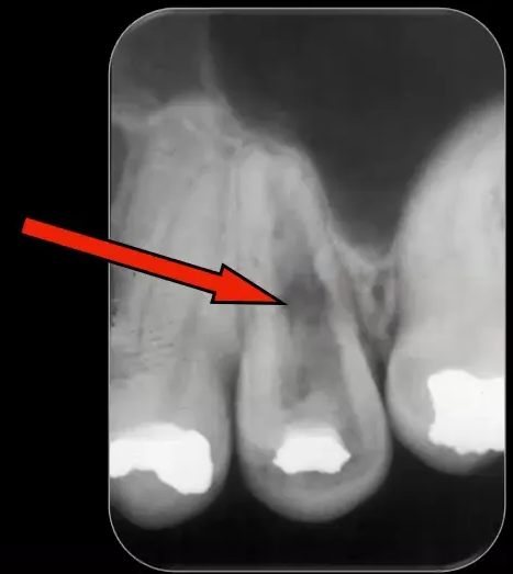
Case: Internal resorption with ankylosis, serial radiographs
Question
Scenario: A patient presented with a clear case of internal resorption. A bitewing radiograph from three years prior was available for comparison with the current presentation.
What’s shown: A bitewing radiograph from three years earlier and a current radiograph showing extensive resorption.
Consider: The progression of the resorption over time and the appropriate management for the tooth.
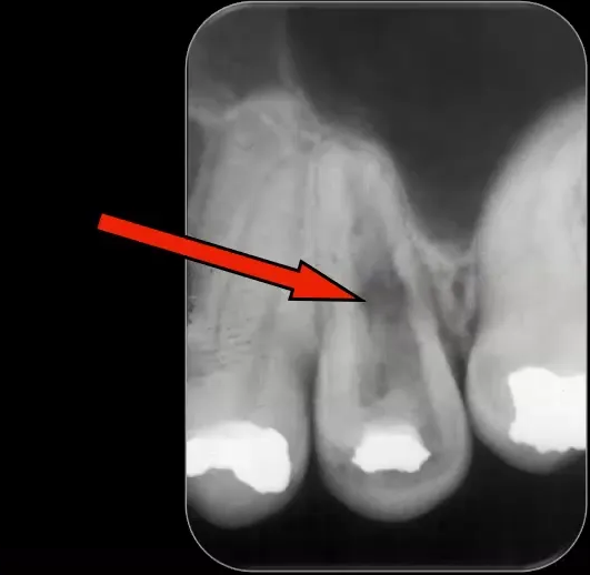
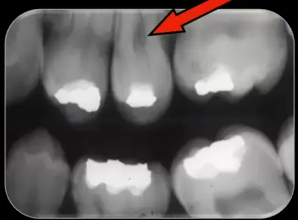
Answer
Observations:
- The bitewing radiograph from three years prior shows the resorption developing and expanding, particularly in the coronal part of the tooth.
- The current radiograph shows extensive resorption and signs of ankylosis.
Reasoning: The resorption has progressed significantly over the three-year period. By the time internal replacement resorption is large enough to be noticed radiographically and shows signs of ankylosis, it is usually too extensive to attempt treatment.
Outcome: The tooth needed to be extracted due to the extensive resorption and ankylosis.
Takeaway: Internal replacement resorption is often only detected incidentally when it is already too extensive for conservative treatment, frequently necessitating extraction.
Case: Asymptomatic incidental internal resorption, serial radiographs
Question
Scenario: An asymptomatic patient had internal resorption noted incidentally on a radiograph. An initial radiograph from two years prior was available for comparison.
What’s shown: An initial radiograph from two years prior and a current radiograph of the affected tooth.
Consider: The progression of the condition over time and the management advice for the patient.
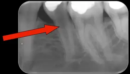
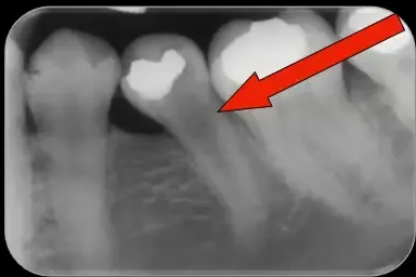
Answer
Observations:
- The patient is completely asymptomatic.
- Comparison of the initial radiograph from two years prior and the current radiograph shows very little change in the resorptive defect, despite slightly different angulations.
Reasoning: The resorption has not progressed significantly over the two-year period. However, because the entire root will eventually be replaced by bone if left untreated, the tooth will ultimately require extraction.
Outcome: The patient was advised to have their dentist monitor the tooth radiographically, with the understanding that it might need to be extracted at some stage. The patient was in the army and relocated frequently, so long-term follow-up was not possible.
Takeaway: Asymptomatic internal replacement resorption found incidentally can be monitored radiographically, with extraction planned for when complications arise, though long-term follow-up may be difficult.
Footnotes
-
Original PDF page 1: L2 Resorption 1 - Introduction, General Internal Resorption, p.1 ↩
-
Original PDF page 2: L2 Resorption 1 - Introduction, General Internal Resorption, p.2 ↩
-
Original PDF page 3: L2 Resorption 1 - Introduction, General Internal Resorption, p.3 ↩
-
Original PDF page 4: L2 Resorption 1 - Introduction, General Internal Resorption, p.4 ↩
-
Original PDF page 5: L2 Resorption 1 - Introduction, General Internal Resorption, p.5 ↩
-
Original PDF page 6: L2 Resorption 1 - Introduction, General Internal Resorption, p.6 ↩
-
Original PDF page 7: L2 Resorption 1 - Introduction, General Internal Resorption, p.7 ↩
-
Original PDF page 8: L2 Resorption 1 - Introduction, General Internal Resorption, p.8 ↩
-
Original PDF page 11: L2 Resorption 1 - Introduction, General Internal Resorption, p.11 ↩
-
Original PDF page 9: L2 Resorption 1 - Introduction, General Internal Resorption, p.9 ↩
-
Original PDF page 10: L2 Resorption 1 - Introduction, General Internal Resorption, p.10 ↩
-
Original PDF page 23: L2 Resorption 1 - Introduction, General Internal Resorption, p.23 ↩
-
Original PDF page 14: L2 Resorption 1 - Introduction, General Internal Resorption, p.14 ↩
-
Original PDF page 16: L2 Resorption 1 - Introduction, General Internal Resorption, p.16 ↩
-
Original PDF page 15: L2 Resorption 1 - Introduction, General Internal Resorption, p.15 ↩
-
Original PDF page 17: L2 Resorption 1 - Introduction, General Internal Resorption, p.17 ↩
-
Original PDF page 18: L2 Resorption 1 - Introduction, General Internal Resorption, p.18 ↩
-
Original PDF page 19: L2 Resorption 1 - Introduction, General Internal Resorption, p.19 ↩
-
Original PDF page 20: L2 Resorption 1 - Introduction, General Internal Resorption, p.20 ↩
-
Original PDF page 13: L2 Resorption 1 - Introduction, General Internal Resorption, p.13 ↩
-
Original PDF page 21: L2 Resorption 1 - Introduction, General Internal Resorption, p.21 ↩
-
Original PDF page 22: L2 Resorption 1 - Introduction, General Internal Resorption, p.22 ↩
-
Original PDF page 24: L2 Resorption 1 - Introduction, General Internal Resorption, p.24 ↩
-
Original PDF page 25: L2 Resorption 1 - Introduction, General Internal Resorption, p.25 ↩
-
Original PDF page 30: L2 Resorption 1 - Introduction, General Internal Resorption, p.30 ↩
-
Original PDF page 31: L2 Resorption 1 - Introduction, General Internal Resorption, p.31 ↩
-
Original PDF page 32: L2 Resorption 1 - Introduction, General Internal Resorption, p.32 ↩
-
Original PDF page 33: L2 Resorption 1 - Introduction, General Internal Resorption, p.33 ↩
-
Original PDF page 34: L2 Resorption 1 - Introduction, General Internal Resorption, p.34 ↩
-
Original PDF page 35: L2 Resorption 1 - Introduction, General Internal Resorption, p.35 ↩
-
Original PDF page 37: L2 Resorption 1 - Introduction, General Internal Resorption, p.37 ↩
-
Original PDF page 39: L2 Resorption 1 - Introduction, General Internal Resorption, p.39 ↩
-
Original PDF page 38: L2 Resorption 1 - Introduction, General Internal Resorption, p.38 ↩
-
Original PDF page 40: L2 Resorption 1 - Introduction, General Internal Resorption, p.40 ↩