L7 Resorption 4 - External Inflammatory
External Inflammatory Resorption: Treatment Effects1
Infection-related (Inflammatory) Root Resorption — Treatment Effects
Prof. Paul V. Abbott AO BDSc, MDS, FRACDS(Endo), FPFA, FADI, FICD, FACD, FIADT
- Specialist Endodontist
- Winthrop Professor of Clinical Dentistry, UWA Dental School, The University of Western Australia
- Oral Health Centre of Western Australia
DMD 3rd Year 2020
 |  |
 |  |
References and Further Reading2
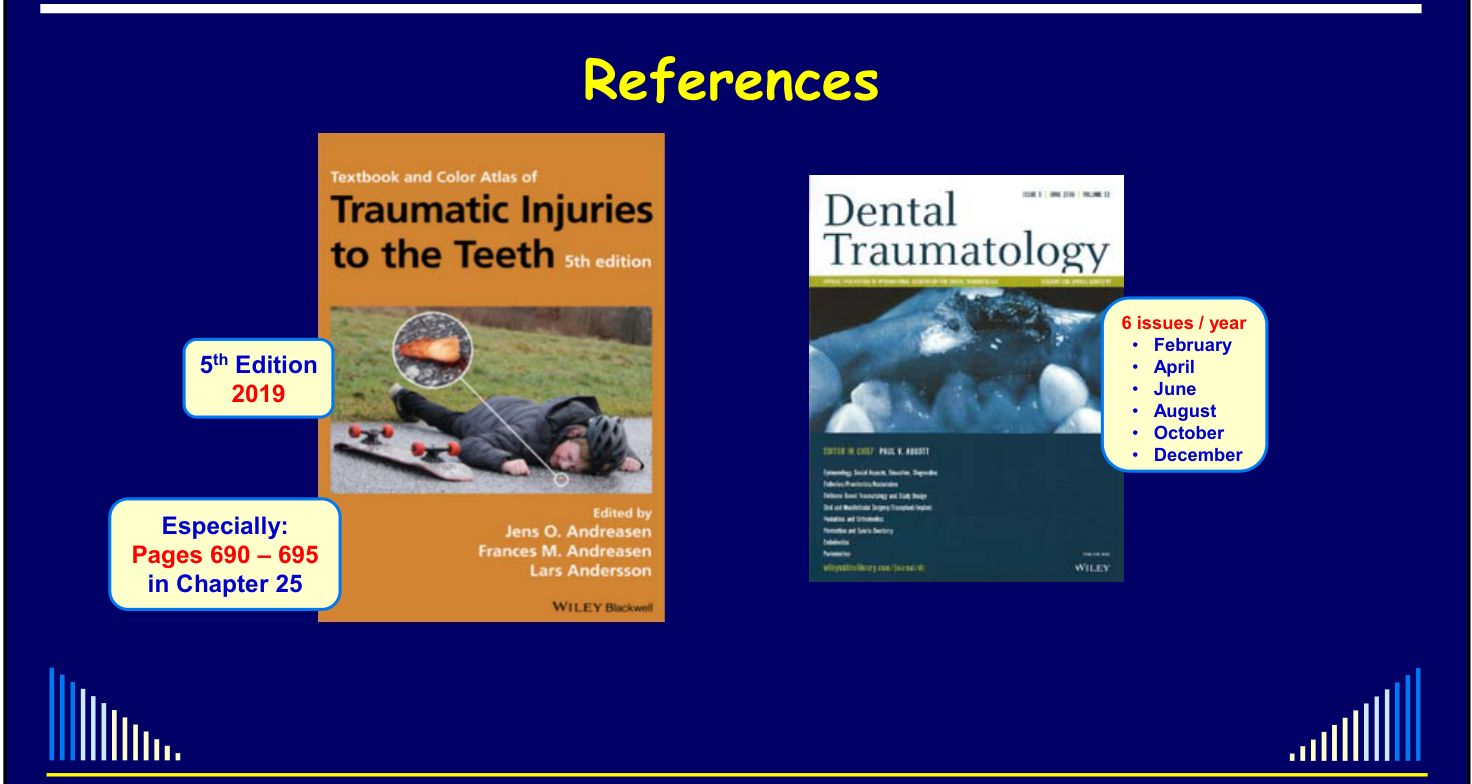
Textbook and Color Atlas of Traumatic Injuries to the Teeth, 5th edition (2019). Edited by Jens O. Andreasen, Frances M. Andreasen, Lars Andersson. WILEY Blackwell.
-
Especially: Pages 690–695 in Chapter 25
-
The lecturer contributed to a section of this chapter. Abbott PV, Castro Salgado J. Strategies to Minimise the Consequences of Trauma to the Teeth. J Oral Health & Dental Management 2014; 13: 229-42.
-
The article also addresses replacement resorption. Dental Traumatology — 6 issues / year:
-
February
-
April
-
June
-
August
-
October
-
December
Abbott PV. Prevention and Management of External Inflammatory Resorption following Trauma to Teeth. Aust Dent J 2016; 61 (1 Suppl): 82-94.
Outline
-
Definition and Terminology
-
Pathogenesis and Diagnosis
-
When Does Inflammatory Resorption Occur?
-
Treatment - Review of the Literature
- The treatment literature review includes systemic antibiotics, tetracyclines, dexamethasone, corticosteroid-antibiotic pastes and calcium hydroxide.
-
Management Strategies
- Preventive approach
- Interceptive approach
-
Clinical cases and treatment outcomes.
-
It also produces a radiolucency in the bone next to the area where tooth structure has been lost.
Resorption navigation: Resorption Index
Definition and Terminology3456
- Important to define what we are talking about
- This will lead to the use of appropriate terminology
- Medical / Dental Terminology for ALL Diseases and Conditions should be:
- Based on what is happening in the tissues
- NOT on the cause of the problem
For example: “a pulpless infected root canal system with chronic apical periodontitis” NOT: “caries-related apical periodontitis”
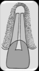
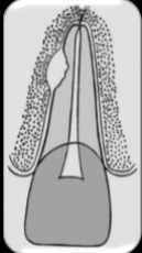
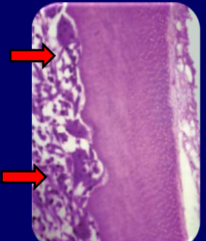
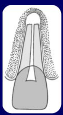
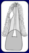
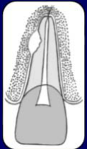
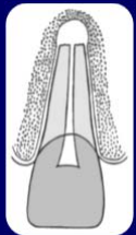
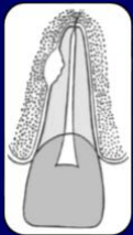
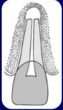
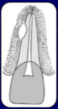
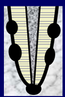
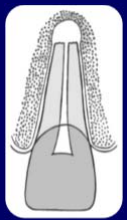
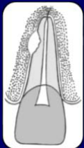
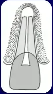
An inflammatory process that results in loss of tooth structure and loss of the adjacent bone.
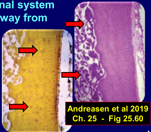
Pierce & Lindskog, Oral Surgery, Oral Medicine, Oral Pathology, 1987. Andreasen et al 2019, Ch. 25 - Fig 25.60.
An inflammatory process that results in loss of tooth structure and loss of the adjacent bone:
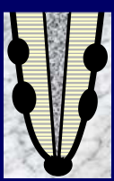
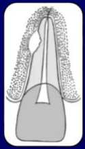
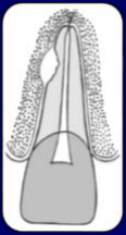
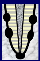
- Requires an infected root canal system PLUS a communication pathway from the root canal to the PDL
- Bacteria / endotoxins diffuse through the dentine tubules
- Leukocytes cannot reach the bacteria in the canal / tubules
- Clastic cells are activated to resorb bone and dentine
An inflammatory process that results in loss of tooth structure and loss of the adjacent bone:
- Requires an infected root canal system PLUS a communication pathway from the root canal to the PDL
- Can occur after trauma where the pulp necroses and becomes infected PLUS there has been damage to the PDL and cementum
- Can also occur with long-standing infected root canal systems - via the apical and / or lateral canal foramina
An inflammatory process that results in loss of tooth structure and loss of the adjacent bone:
- Requires an infected root canal system PLUS a communication pathway from the root canal to the PDL
- External Lateral Inflammatory Resorption
- External Apical Inflammatory Resorption
Which Terminology?
External Infection-related Resorption
OR
Pierce & Lindskog, Oral Surgery, Oral Medicine, Oral Pathology, 1987.
- External Inflammatory Resorption
- Lateral
- Apical
Outline
- Definition and Terminology
- Pathogenesis and Diagnosis
- When Does Inflammatory Resorption Occur?
- Treatment - Review of the Literature
- Management Strategies
- Preventive approach
- Interceptive approach
An inflammatory process that results in loss of tooth structure and loss of the adjacent bone:
- Requires an infected root canal system PLUS a communication pathway from the root canal to the PDL
- → Bacteria / endotoxins diffuse through the dentine tubules
- → Leukocytes cannot reach the bacteria in the canal / tubules
- → Clastic cells activated to resorb bone and dentine
Andreasen et al 2019, Ch. 25 - Fig 25.60.
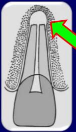
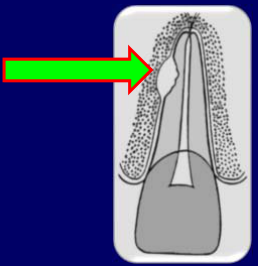
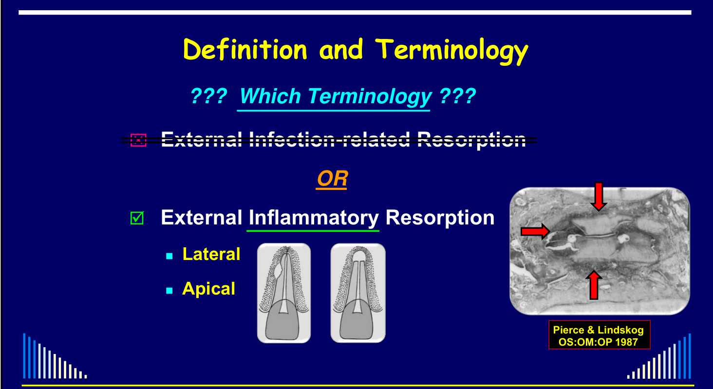
Requirements7
- Dentinal tubules exposed by external root surface damage or by surface resorption
- PLUS: bacteria in the root canal system
 | 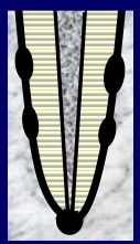 |
 |
Typical Occurrence
- After trauma - e.g. luxation, avulsion
- With long-standing infected root canals
- With concurrent Endo. & Perio. Diseases
Lecturer — Typical Occurrence
The site and frequency of external inflammatory resorption vary with the source of the communication.
- Following trauma, pulp necrosis and infection occur alongside periodontal ligament and cementum damage, and resorption most commonly develops on the lateral aspect of the root.
- With a long-standing infected root canal system, communication is usually through the apical foramen or lateral canal foramina, so resorption typically develops apically.
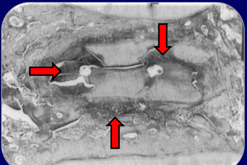
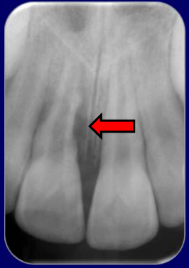
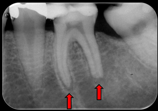
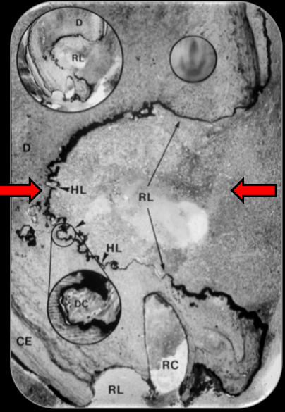
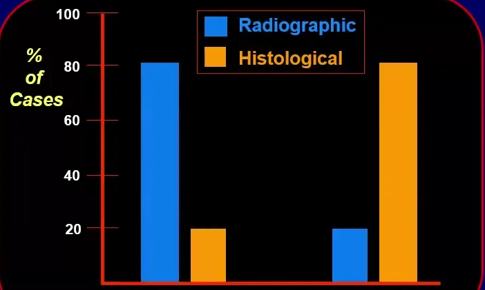
- Histological examination found external apical inflammatory resorption in approximately 80% of teeth with a periapical radiolucency, although it was visible radiographically in only approximately 20% of cases. Laux et al 2000 - Apical Inflammatory Resorption Detection
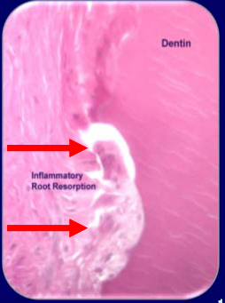
Dentin — Inflammatory Root Resorption
Pathogenesis and Diagnosis8
 |  |
 |
Clinical Features9
- History - trauma, infected RCS, etc.
- Pulp Sensibility Tests - no responses
- Other signs/symptoms of an infected RCS
Lecturer — Inflammatory Resorption
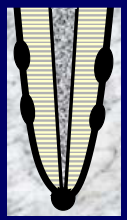
The inflammatory process begins when bacterial products from the infected canal reach the periodontal ligament.
- Leukocytes and other defense mechanisms cannot directly reach bacteria within the root canal and dentine tubules.
- Clastic cells are activated in the periodontal ligament and resorb dentine and adjacent bone.
- A restoration that has broken down or caries may be part of the history.
- Apical periodontitis may be acute or chronic.
 | 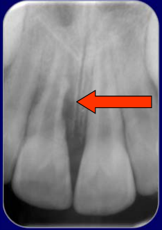 |
Radiographic Features
-
Loss of external tooth substance PLUS
-
Radiolucency in the adjacent bone
-
Diagnosis and differentiation between types of resorption depend primarily on the radiographic appearance.
-
In lateral inflammatory resorption, the radiolucency is alongside the root.
-
In apical inflammatory resorption, the radiolucency is at the root apex. Outline
-
Definition and Terminology
-
Pathogenesis and Diagnosis
-
When Does Inflammatory Resorption Occur?
-
Treatment - Review of the Literature
-
Management Strategies
- Preventive approach
- Interceptive approach
Laux M, Abbott PV, Nair PRN. Apical Inflammatory Root Resorption: A Correlative Radiographic and Histological Assessment. Int Endod J 2000; 33: 483-93.
When Does Inflammatory Resorption Occur?10
Little clear data available in the Literature.
Lecturer — Principal Settings
Inflammatory resorption occurs principally when trauma is associated with pulp necrosis, infection, and damage to the periodontal ligament and cementum. It can also develop from a long-standing infected root canal system.
Some studies have noted “progressive root resorption”:
- → Ranges from 6% up to 96%
- → But this is Inflammatory and Replacement Resorption combined
- → Often also no separation of immature and mature teeth
Need to consider how likely is it to occur for each individual tooth after trauma.
External Inflammatory Resorption following trauma depends on several factors:
- The type of injury - especially luxations, avulsion
- The severity of the injury
- Stage of root development
- How likely is pulp necrosis and infection
- Any concurrent injury
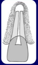 | 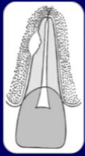 |
Key Factors: How likely is damage to the PDL and cementum?11
Depends on the type of injury:
- Concussion } Least / Nil
- Subluxation
- Extrusion
- Lateral Luxation
- Intrusion } Worst
- Avulsion
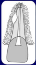
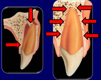
Lecturer — Luxation Damage
Lateral luxation may damage both the palatal root surface and the apical region. Further damage may occur during repositioning if the tooth is forcibly pushed back instead of first being disengaged from the bone; intrusion may damage almost the entire root surface and crush the periodontal ligament and surrounding bone.
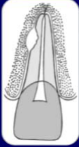 |  |
 |
How likely is pulp necrosis?12
Depends on the type of injury:
- Luxations, avulsions, root fractures
- Severe the blood supply apically or at the fracture line
- Depends on the pulp’s ability to revascularize
Depends on the type of injury:
- Subluxation, concussion, crown fractures
- Apical blood supply is intact
- Depends on the pulp’s ability to resist bacterial invasion
- AND whether any pathway(s) exist for bacteria to enter the tooth
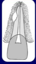
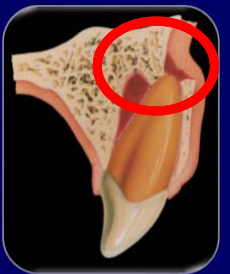
Lecturer — Bacterial Entry
An infraction can provide a bacterial pathway when it extends into dentine. Other possible routes include caries, cracks, and failing restorations; once bacteria enter the pulp and root canal system, pulp necrosis and infection may follow.
 |  |
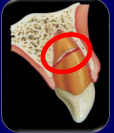 |
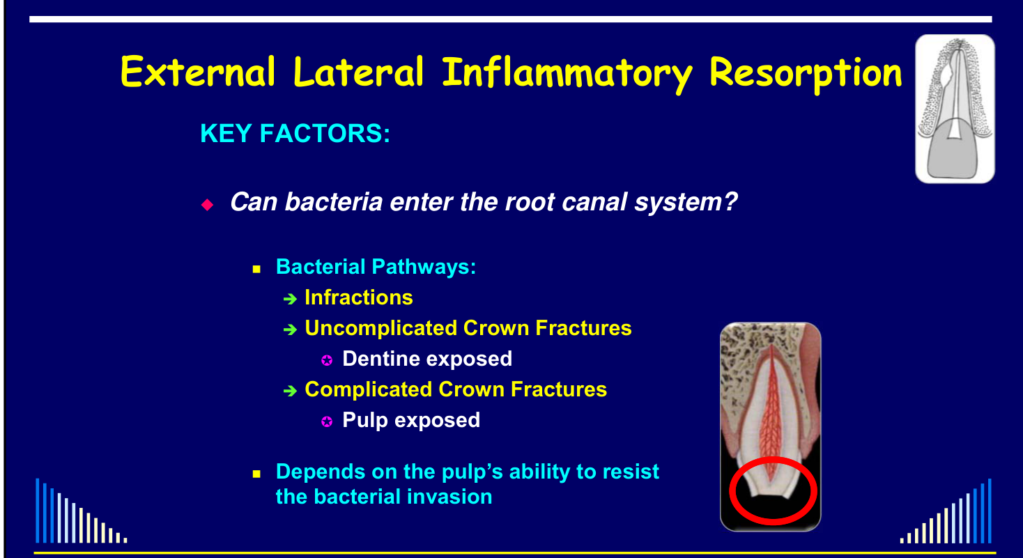
Can bacteria enter the root canal system?13
Bacterial Pathways:
- → Infractions
- → Uncomplicated Crown Fractures
- Dentine exposed
- → Complicated Crown Fractures
- Pulp exposed
Depends on the pulp’s ability to resist the bacterial invasion.
 | 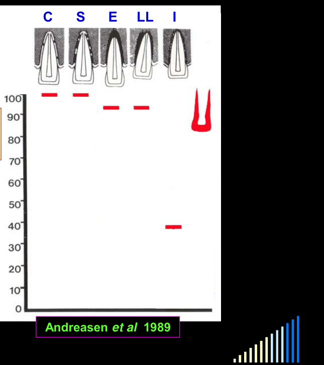 |
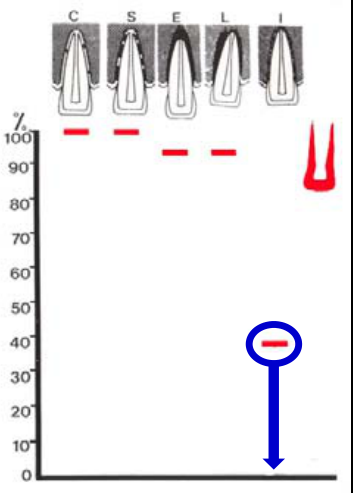
Relationship between Luxation Injury, Pulp Healing and Stage of Development
Graph: % Teeth with Pulp Healing (C S E LL I; scale 0–100) — Andreasen et al 1989
Lecturer — Luxation Survival Rates
The figures show different pulp-survival rates for immature and mature teeth.
- In immature teeth, approximately 5–10% of pulps may not survive extrusion or lateral luxation, while approximately 35% may survive intrusion.
- In mature teeth, approximately 90–95% may survive concussion, 75% subluxation, 40% extrusion, and 25% lateral luxation; no pulps survived intrusion in the figures presented.
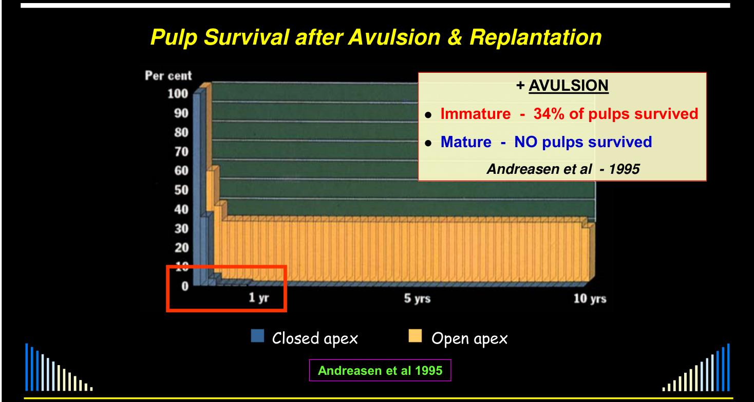
+ AVULSION (Andreasen et al - 1995):
- Immature - 34% of pulps survived
- Mature - NO pulps survived
Graph: Per cent (100–0) over 1 yr / 5 yrs / 10 yrs — Closed apex vs Open apex — Andreasen et al 1995
+ AVULSION (Andreasen et al - 1995):
- Immature - 34% of pulps survived
- Mature - NO pulps survived
Teeth with Open Apices14
Graph: % (100%, 75%, 50%, 25%, 0%) across None / Conc. / Sublux. / Extrus. / Lat.lux. / Intrusion — PCC, Pulp Survival, Pulp Necrosis — C S E L I; scale 0–100
Andreasen et al 1989, 1992
 |  |
Pulp Survival After Crown Fracture — Teeth with Closed Apices15
Graph: 100%, 75%, 50%, 25%, 0% across None / Conc. / Sublux. / Extrus. / Lat.lux. / Intrusion — PCC, Pulp Survival, Pulp Necrosis
Andreasen et al 1989, 1992
Lecturer — Open Apex Findings
With an open apex, most pulps survive an isolated crown fracture, but survival declines when concussion, subluxation, extrusion, or lateral luxation is added. No pulps survived intrusion combined with a crown fracture in the data presented.
Lecturer — Closed Apex Findings
In mature teeth, crown fracture combined with luxation also reduces pulp survival, particularly with subluxation, extrusion, and lateral luxation. Intrusion already had a zero pulp-survival rate without a crown fracture, so the added fracture did not reduce it further.
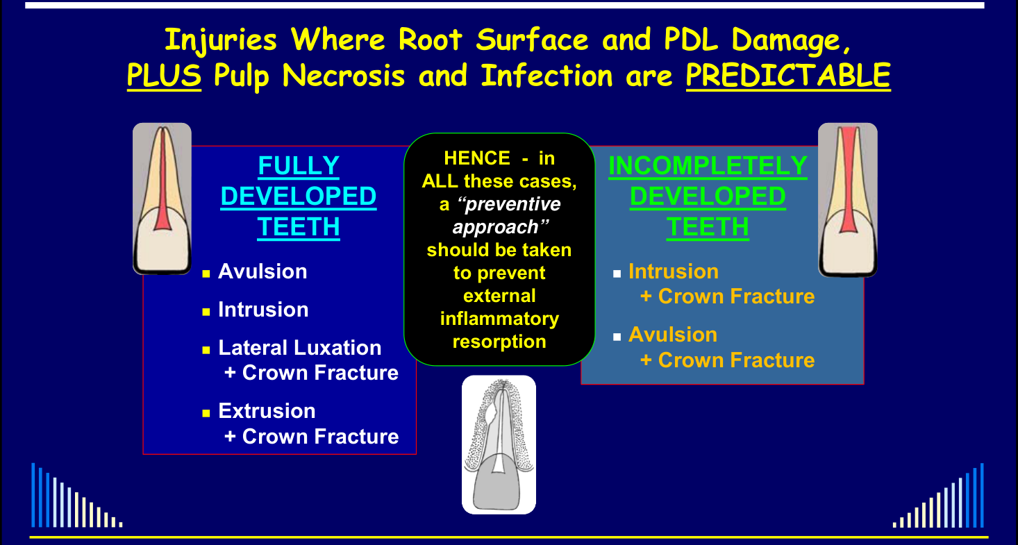
Injuries Where Root Surface and PDL Damage, PLUS Pulp Necrosis and Infection are PREDICTABLE
Fully developed teeth:
- Avulsion
- Intrusion
- Lateral Luxation + Crown Fracture
- Extrusion + Crown Fracture
Incompletely developed teeth:
- Intrusion + Crown Fracture
- Avulsion + Crown Fracture
HENCE - in ALL these cases, a “preventive approach” should be taken to prevent external inflammatory resorption
Lecturer — Established Resorption
The study distinguished prevention from treatment of established resorption.
- Immediate systemic or intracanal antibiotics prevented inflammatory resorption.
- Intracanal antibiotics could stop established resorption from progressing, whereas systemic antibiotics could not.
Treatment - Review of the Literature
Systemic Antibiotic Studies16
Hammarström L, Blomlöf L, Feiglin B, Andersson L, Lindskog S. Replantation of teeth and antibiotic treatment. Endod Dent Traumatol 1986; 2: 51-7.
Replantation of teeth and antibiotic treatment — Hammarström et al, EDT 198617
In replanted teeth with infected canals after 1 hour out of socket, immediate streptomycin–penicillin prevented inflammatory resorption by either systemic or intracanal delivery; after 3 weeks, only intracanal treatment nearly eliminated it. Hammarstrom et al 1986 - Replantation and Antibiotic Treatment
Sae-Lim V, Wang CY, Trope M. Effect of systemic tetracycline and amoxycillin on inflammatory root resorption of replanted dogs’ teeth. Endod Dent Traumatol 1998; 14: 216-20.
Effect of systemic tetracycline and amoxycillin on inflammatory root resorption of replanted dogs’ teeth — Sae-Lim et al, EDT 1998; 14: 216-2018
Results1920
In replanted dog teeth with infected canals and damaged cementum, tetracycline and amoxycillin had similar outcomes; tetracycline differed from the no-antibiotic control. Sae Lim et al 1998 - Systemic Antibiotics Pages 216-220
Sae-Lim V, Wang CY, Choi GW, Trope M. The effect of systemic tetracycline on resorption of dried replanted dogs’ teeth. Endod Dent Traumatol 1998; 14: 127-32.
Effect of systemic tetracycline on resorption of dried replanted dogs’ teeth — Sae-Lim et al, EDT 1998; 14: 127-322122
In a separate dog replacement-resorption model, root-filled teeth bench-dried for 1 hour before replantation showed about 35% complete root-surface healing with systemic tetracycline versus about 11% with amoxicillin and control groups. Sae Lim et al 1998 - Tetracycline Pages 127-132
Topical Doxycycline and Tetracycline Properties
Cvek M, Cleaton-Jones P, Austin J, Kling M, Lownie J, Fatti P. Effect of topical application of doxycycline on pulp revascularization and periodontal healing in reimplanted monkey incisors. Endod Dent Traumatol 1990; 6: 170-6.
Topical application of doxycycline on pulp revascularization and periodontal healing in replanted monkey incisors — Cvek et al, EDT 1990; 6: 170-623
In replanted monkey incisors kept in doxycycline solution, pulp revascularization increased while pulpal microorganisms, ankylosis, replacement resorption, and inflammatory resorption decreased. Cvek et al 1990 - Topical Doxycycline in Monkey Incisors
Vernillo et al. Non-antimicrobial action of tetracyclines. Curr Opin Perio 1994; 2: 111-8.
Non-antimicrobial properties of tetracyclines — Vernillo et al 199424
Tetracyclines modulate host responses
- Inhibit osteoclast function
- Synthetic tetracyclines more potent than tetracycline
- Bind to bone and teeth
- Slows release, prolongs action
Lecturer — Tetracycline Targets
Osteoclasts, cementoclasts and dentinoclasts were described as very similar clastic cells acting on different tissues. Synthetic tetracyclines, including doxycycline and demeclocycline, were described as more potent than tetracycline.
Local Dexamethasone Studies25
Sae-Lim V, Metzger Z, Trope M. Local dexamethasone improves healing of replanted dogs’ teeth. Endod Dent Traumatol 1998; 14: 232-6.
Local dexamethasone improves periodontal healing of replanted dogs’ teeth — Sae-Lim et al, EDT 1998; 14: 232-626
In root-filled replanted dog teeth treated topically for 48 hours, topical dexamethasone was associated with more complete periodontal healing and fewer resorption complications than systemic dexamethasone (ViaSpan control). Sae Lim et al 1998 - Dexamethasone Pages 232-236
Corticosteroid-Antibiotic Paste
Pierce A, Lindskog S. The effect of an antibiotic/corticosteroid paste on inflammatory root resorption in vivo. OS : OM : OP 1987; 64: 216-20.
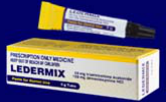
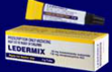
Ledermix Paste27
- Triamcinolone — 1%
- Demeclocycline — 3%
In a water soluble paste of:
- Triethanolamine NF
- Calcium chloride USP
- Zinc oxide
- Sodium sulphite (anhydrous)
- Polyethylene glycol 4,000 USP
- Distilled water
Topic reference: Ledermix Paste in Resorption
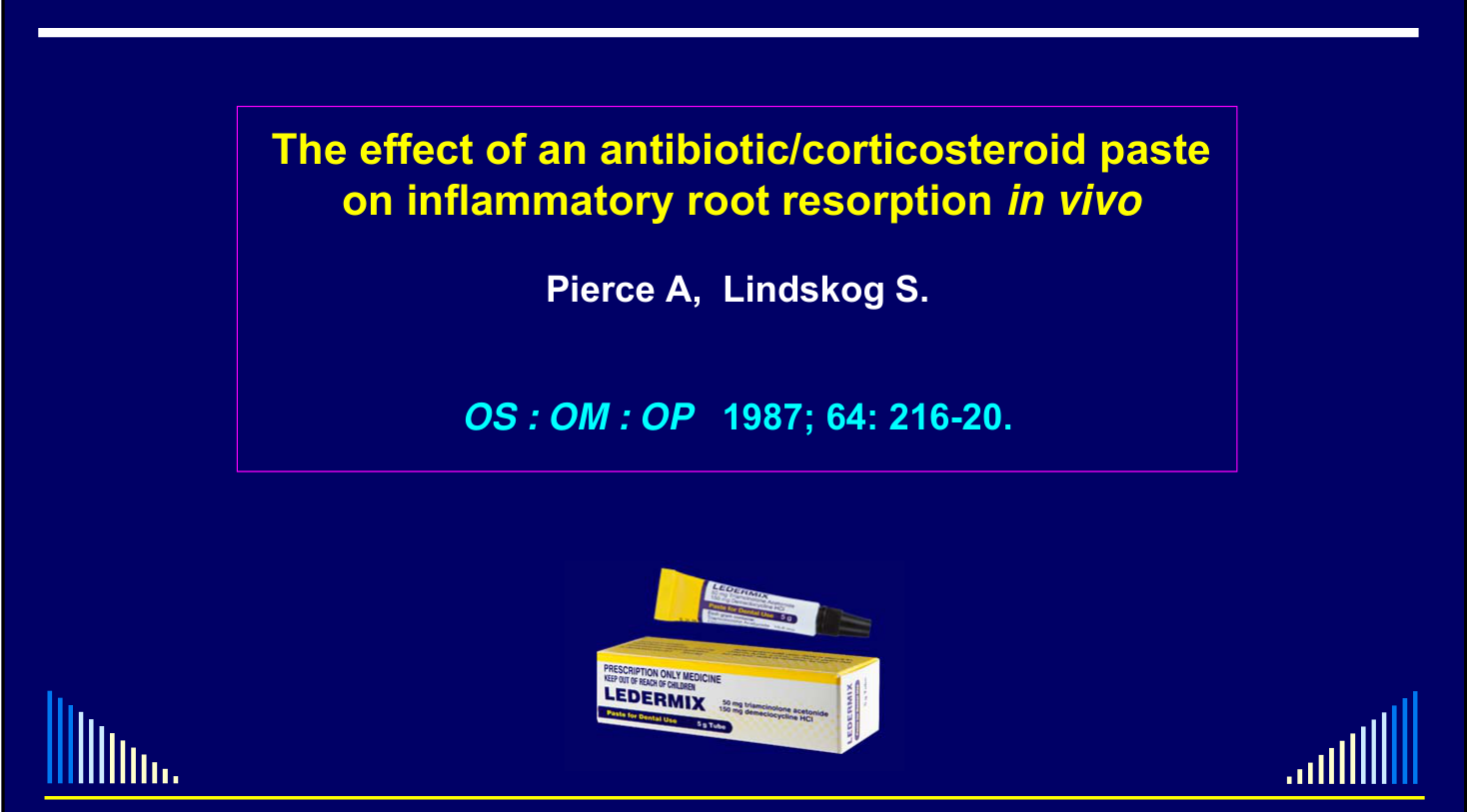 |  |
CS-Ab Paste & Inflammatory Resorption — Pierce & Lindskog, OS:OM:OP 19872829
In dog teeth bench-dried for 1 hour before replantation, Ledermix prevented inflammatory resorption but did not prevent replacement resorption. Pierce and Lindskog 1987 - Antibiotic Corticosteroid in Vivo
Pierce A, Heithersay G, Lindskog S. Evidence for direct inhibition of dentinoclasts by a corticosteroid / antibiotic endodontic paste. Endod Dent Traumatol 1988; 4: 44-5.
Direct inhibition of dentinoclasts by a CS-Ab paste — Pierce et al, EDT 1988; 4: 44-530
In the rat dentinoclast assay, Ledermix-exposed cells became spherical and were absent by 18 hours, supporting direct inhibition under laboratory conditions. Pierce et al 1988 - Dentinoclast Inhibition Assay
The lecture interprets the components as follows:
- Triamcinolone directly inhibits resorbing cells, consistent with steroid effects on osteoclasts (Suda et al 1983).
- Demeclocycline kills bacteria in the root canal and dentine tubules and also has some anti-resorptive activity.
The effect of intracanal anti-inflammatory medicaments on external root resorption of replanted dogs’ teeth after extended extra-oral dry time — Chen, Teixira, Ritter, Levin, Trope, AAE 20053132
In dog teeth kept dry for 60 minutes before replantation, immediate intracanal medicaments produced different periodontal outcomes measured after 4 weeks; the corticosteroid–antibiotic combination was most favorable in the lecture comparison. Chen et al 2005 - Intracanal Medicaments After Dry Storage
Diffusion attribution hold
Abbott 1988–1989 diffusion results are bundled across citations and shared figures; preserve the measurements and image captions as a combined source block pending exact attribution.
Abbott PV, Hume WR, Heithersay GS. Endod Dent Traumatol 1988; 4: 55-62; 1989; 5: 92-7; 1989; 5: 98-104; 1989; 5: 188-92.
 | |
 |
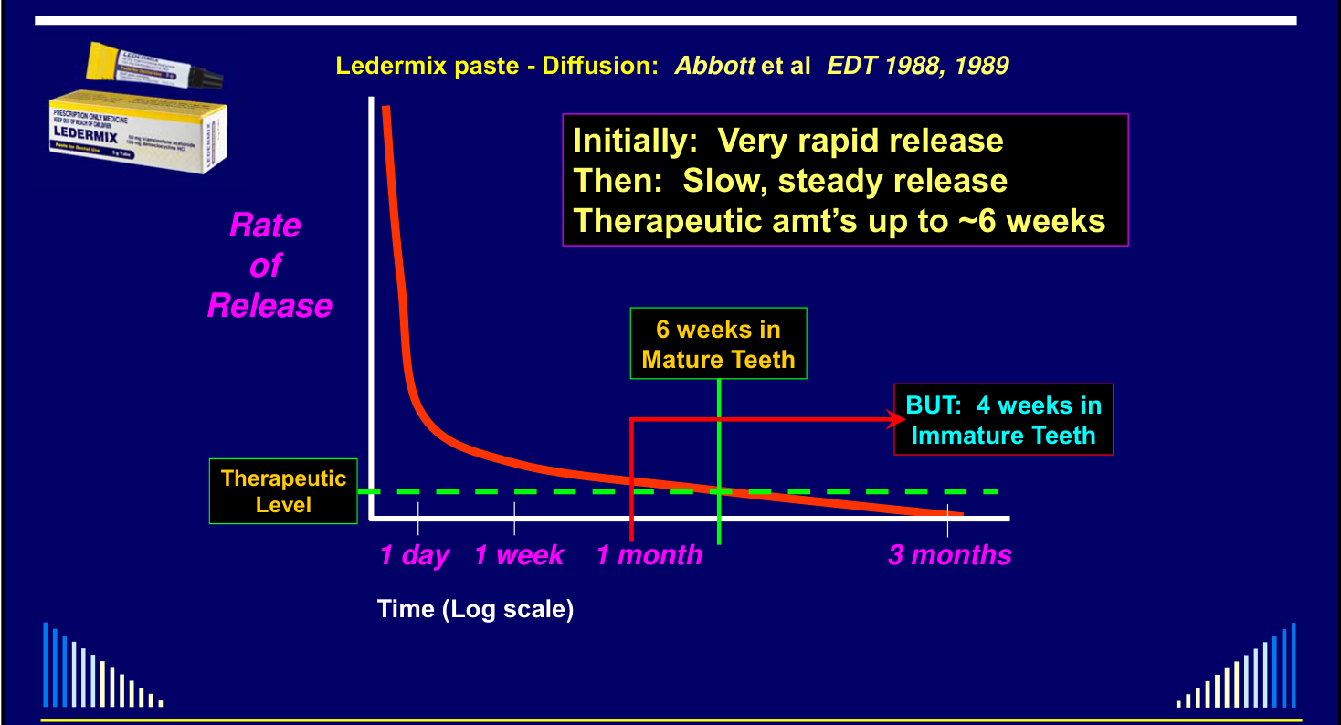
Rate of Release33
- Initially: very rapid release
- Then: slow, steady release
- Therapeutic amounts up to ~6 weeks
6 weeks in mature teeth, but 4 weeks in immature teeth.
Lecturer — Drug Delivery System
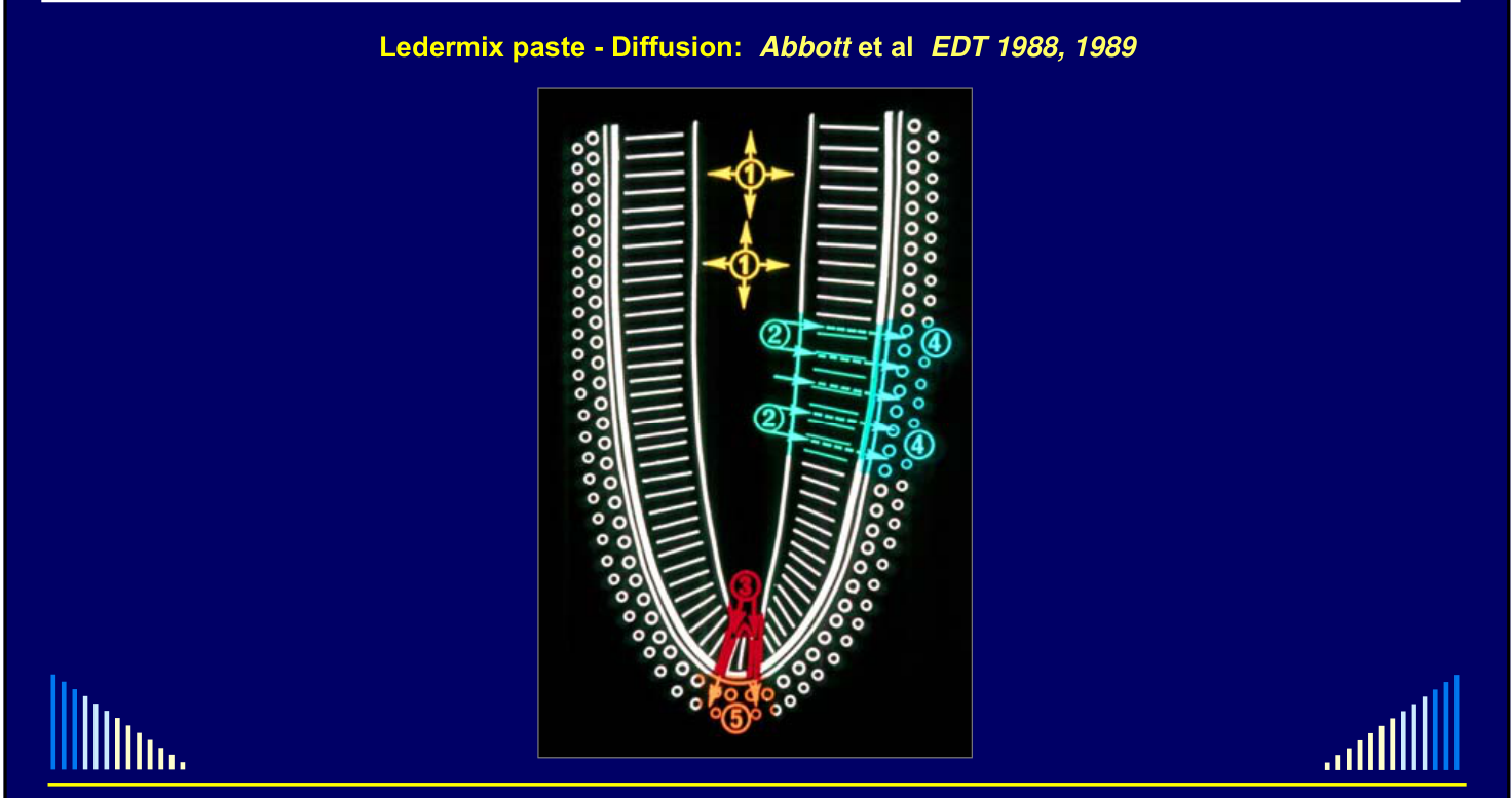
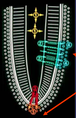
The root canal functions as a drug-delivery system.
- Active components act directly on bacteria and tissue inside the canal.
- They diffuse through dentine tubules and can also be released through the apical foramen into periapical tissues.
Therapeutic level over time (1 day, 1 week, 1 month, 3 months; time on log scale).
 |  |
Triamcinolone (Corticosteroid)
Measured peri-radicular concentration:
- Detected in the nanomolar range
- Sufficient for anti-inflammatory action
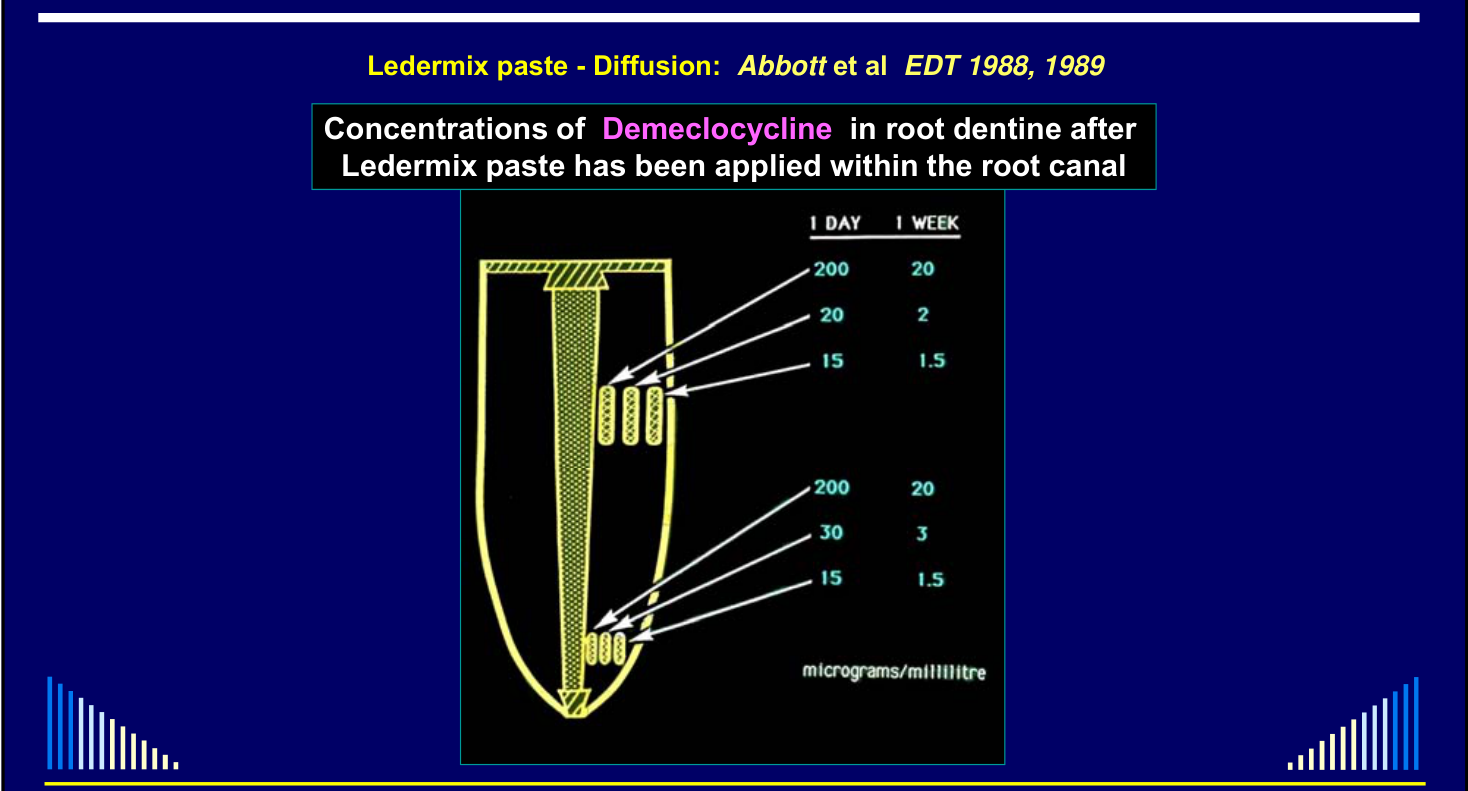
Ledermix paste — Diffusion: Abbott et al, EDT 1988, 198934
| 1 day | 1 week | |
|---|---|---|
| 200 | 20 | |
| 20 | 2 | |
| 15 | 1.5 | |
| 200 | 20 | |
| 30 | 3 | |
| 15 | 1.5 |
micrograms/millilitre
Bryson E, Levin L, Banchs F, Abbott P, Trope M. Dent Traumatol 2002; 18: 316-21.
 | |
Bryson, Levin, Banchs, Abbott & Trope — Dent Traumatol 200235
Immediate Placement of Ledermix Paste — Bryson, Levin, Banchs, Abbott & Trope, Dent Traumatol 200236
In a dog replantation model after 1 hour dry, immediate Ledermix was associated with less resorption and greater residual root mass than calcium hydroxide at 4 months. Bryson et al 2002 - Immediate Ledermix After Replantation
Lecturer — Timing of Treatment
The findings challenged waiting 10–14 days before beginning root canal treatment after avulsion and replantation. Because inflammation begins immediately after injury, delaying treatment may allow inflammatory resorption to become established.
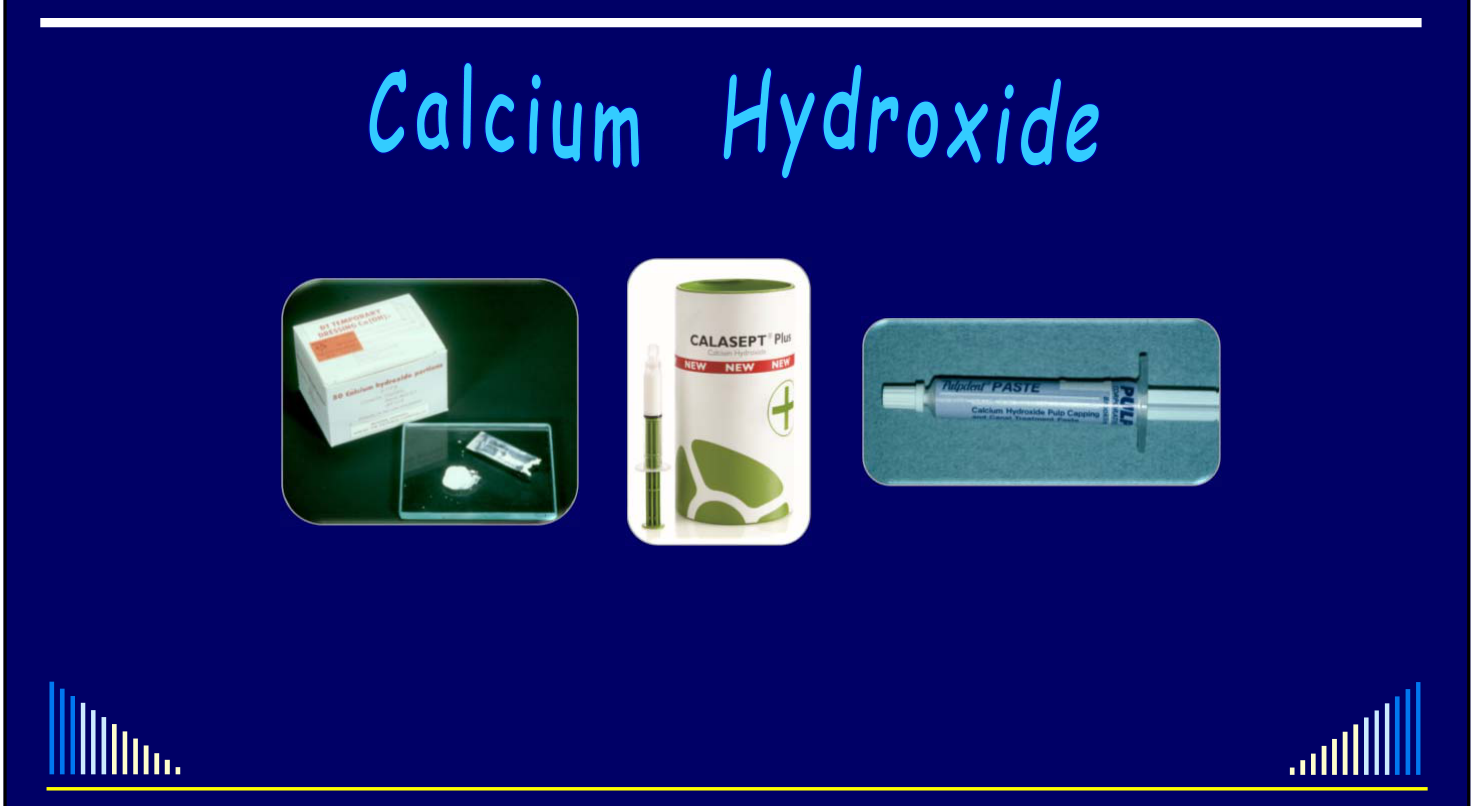
Calcium Hydroxide373839
Source attribution hold
The lecture attributes calcium-hydroxide-induced cell necrosis to Hammarström et al. 1986; retain this claim as presented pending source verification.
Replantation of teeth and antibiotic treatment — Hammarström et al, EDT 1986
- Calcium hydroxide in experimentally-induced inflammatory resorption
- Induced necrosis of ALL cells
- i.e. both the resorbing and the reparative cells
- Necrosis of the reparative cells favours ankylosis and replacement resorption
- Induced necrosis of ALL cells
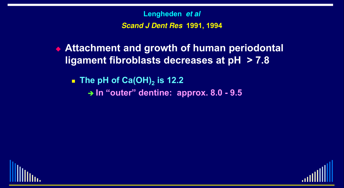
Lengheden et al, Scand J Dent Res 1991, 199440
- Attachment and growth of human periodontal ligament fibroblasts decreases at pH > 7.8
- The pH of Ca(OH)₂ is 12.2
- In “outer” dentine: approx. 8.0 – 9.5
 |
Average pH values
The study compared pH profiles across four calcium hydroxide products and controls over 0–84 days. Plataniotis and Abbott 1999 - Calcium Hydroxide Product pH
The dentine model tracked cervical and apical inner and outer pH over 0–28 days after calcium hydroxide placement. Nerwich et al 1993 - Calcium Hydroxide Dentine pH
-
Attachment and growth of human periodontal ligament fibroblasts decreases at pH > 7.8
- The pH of Ca(OH)₂ is 12.2
- In “outer” dentine: approx. 8.0 – 9.5
- The pH of Ca(OH)₂ is 12.2
-
Hence Ca(OH)₂ inhibits fibroblasts — so:
- The bone healing response dominates
- Favours ankylosis and replacement resorption
- The bone healing response dominates
-
Calcium hydroxide almost entirely eliminated established inflammatory resorption elicited by bacterial contamination in the replanted teeth with a necrotic PDL
-
BUT the resultant healing was characterised by ankylosis which gradually developed into replacement resorption
In traumatised teeth with infected root canals:
“The duration of use of calcium hydroxide should be minimised”
“Furthermore, the use of less toxic and more biocompatible medicaments, such as the antibiotic-corticosteroid combinations, should be encouraged”
Calcium Hydroxide41
- Ca(OH)₂ is the most widely-used endodontic medicament throughout the world
- Mainly because of its anti-bacterial activity
- Other very useful properties:
- Stimulates hard tissue formation
- Helps dissolve necrotic tissue
- Detoxifies bacterial endotoxin (LPS)
- Safavi & Nicholls 1993, 1994; Olsen et al 1999, Nelson-Filho et al 2002a, 2002b, Silva et al 2002, Jiang et al 2003, Tanomaru et al 2003
Tanomaru et al. Effect of different irrigating solutions and calcium hydroxide on bacterial LPS. Int Endo J 2003; 36: 733-9.
- LPS → inflammation and bone resorption
- Stimulates macrophages to release cytokines
- e.g. tumour necrosis factor, interleukins 1, 6, 8 In the reported assay, saline, 2% chlorhexidine, and 1–5% sodium hypochlorite did not inactivate LPS, whereas Ca(OH)₂ did. Tanomaru et al 2003 - Irrigants Calcium Hydroxide and LPS
- Stimulates macrophages to release cytokines
Lecturer — Calcium Hydroxide Timing
Calcium hydroxide was effective against established inflammatory resorption but could produce ankylosis and replacement resorption. It was therefore described as better used as a secondary medicament rather than as the initial medicament in these cases.
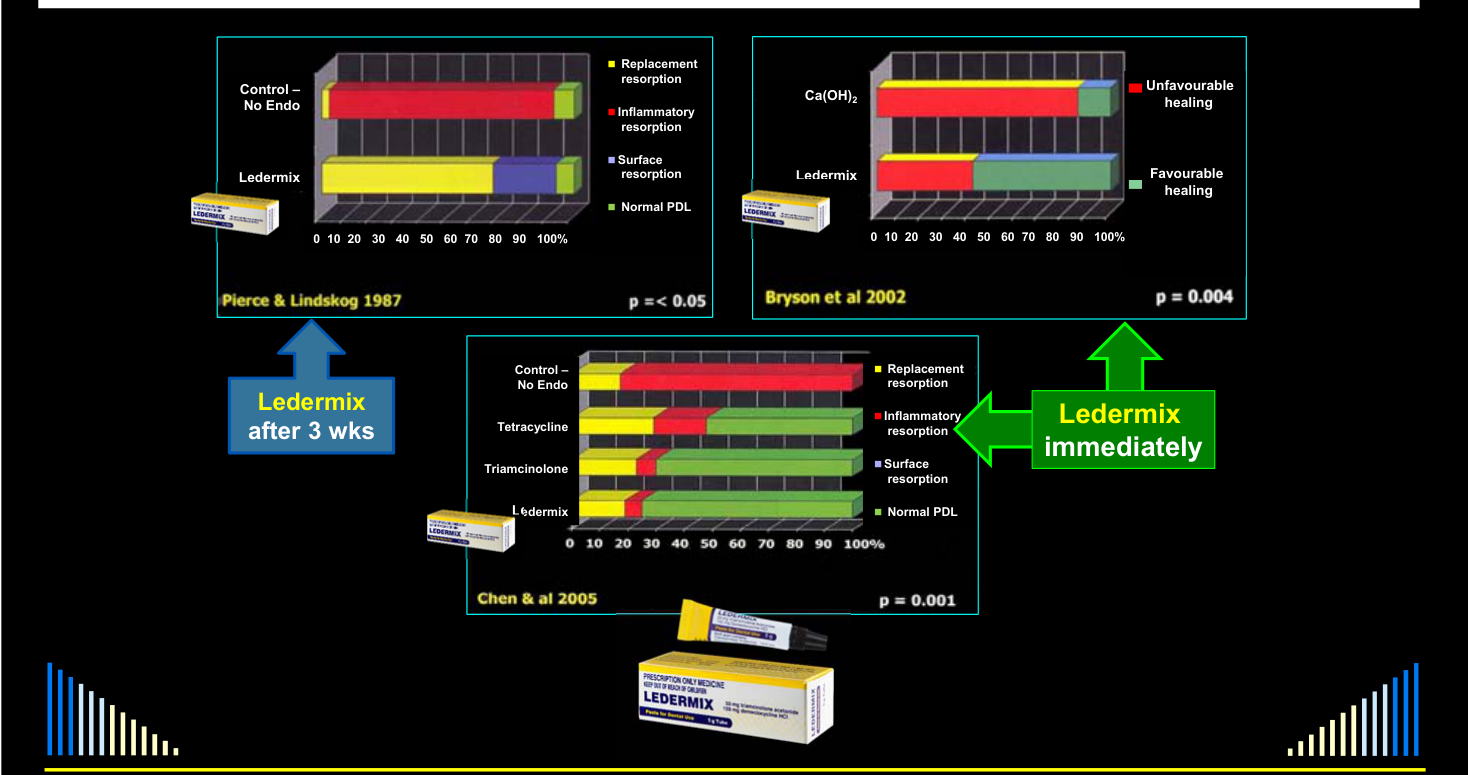
Comparative studies (scale: unfavourable healing → favourable healing; outcomes: replacement resorption, inflammatory resorption, surface resorption, normal PDL):
- Control (no endo) versus Ledermix — Pierce & Lindskog 1987, p = < 0.05
- Ca(OH)₂ versus Ledermix — Bryson et al 2002, p = 0.004
- Ledermix after 3 wks versus Ledermix immediately
- Control (no endo), tetracycline, triamcinolone, Ledermix — Chen et al 2005, p = 0.001
LEDERMIX
Management Strategies42
- Definition and Terminology
- Pathogenesis and Diagnosis
- When Does Inflammatory Resorption Occur?
- Treatment — Review of the Literature
- Management Strategies
- Preventive approach
- Interceptive approach
Management Approaches43
- Preventive → After injuries where pulp necrosis and infection PLUS root surface and PDL damage are likely
- Interceptive → When inflammatory resorption is already present
Aims of Prevention vs Aims of Interception
| Aims of Prevention | Aims of Interception |
|---|---|
| Reduce PDL inflammation | Reduce PDL inflammation |
| Inhibit clastic cells | Inhibit clastic cells |
| Stop bacteria entering the root canal | Kill all bacteria that are already in the root canal |
| Kill any bacteria that entered the canal during the injury | Encourage healing with hard tissue |
Preventive Management
How to Prevent44
Consider: When does PDL inflammation start?
- IMMEDIATELY !!!
- i.e. as soon as the injury has occurred
Preventive Management
Based on TWO important principles:
- Immediate systemic antibiotics for 1 week
- Immediate pulp removal and placement of a CS-AB intracanal medicament
Prevention after Trauma45
- Systemic antibiotics — IMMEDIATELY and for 1 week
- → Penicillin (Hammarström et al 1986)
- → Tetracycline (Sae-Lim et al 1998 a, b)
- Pulp removal — IMMEDIATELY after the trauma
- → CS-AB paste dressing (Hammarström et al 1986, Pierce & Lindskog 1987, Pierce et al 1988, Bryson et al 2002, Chen et al 2005)
- → If pulp not removed immediately, then do it AS SOON AS POSSIBLE
 |  |
Preventive Management — Mature and Immature Teeth
- IMMEDIATELY after the emergency treatment
- → Start systemic antibiotics — Tetracycline preferred
- e.g. Doxycycline 100 mg, 2 tabs. on 1st day, then 1 tab. daily for 1 week
- → Start systemic antibiotics — Tetracycline preferred
- IMMEDIATELY after replanting / repositioning & splinting
- → Remove the pulp
- → Prepare the canal if time available — otherwise do it at the next visit
- → Place a CS-AB paste dressing
Mature Teeth
- MATURE teeth — After 6 weeks
- Re-dress canal with the CS-AB paste
- MATURE teeth — After another 6 weeks
- Take PA radiograph
- If resorption present: Re-dress canal with the CS-AB paste
- If no resorption: Place new dressing using 50:50 CS-AB + Ca(OH)₂
- Take PA radiograph
- MATURE teeth — After a further 2 months
- Take PA radiograph
- If no resorption: Complete the Root Canal Filling (GP + cement)
- Take PA radiograph
Preventive Management — Immature Teeth
- IMMATURE teeth — After 4 weeks
- Re-dress canal with the CS-AB paste
- IMMATURE teeth — After a further 4 weeks
- Re-dress canal with the CS-AB paste
- IMMATURE teeth — After another 4 weeks
- Take PA radiograph
- If resorption present: Re-dress canal with the CS-AB paste
- If no resorption: Place new dressing using 50:50 CS-AB + Ca(OH)₂
- Take PA radiograph
Immature Teeth (continued)
- IMMATURE teeth — After a further 2 months
- Take PA radiograph
- If no resorption:
- An apical hard tissue barrier is desirable
- Re-dress canal with Ca(OH)₂ paste
- Change Ca(OH)₂ dressing every 3 months
- Until hard tissue repair / apexification evident
- Complete the Root Canal Filling (GP + cement)
- An apical hard tissue barrier is desirable
- If no resorption:
- Take PA radiograph
Lecturer — Immature Tooth Reviews
Immature teeth require more frequent review because medicament diffusion is faster through the wider open apex and wider dentine tubules.
- The corticosteroid-antibiotic dressing is changed at approximately four, eight and twelve weeks.
- At approximately three months, a radiograph assesses for resorption; if none is present, a 50:50 corticosteroid-antibiotic and calcium hydroxide mixture is used for a further two months.
- Traditional calcium hydroxide apexification was described as the most predictable approach when an immature tooth requires root canal treatment.
Preventive Management — All Cases
- Clinical and radiographic reviews are essential
- At 6 months after the RCF is completed
- Then every 3–4 years
- For as long as possible !!!
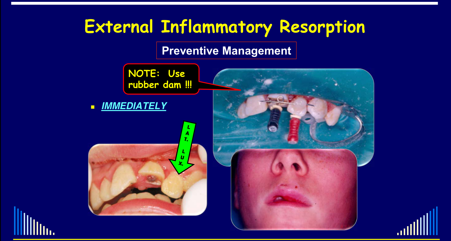
NOTE: Use rubber dam !!! — IMMEDIATELY
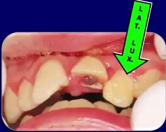
Figure labels: L A T. L U X
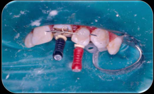
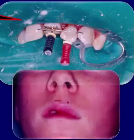
Use RUBBER DAM — Cuff Technique46
- Excellent management tool — esp. if after hours
- Controls lips, tongue, moisture, etc.
- Easier to reposition / replant teeth
- Easier to splint teeth
- Essential for Endodontic treatment
- And many other reasons…
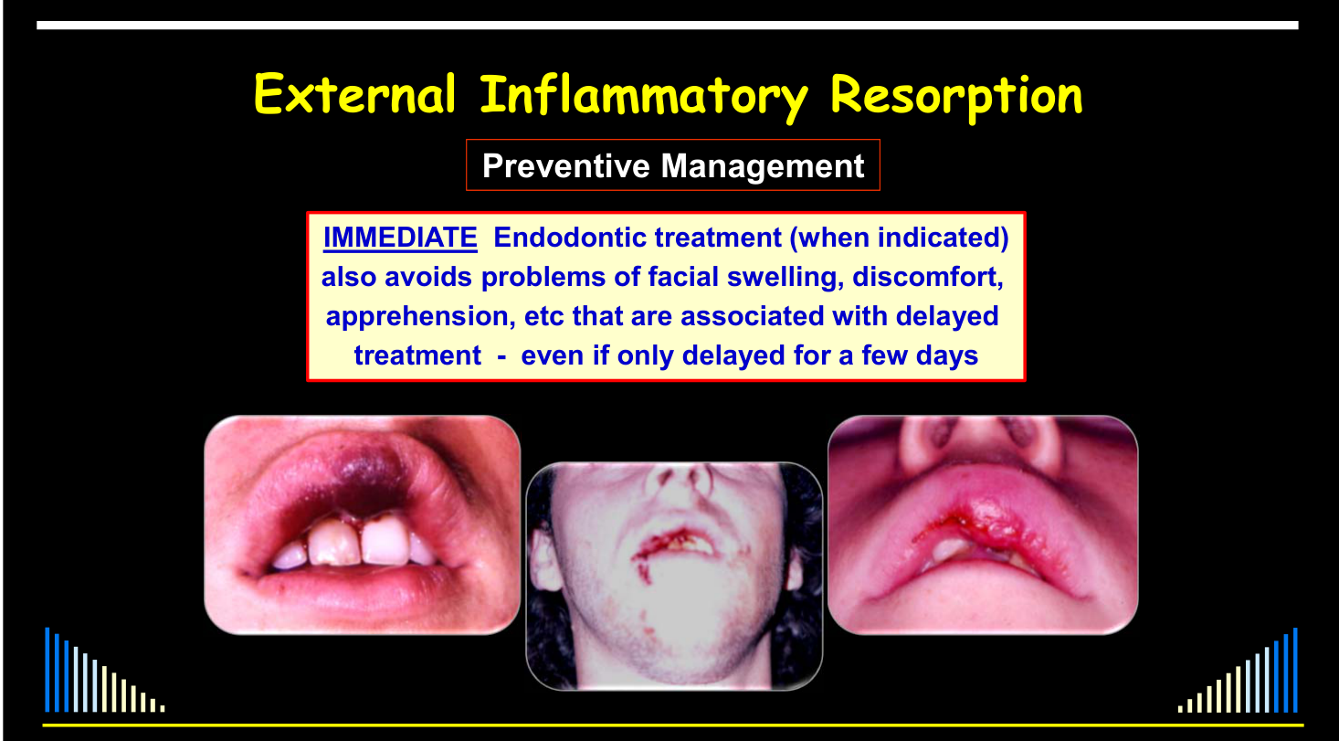
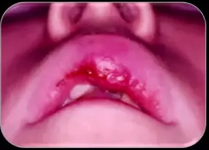
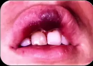
IMMEDIATE Endodontic treatment (when indicated) also avoids problems of facial swelling, discomfort, apprehension, etc that are associated with delayed treatment — even if only delayed for a few days.
Traumatic Injuries to the Teeth, 4th edition, Chapter 25, Page 695.
 |  |
Fully Developed Teeth — Preventive Management47
- Systemic antibiotics — start IMMEDIATELY
- Check the patient’s age, weight, allergies etc. to determine appropriate systemic antibiotic and doses
- Tetracycline preferred — e.g. doxycycline 100 mg — 2 tablets on the 1st day, and then 1 tablet daily for 1 week
- If allergic to tetracycline, use phenoxymethyl penicillin 1000 mg immediately and then 500 mg every 6 hours (1 hour before meals) for 1 week
- Another alternative if allergy to tetracycline: use amoxicillin 1000 mg immediately, then 500 mg every 8 hours for 1 week
- IMMEDIATELY after replantation/repositioning and stabilization with a splint
- Remove the pulp, clean the root canal — if time: measure, file, irrigate, dry
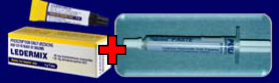
- Place a CS-AB paste intracanal dressing, e.g. Ledermix paste
- After 6 weeks — complete canal preparation, place a new CS-AB paste intracanal dressing
- After another 6 weeks — take a periapical radiograph
- If no inflammatory resorption evident — place an intracanal dressing using a 50:50 mixture of CS-AB & Ca(OH)₂
- After 2–3 months — take a periapical radiograph
- If no inflammatory resorption evident — place the root canal filling using gutta percha and cement
- Perform internal bleaching if required and then restore the access cavity
- Arrange to review after 6 months and then annually for at least 5 years.
NOTES: 1) The root canal treatment must be commenced as soon as the tooth has been replanted/repositioned and stabilised with a splint. 2) CS-AB = corticosteroid/antibiotics. 3) Working length and canal preparation can be deferred until the second appointment if insufficient time available at the emergency appointment. 4) Rubber dam must be used for all endodontic procedures.
Source: Reproduced from (221) with permission.
Table 25.3 – Recommended preventive treatment protocol for incompletely developed teeth to prevent external inflammatory root resorption after suffering the injuries listed in Table 25.1.
 |  |
Incompletely Developed Teeth — Preventive Management
- Systemic antibiotics — start IMMEDIATELY
- Check the patient’s age, weight, allergies etc. to determine appropriate systemic antibiotic and doses
- Tetracycline preferred — e.g. doxycycline 100 mg — 2 tablets on the 1st day, and then 1 tablet daily for 1 week
- Alternatively use phenoxymethyl penicillin 1000 mg immediately and then 500 mg every 6 hours (1 hour before meals) for 1 week
- Or use amoxicillin 1000 mg immediately, then 500 mg every 8 hours for 1 week
- IMMEDIATELY after replantation/repositioning and stabilization with a splint
- Remove the pulp, clean the root canal — if time: measure, file, irrigate, dry
- Place a CS-AB paste intracanal dressing, e.g. Ledermix paste
- After 4 weeks — complete canal preparation, place a new CS-AB paste intracanal dressing
- After another 4 weeks — place a new CS-AB paste intracanal dressing
- After another 4 weeks — take a periapical radiograph
- If no inflammatory resorption evident — place a new intracanal dressing using a 50:50 mixture of CS-AB & Ca(OH)₂
- After 2–3 months — take a periapical radiograph
- If no inflammatory resorption evident — place a Ca(OH)₂ intracanal dressing to induce formation of an apical hard tissue barrier
- Change the Ca(OH)₂ intracanal dressing every 3 months until hard tissue repair (e.g. apexification) is evident. Periapical radiograph every 6–9 months to ensure healing
- Place the root canal filling using gutta percha and cement
- Perform internal bleaching if required and then restore the access cavity
- Arrange to review after 6 months and then annually for at least 5 years.
Lecturer — Disease Requirements
External inflammatory resorption requires both an infected root canal system and a pathway between the canal and the periodontal ligament. It is an inflammatory process involving loss of tooth structure and adjacent bone, which explains why treatment must first control the infection and then promote hard-tissue repair.
NOTES: 1) The root canal treatment must be commenced as soon as the tooth has been replanted/repositioned and stabilised with a splint. 2) CS-AB = corticosteroid/antibiotics. 3) Working length and canal preparation can be deferred until the second appointment if insufficient time available at the emergency appointment. 4) Rubber dam must be used for all endodontic procedures.
Source: Reproduced from (221) with permission.
Clinical Case Illustrations48
 |  |
 |
Two Cases to Illustrate …49
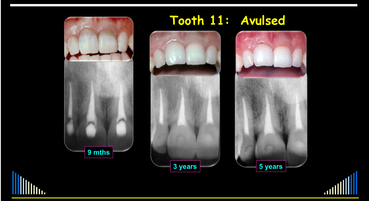
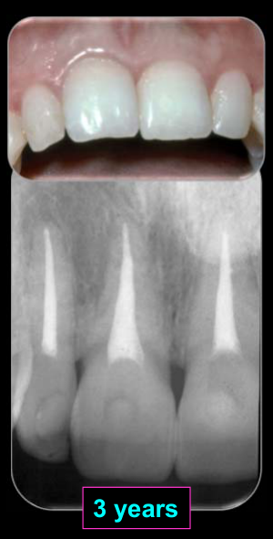
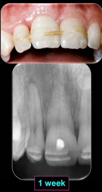
Tooth 11: Avulsed
- Replanted within 5 minutes
- Flexible splint
- Immediate Endodontics — Ledermix
- Systemic Antibiotics — 1 week
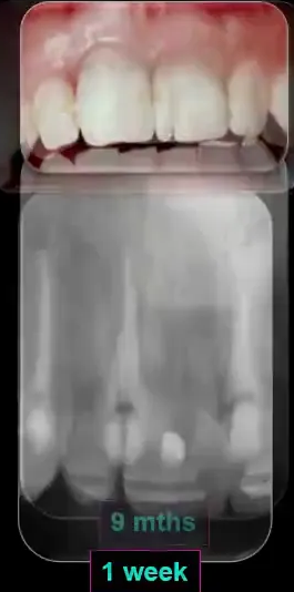
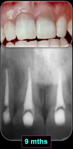
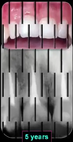
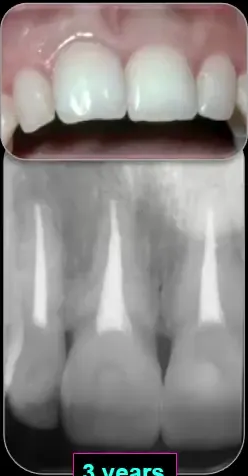
Tooth 11: Avulsed — 9 mths, 3 years, 5 years
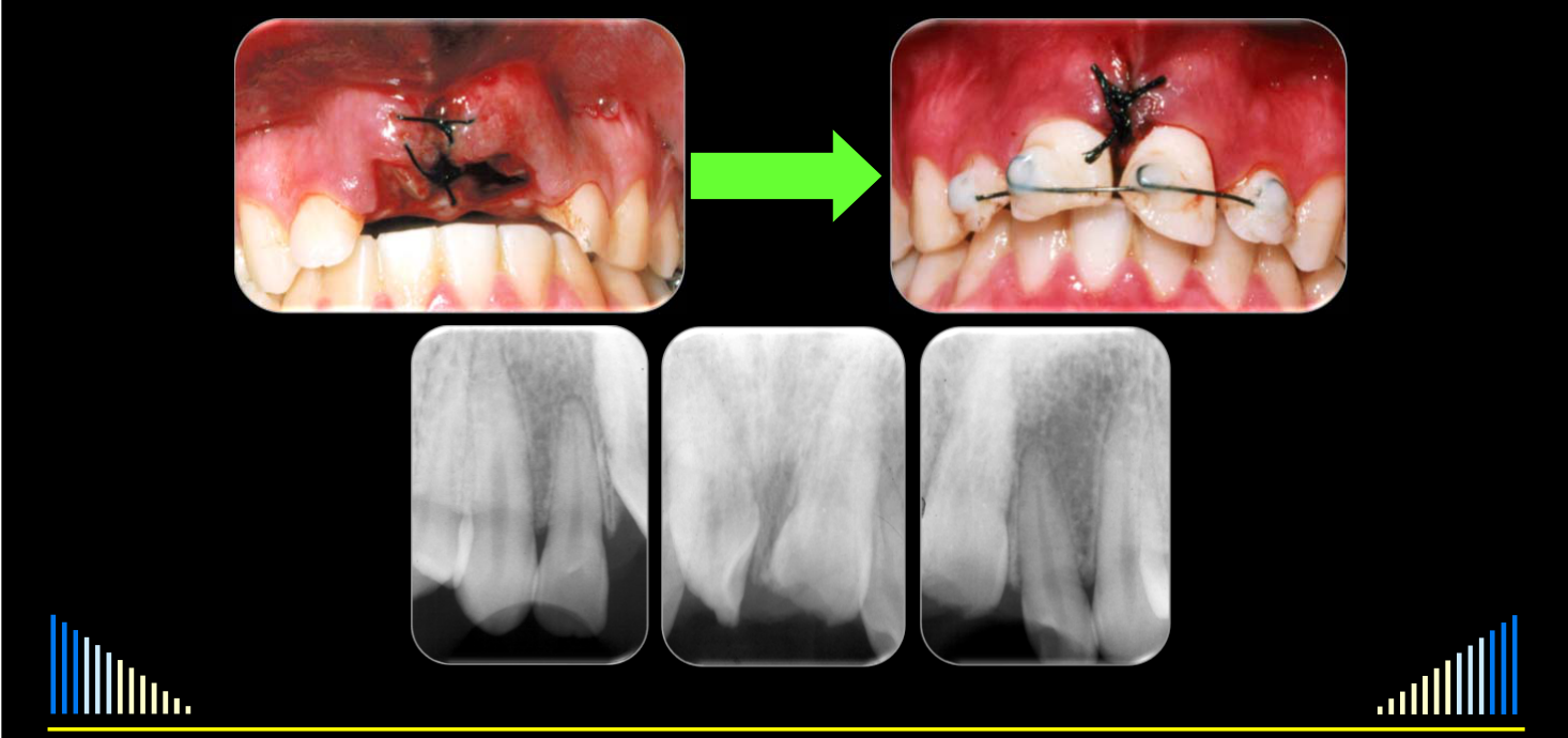
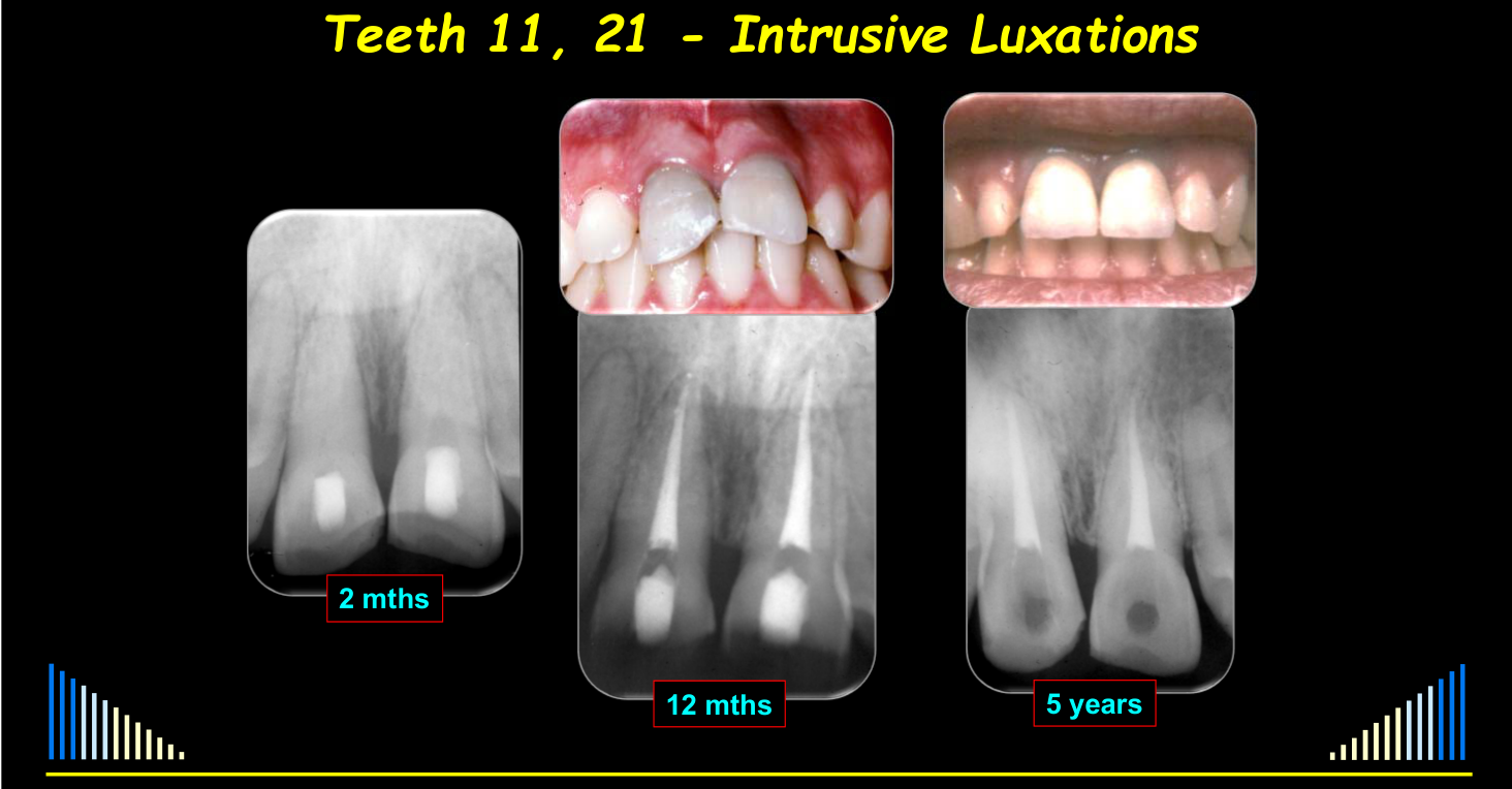
Teeth 11, 21 — Intrusive Luxations
Teeth 11, 21 — Intrusive Luxations — 2 mths, 12 mths, 5 years
Immediate Trauma Cases
Several clinical cases demonstrated the feasibility and value of immediate treatment under rubber dam.
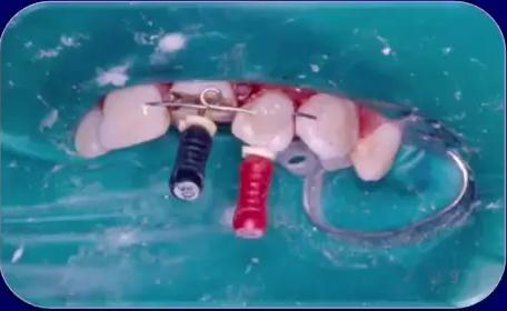
- A young boy sustained multiple football-related injuries: one lateral incisor was laterally luxated, repositioned, stabilized and treated endodontically on the day of injury. The lip swelling that developed later illustrated the difficulty of delayed treatment.
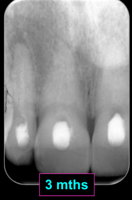
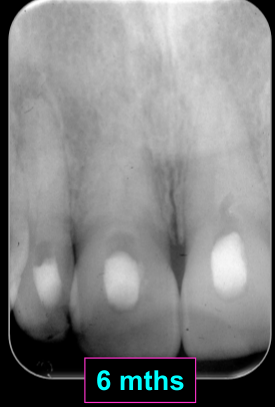
- A dental therapist avulsed an upper central incisor in a car accident, cleaned it with saliva, replanted it and applied a flexible nylon fishing-line splint. Immediate root canal treatment, Ledermix and systemic antibiotics were provided; no external inflammatory resorption was seen at one week, nine months, three years or five years. The tooth was eventually extracted after 15 years because of replacement resorption.
- Two intruded teeth with uncomplicated crown fractures were initially missed by the dentist, then repositioned, stabilized and treated endodontically approximately 12 hours after injury. Follow-up showed no external inflammatory or replacement resorption.
 |  |
 |
Interceptive Management
Aims of Interceptive Management50
- Reduce PDL inflammation
- Inhibit clastic cells
- Kill all bacteria in the root canal system
- Encourage healing with hard tissue
Two Differences to the Preventive Management Approach5152
- Systemic antibiotics are NOT indicated and they will not stop the inflammatory resorption
- Only prescribe if the patient presents with an acute apical abscess with systemic signs, or if facial cellulitis / spreading infection
- Long-term Ca(OH)₂ intracanal dressings WILL BE required to stimulate hard tissue repair
- Since hard tissue (bone, cementum, dentine) has been lost
- e.g. may be an open apical foramen
- Since hard tissue (bone, cementum, dentine) has been lost
 |  |
 |
Treatment Protocol
-
Commence root canal treatment
-
Remove the cause(s) of the disease
- i.e. remove the caries, cracks, old restorations
-
Prepare & disinfect the root canal system
- i.e. WL, instrumentation, irrigants, etc.
-
Place a CS-AB paste dressing
-
After 6 weeks — Re-dress canal with the CS-AB paste
-
After another 6 weeks
- → Take PA radiograph
- If resorption has not stopped: Re-dress canal with the CS-AB paste
- If no further resorption: Place new dressing of 50:50 CS-AB + Ca(OH)₂
- → Take PA radiograph
-
After a further 2 months
- → Take PA radiograph
- If repair evident / no further resorption: Dress canal with Ca(OH)₂
- Since hard tissues (bone, cementum, dentine) have been lost
- If repair evident / no further resorption: Dress canal with Ca(OH)₂
- → Take PA radiograph
Lecturer — Interceptive Timing
In interceptive cases, root canal treatment can begin when convenient unless acute apical periodontitis or an acute apical abscess requires more urgent treatment.
- The tooth should first be assessed for suitability for root canal treatment and restoration.
- Canal preparation includes working-length determination, instrumentation and irrigation with agents such as sodium hypochlorite and EDTA.
- Root canal filling is usually possible after approximately 9–12 months, although some cases require longer treatment.
All Cases53545556
- Clinical and radiographic reviews are essential
- At 6 months after the RCF is completed
- Then every 3–4 years
- → For as long as possible !!!
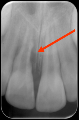
Pre-operative Dressings — 3 mths — RCF — 18 mths — 4 yrs — Review*
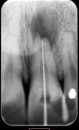
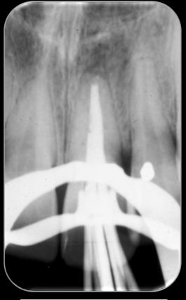
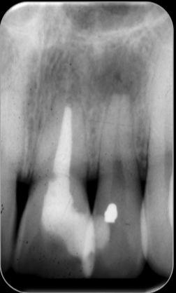
Interceptive Management — Pre-operative — WL — RCF — 15 mths — 4 yrs — Review
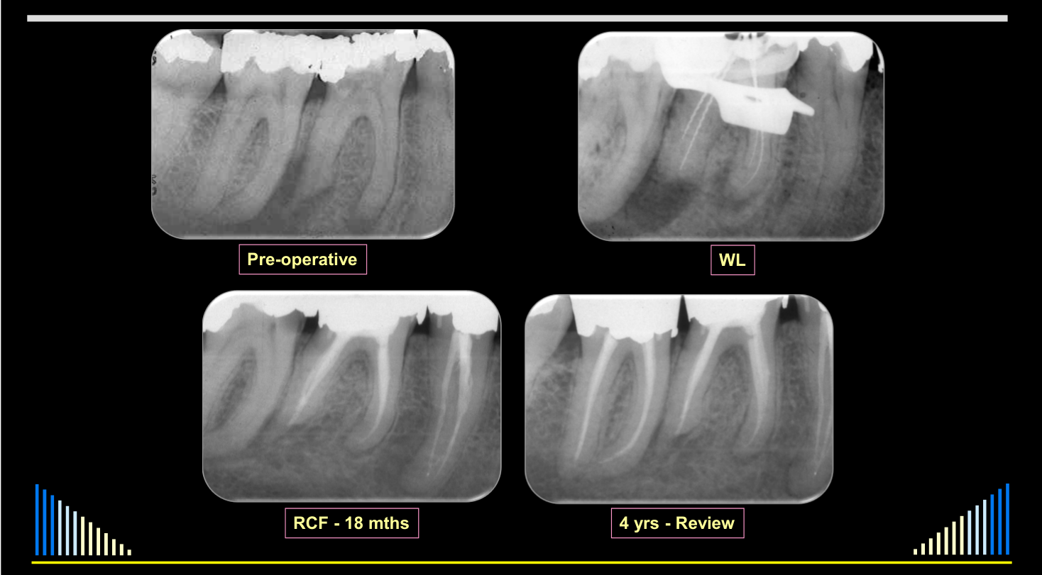
Pre-operative — WL — RCF — 18 mths — 4 yrs — Review
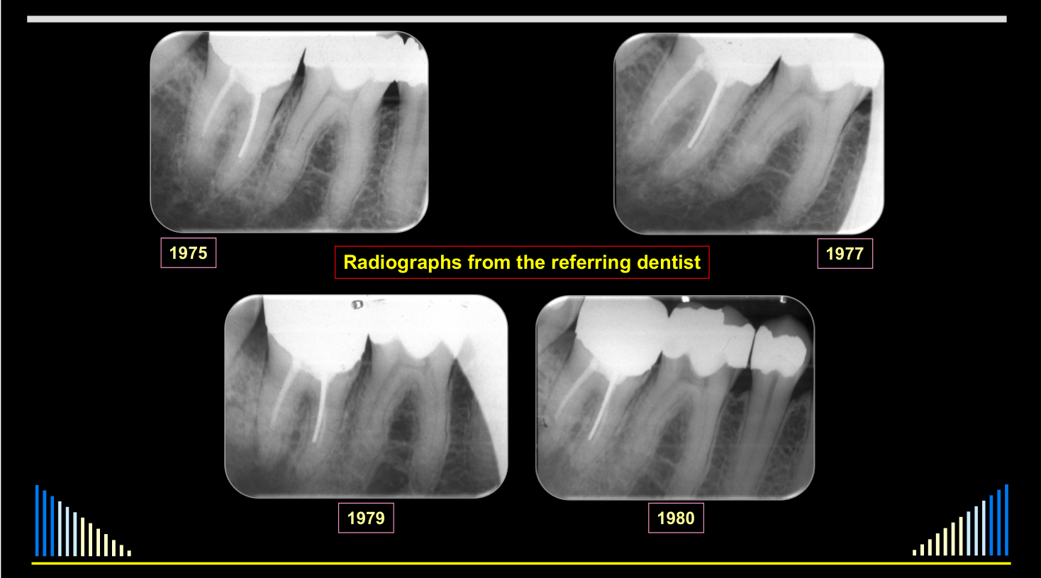
Radiographs from the referring dentist: 1975, 1980, 1979, 1977
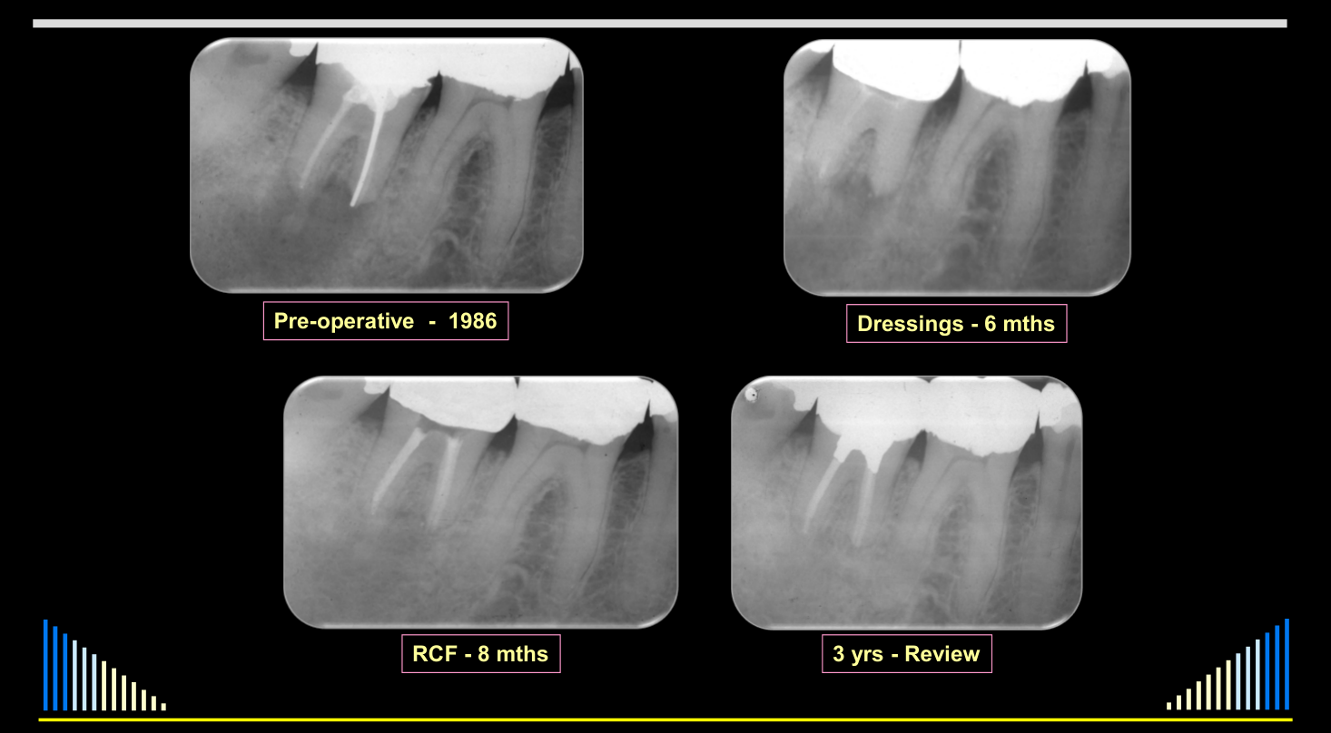
Pre-operative — 1986 — Dressings — 6 mths — RCF — 8 mths — 3 yrs — Review
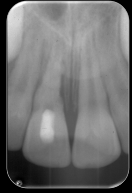
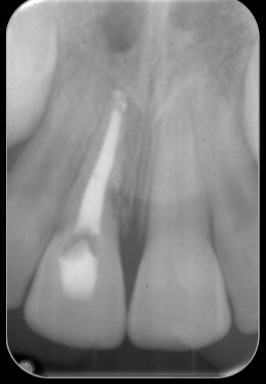
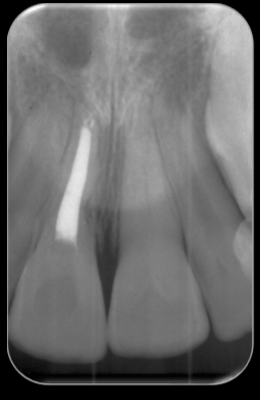
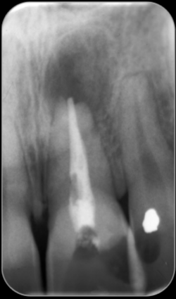
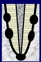
Outcomes of Managed Cases57
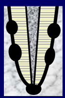 |  |  |
 |  |
Cohort context
This is an uncontrolled descriptive series of 155 patients and 178 teeth; it reports outcomes under the combined management strategies and does not establish causation by a single intervention.
A Review of 178 Cases Managed with these Strategies58
155 patients with 178 teeth over 15 years:
- 52 patients with 60 teeth — Preventive management after trauma
- 103 patients with 118 teeth — Interceptive management — established lesions
- 43 teeth — Lateral
- 75 teeth — Apical
Outcomes59
Using the management regimes outlined:
- Average total treatment time: 12 months
- Range: 8–18 months
 |  | |
 |  |  |
Preventive Group60
- 52 patients with 60 teeth — Prevention after trauma
- No teeth lost due to ongoing inflammatory root resorption
- 100% favourable outcome
 | |  |
 |  |  |
 |
Interceptive Group
-
103 patients with 118 teeth — Interceptive treatment — established resorption
-
43 teeth — Lateral inflammatory resorption
- Three teeth lost due to periodontal problems and external inflammatory resorption
- All from the ONE patient
- Three teeth lost due to periodontal problems and external inflammatory resorption
-
Three teeth lost due to periodontal problems and external inflammatory resorption
- All from the ONE patient
- Avulsed & replanted after 90 mins. in milk
- Inadequate splinting
- No systemic antibiotics
- No early endodontics
- Rapid bone loss
- Periodontal pockets >10 mm
- Very poor patient compliance
- All from the ONE patient
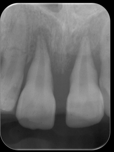
On referral — 3 months after injury
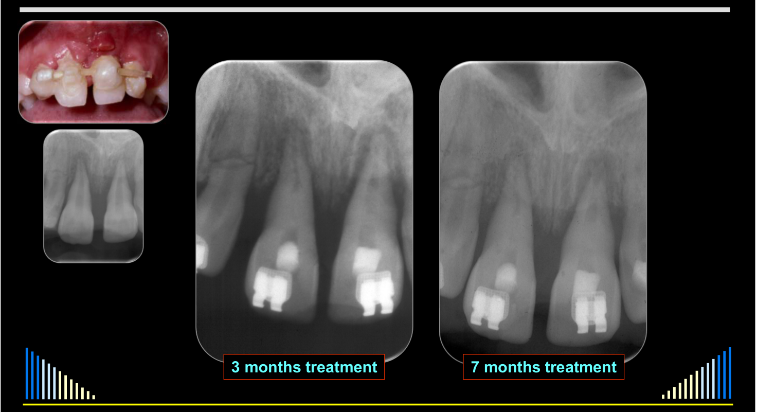
Lateral Inflammatory Resorption — 43 Teeth61
Three teeth lost due to periodontal problems and external inflammatory resorption.
Favourable outcomes:
- 97.7% of patients
- 93.0% of teeth
NB — the % of teeth is skewed by the three teeth with unfavourable outcomes being from the same patient
 |  | |
 |  |
Apical Inflammatory Resorption — 75 Teeth62
103 patients with 118 teeth — interceptive treatment for established resorption:
- 75 teeth — Apical inflammatory resorption
- 73 successfully managed — 97.3%
- 2 teeth required periapical surgery — 2.6%
- Overall — 100% of the teeth were retained
 |  |  |
| |  |
Overall Outcome of Management Regimes
- Patients: only 1 patient lost teeth — 99.4% favourable
- Teeth: 3 teeth lost from one patient — 98.3% favourable
- However: 2 teeth required periapical surgery — but they healed & were retained
Lecturer — Calcium Hydroxide Timing
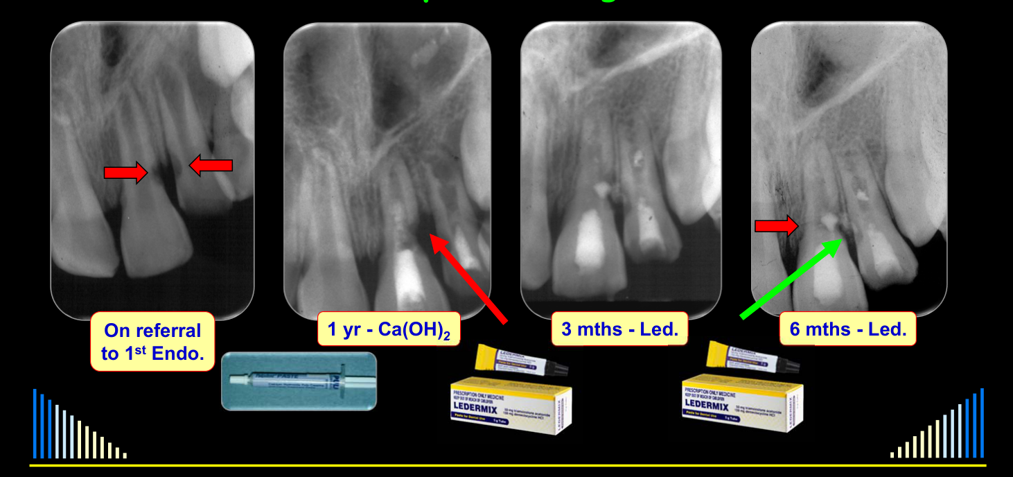
Calcium hydroxide should not be used as the initial medicament in these traumatized teeth because its high pH may damage periodontal-ligament fibroblasts and encourage ankylosis and replacement resorption.
- In a case involving two immature avulsed incisors, calcium hydroxide used for one year was associated with worsening resorption and interproximal bone loss.
- After replacement with Ledermix, three months of treatment produced progressive repair, although the patient later sustained a root fracture.
Conclusions and Summary
Conclusions
- Treat with a CS-AB paste initially
- Use Ca(OH)₂ as a secondary medicament
One more example…
On referral to 1st Endo. — 3 mths — Led. — 6 mths — Led. — 1 yr — Ca(OH)₂*
Summary63
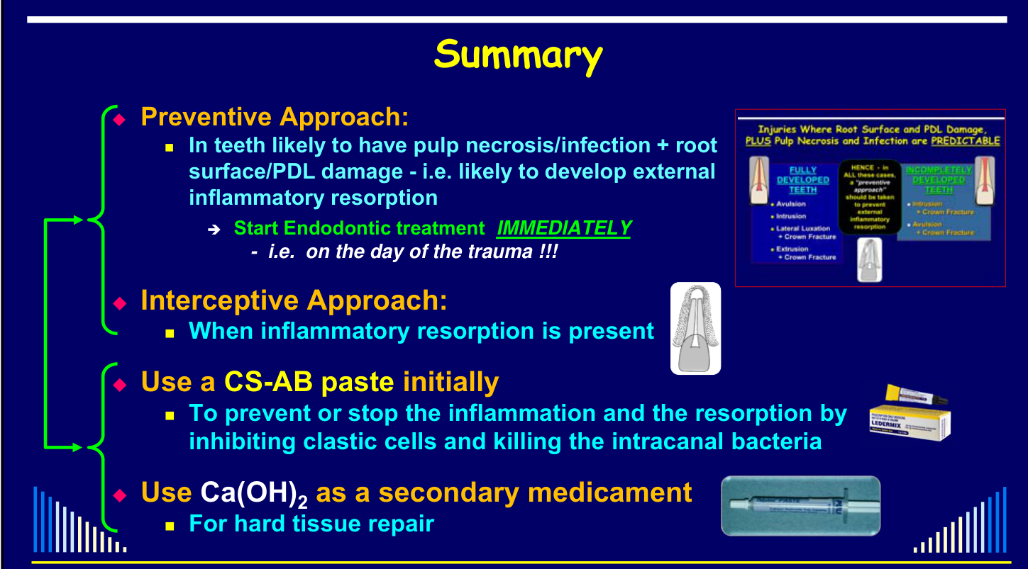
- Preventive Approach:
- In teeth likely to have pulp necrosis/infection + root surface/PDL damage — i.e. likely to develop external inflammatory resorption
- → Start Endodontic treatment IMMEDIATELY — i.e. on the day of the trauma !!!
- In teeth likely to have pulp necrosis/infection + root surface/PDL damage — i.e. likely to develop external inflammatory resorption
- Interceptive Approach:
- When inflammatory resorption is present
- Use a CS-AB paste initially
- To prevent or stop the inflammation and the resorption by inhibiting clastic cells and killing the intracanal bacteria
- Use Ca(OH)₂ as a secondary medicament
- For hard tissue repair
Thank you
Clinical Cases
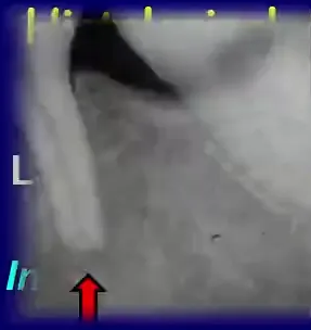
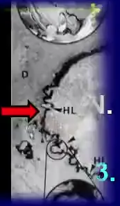
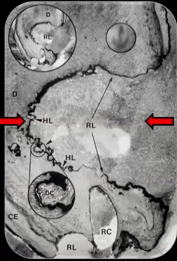
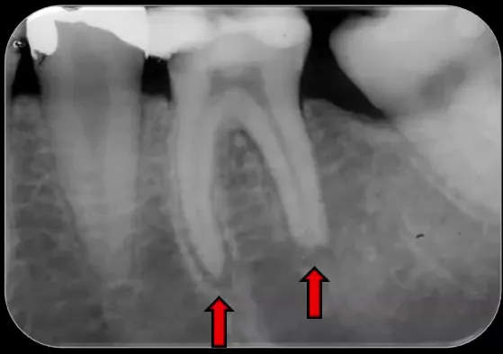
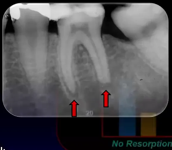
Case: Extracted tooth with apical inflammatory resorption, radiograph and electron microscopy
Question
Scenario: An extracted tooth from a study investigating the correlation between radiographic and histological findings in teeth with periapical radiolucencies.
What’s shown: A radiograph and an electron microscopy slide of the extracted tooth.
Consider: What specific findings are visible on the radiograph and the electron microscopy slide, and what do they demonstrate about the detection of external apical inflammatory resorption?
Answer
Observations:
- The radiograph shows obvious signs of apical inflammatory resorption of both tooth roots, with the distal root being more advanced than the mesial root.
- The electron microscopy slide reveals fairly extensive resorption lacunae at the end of the tooth root.
Reasoning: The lecturer points out these specific findings to illustrate the study’s methodology of correlating radiographic signs with histological reality. By showing that the radiograph displays obvious resorption while the electron microscopy confirms extensive resorption lacunae, the lecturer demonstrates how histological examination reveals the true extent of the disease that radiographs may miss.
Outcome: The study found that radiographs only detect external apical inflammatory resorption in about 20% of cases, whereas histological examination shows it is present in about 80% of cases. This indicates that four out of five teeth with a periapical radiolucency likely have external inflammatory resorption.
Takeaway: Clinicians must keep in mind that external apical inflammatory resorption is likely present in the majority (approximately 80%) of teeth with a periapical radiolucency, even if it is not always clearly visible on radiographs, when treating an infected root canal system with chronic apical periodontitis.
Case: Replanted monkey tooth, no medicament group histology
Question
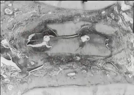
Scenario: A histology picture from a study on replanted monkey teeth is presented, showing a tooth from the group that received no intracanal medicament after being left dry for an hour.
What’s shown: A micrograph of a tooth root and adjacent teeth.
Consider: Identify the findings visible in the root structure and surrounding tissues.
Answer
Observations:
- Areas of intense inflammatory resorption and inflammation, appearing as dark areas.
- Significant loss of tooth root structure.
- Adjacent teeth visible at the top and bottom of the image.
Reasoning: The extensive dark areas and severe loss of tooth structure indicate intense external inflammatory root resorption, which occurs when no medicament is used to control inflammation after prolonged dry time.
Outcome: None stated for this specific exhibit.
Takeaway: Without intracanal medicament, replanted teeth left dry for extended periods undergo severe inflammatory root resorption, destroying much of the root structure.
Case: Replanted monkey tooth, Ledermix group histology
Question
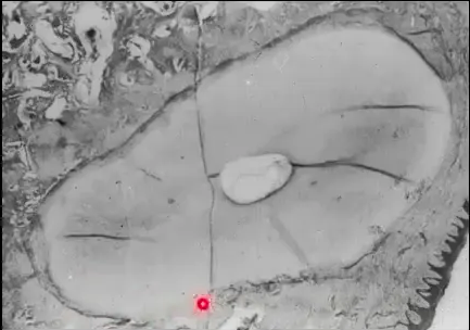
Scenario: A representative histology picture from the same study is presented, showing a tooth root from the group treated with Ledermix paste.
What’s shown: A micrograph of a tooth root.
Consider: Identify the findings visible in the root structure and surrounding tissues and compare them to the untreated group.
Answer
Observations:
- No areas of inflammation or inflammatory resorption.
- Presence of some normal periodontal ligament.
- Areas of ankylosis and replacement resorption.
Reasoning: The absence of inflammation demonstrates that the Ledermix paste effectively inhibited inflammatory resorption. However, the presence of ankylosis and replacement resorption reflects the underlying damage to the periodontal ligament cells from the extended dry time before replantation, which the medicament cannot reverse.
Outcome: None stated for this specific exhibit.
Takeaway: Ledermix paste effectively prevents inflammatory root resorption but does not prevent replacement resorption caused by periodontal ligament cell death during extended extra-oral dry times.
Case: Young boy with multiple dental injuries, clinical photo
Question
Scenario: A young boy presents after being kicked in the mouth during a football game, resulting in multiple dental injuries.
What’s shown: A clinical photograph showing the repositioned lateral incisor with a splint in place, rubber dam isolation, and noticeable swelling of the lip a couple of hours post-injury.
Consider: The immediate management of the traumatized teeth and the advantages of the clinical techniques demonstrated.
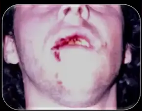
Answer
Observations:
- Multiple injuries are present, with lateral luxation of the lateral incisor highlighted.
- The lateral incisor has been repositioned and splinted, though the splint is not yet finalized on the central incisor.
- Rubber dam is placed for the emergency endodontic procedure.
- The patient’s lip is already quite swollen a couple of hours after the injury.
Reasoning: Immediate repositioning, splinting, and endodontic intervention (pulp removal and medicament placement) are critical on the day of injury to prevent inflammatory resorption. Placing the rubber dam before any procedure other than local anesthesia provides excellent isolation and holds the rapidly swelling lip out of the way, making the emergency management much easier to perform.
Outcome: The emergency management, including access, pulp removal, and placement of a corticosteroid antibiotic dressing, was completed on the day of the injury under rubber dam. The splint was placed, and further endodontic treatment for the central incisor was planned.
Takeaway: Initiating endodontic treatment immediately on the day of trauma, utilizing rubber dam isolation, effectively manages the teeth before facial swelling exacerbates and helps prevent external inflammatory resorption.
Case: Upper right central incisor following a car accident
Question
Scenario: A dental therapist was involved in a car accident where her face hit the steering wheel, resulting in the avulsion of her upper right central incisor. She reimplanted the tooth herself after cleaning it with saliva, and her dentist placed a flexible splint and started immediate root canal treatment.
What’s shown: A photograph and radiograph at one week, followed by radiographs at 9 months, 3 years, and 5 years.
Consider: Evaluate the long-term outcome of the immediate preventive endodontic treatment protocol.
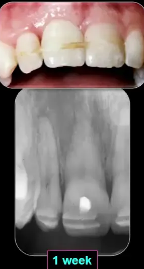
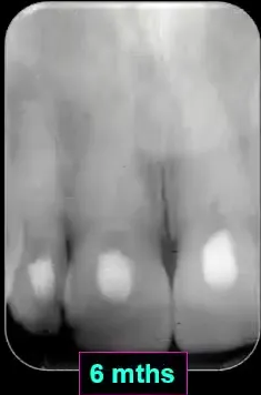
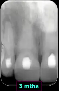
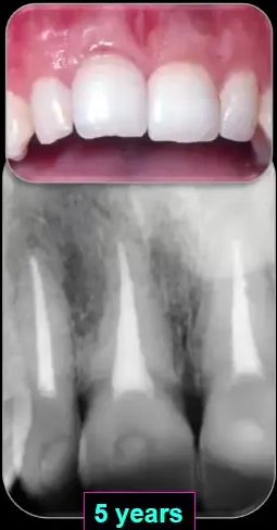
Answer
Observations:
- At 1 week, 9 months, 3 years, and 5 years: no signs of external inflammatory resorption of the central incisor.
- Some discoloration of the teeth.
- At 5 years: signs of ankylosis in one area, with persistent percussion sounds.
Reasoning: Immediate treatment and the preventive protocol (flexible splint, Linimex paste, systemic antibiotics) successfully stopped external inflammatory resorption. However, the development of ankylosis eventually led to external replacement resorption.
Outcome: Internal bleaching was performed for tooth discoloration. The central incisor lasted 15 years before it had to be extracted due to external replacement resorption.
Takeaway: The preventive treatment protocol can effectively stop external inflammatory resorption and slow down external replacement resorption, though ankylosis may eventually lead to replacement resorption and tooth loss.
Case: Two intruded teeth following trauma
Question
Scenario: A patient presented with two intruded teeth and uncomplicated crown fractures. The initial dentist did not recognize the intrusion, thought the teeth were lost, and only placed sutures. The patient was seen 12 hours later.
What’s shown: Photographs and radiographs at 12 hours post-injury, plus a follow-up radiograph and photograph from a new dentist after the patient relocated.
Consider: Evaluate the outcome of repositioning and immediate root canal treatment for the intruded teeth.
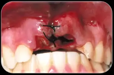
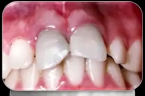
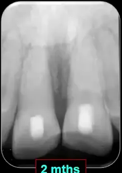
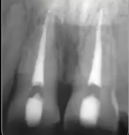
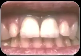
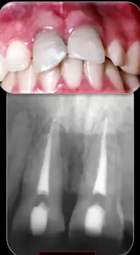
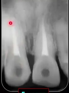
Answer
Observations:
- The teeth were intruded with uncomplicated crown fractures.
- Follow-up images from the new dentist show no signs of external inflammatory or replacement resorption.
Reasoning: Repositioning, stabilization, and immediate root canal treatment with Lidomex paste followed the preventive protocol, which prevented resorptive complications.
Outcome: The teeth were repositioned, stabilized, and root canal treatment was started immediately. They were eventually restored, and long-term follow-up confirmed no resorption.
Takeaway: The preventive protocol can successfully manage intruded teeth and prevent resorptive complications, even when initial management was inadequate.
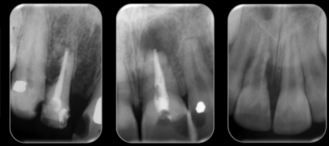
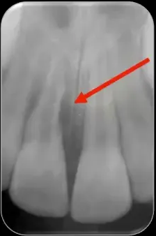
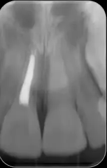
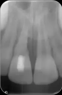
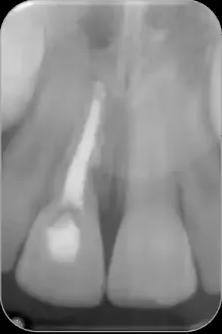
Case: Tooth with lateral inflammatory resorption and an open apex
Question
Scenario: A tooth presenting with lateral inflammatory resorption and an open apex, treated using the interceptive management protocol.
What’s shown: Radiographs at baseline, 3 months, and 4 years post-treatment.
Consider: Evaluate the repair process and treatment duration for a tooth with an open apex.
Answer
Observations:
- At 3 months: good repair occurring.
- At 4 years: the tooth is stable, with periodontal ligament visible in the area where resorption previously occurred.
Reasoning: The interceptive protocol (corticosteroid antibiotic, then calcium hydroxide) stimulated hard tissue repair (cementum, PDL, and bone) despite the loss of tooth structure. The open apex required a longer dressing period.
Outcome: The tooth was dressed for 18 months before completing the root filling. It remained stable at the 4-year follow-up with evidence of periodontal ligament repair.
Takeaway: Interceptive management can achieve good hard tissue repair in lateral inflammatory resorption, even with an open apex, though it may require extended dressing times.
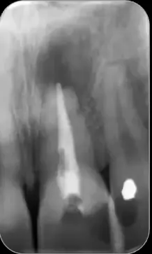
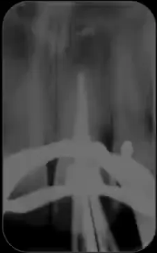
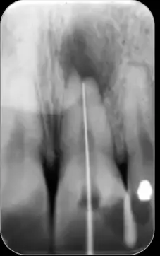
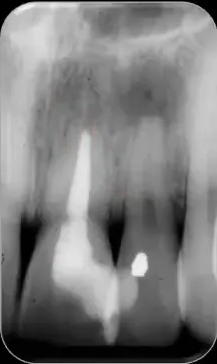
Case: Infected root-filled tooth with apical resorption
Question
Scenario: A tooth with external apical inflammatory resorption associated with an infected root-filled tooth and an open apex.
What’s shown: Radiographs preoperatively, at 15 months, and at 4 years post-treatment.
Consider: Evaluate the formation of a hard tissue barrier and long-term stability.
Answer
Observations:
- At 15 months: able to complete the root canal filling.
- At 4 years: a nice hard tissue barrier formed across the end of the root, and the tooth is stable.
Reasoning: The interceptive protocol successfully halted the inflammatory resorption and stimulated hard tissue repair across the open apex.
Outcome: The root canal filling was completed at 15 months. The tooth was stable at the 4-year review with a hard tissue barrier.
Takeaway: Interceptive management can successfully treat external apical inflammatory resorption in infected root-filled teeth with open apices, leading to a stable hard tissue barrier.
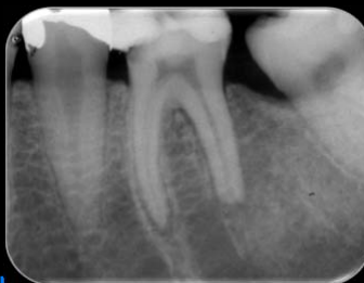
Case: Lower first molar with root resorption and caries
Question
Scenario: A lower first molar with significant inflammatory resorption of the distal root (and some mesial root) associated with a breaking down amalgam restoration and significant caries.
What’s shown: Radiographs preoperatively and at a 4-year review.
Consider: Evaluate the outcome of the interceptive protocol on a posterior tooth with extensive resorption.
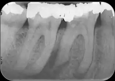
Answer
Observations:
- Preoperative: significant inflammatory resorption of the distal root and some of the mesial root.
- At 4-year review: the tooth is stable, with slight widening of the ligament spaces likely representing scar tissue.
Reasoning: Removing the cause (caries and breaking down restoration) and applying the interceptive protocol halted the resorption and allowed for stabilization.
Outcome: The root canal filling was completed after 18 months. The tooth was stable at the 4-year review.
Takeaway: Interceptive management can successfully stabilize posterior teeth with significant inflammatory resorption secondary to carious exposure.
Case: Tooth with root resorption of both roots, previously root-filled
Question
Scenario: A patient referred with a tooth that had root canal treatment in 1975. Serial radiographs from the referring dentist showed progressive periapical changes over the years, leading to significant inflammatory resorption of both roots by the time the lecturer saw the patient.
What’s shown: Old radiographs from 1975, 1977, 1979, 1980, and at presentation. Follow-up radiographs at 6 months, a couple of months later, and 3 years post-treatment.
Consider: Evaluate the repair process after removing the old root fillings and restoring the tooth.
Answer
Observations:
- Pre-treatment: significant inflammatory resorption of both roots.
- At 6 months: fairly good repair occurring.
- A couple of months later: very good repair.
- At 3 years: the tooth is stable.
Reasoning: Removing the old root fillings, amalgam, and crown, and temporizing the tooth allowed the interceptive protocol to halt resorption and stimulate repair.
Outcome: The tooth was temporized with a stainless steel band and glass ionomer. The root canal filling was completed after a couple of months of dressing. The tooth was stable at the 3-year follow-up.
Takeaway: Even long-standing, significant inflammatory resorption of both roots can be successfully treated and stabilized using the interceptive protocol.
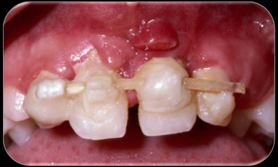
Case: Inadequately splinted avulsed teeth with periodontal complications
Question
Scenario: A patient presented a few months after trauma involving three avulsed teeth and a transverse root fracture of an adjacent lateral incisor. The teeth had been replanted after being stored in milk, but the initial management lacked systemic antibiotics and root canal treatment.
What’s shown: An intraoral view of the splinted teeth, showing the type of splint used and the restorative material applied.
Consider: What are the clinical findings on the splint and teeth, and what was the outcome for these teeth?
Answer
Observations:
- The splint consists of nylon fishing line with no anchorage, binding the three avulsed teeth and the fractured tooth together.
- Massive blobs of composite resin are present over the teeth.
- The patient was unable to clean the area, leading to poor oral hygiene.
- Periodontal pocketing was greater than 10 mm with rapid bone loss.
Reasoning: The inadequate splinting technique and poor oral hygiene led to severe periodontal problems. These periodontal issues contributed to ongoing external inflammatory resorption, which could not be halted despite the interceptive treatment protocol.
Outcome: The three avulsed teeth were ultimately extracted due to the ongoing periodontal problems and inflammatory resorption.
Takeaway: Inadequate splinting and poor oral hygiene can lead to severe periodontal complications, compromising the outcome of interceptive management for inflammatory root resorption.
Case: Avulsed immature incisors treated with calcium hydroxide versus corticosteroid-antibiotic paste
Question
Scenario: A young girl had her central and lateral incisors avulsed. Due to immature roots, revascularization was attempted but failed, and external inflammatory resorption developed. She was treated by a private endodontist with calcium hydroxide for 12 months before being referred to a university clinic.
What’s shown: Radiographs showing the initial resorption defects, the progression after 12 months of calcium hydroxide, the repair after switching medicaments, and a subsequent complication.
Consider: How did the resorption and bone respond to the change in medicament, and what was the final outcome for the teeth?
Answer
Observations:
- Initial radiographs showed external inflammatory resorption on both central and lateral incisors.
- After 12 months of calcium hydroxide, the resorption worsened, and interproximal bone was lost.
- After switching to a corticosteroid-antibiotic paste for three months, the teeth and interproximal bone showed progressive repair.
- A subsequent radiograph revealed a root fracture following further trauma.
Reasoning: Using calcium hydroxide as the initial medicament failed to control the inflammation, leading to worsening resorption and bone loss. Switching to a corticosteroid-antibiotic paste successfully inhibited the clastic cells and bacteria, allowing the periodontal ligament and bone to repair.
Outcome: The resorption defects and interproximal bone progressively repaired after switching to the corticosteroid-antibiotic paste, but the teeth ultimately suffered a root fracture due to further trauma.
Takeaway: Calcium hydroxide should be used as a secondary medicament for hard tissue repair only after initial inflammation is controlled with a corticosteroid-antibiotic paste to prevent ongoing resorption, ankylosis, and replacement resorption.
Footnotes
-
Original PDF page 1: L7 Resorption 4 - External Inflammatory, p.1 ↩
-
Original PDF page 2: L7 Resorption 4 - External Inflammatory, p.2 ↩
-
Original PDF page 3: L7 Resorption 4 - External Inflammatory, p.3 ↩
-
Original PDF page 4: L7 Resorption 4 - External Inflammatory, p.4 ↩
-
Original PDF page 5: L7 Resorption 4 - External Inflammatory, p.5 ↩
-
Original PDF page 6: L7 Resorption 4 - External Inflammatory, p.6 ↩
-
Original PDF page 7: L7 Resorption 4 - External Inflammatory, p.7 ↩
-
Original PDF page 9: L7 Resorption 4 - External Inflammatory, p.9 ↩
-
Original PDF page 8: L7 Resorption 4 - External Inflammatory, p.8 ↩
-
Original PDF page 10: L7 Resorption 4 - External Inflammatory, p.10 ↩
-
Original PDF page 11: L7 Resorption 4 - External Inflammatory, p.11 ↩
-
Original PDF page 12: L7 Resorption 4 - External Inflammatory, p.12 ↩
-
Original PDF page 13: L7 Resorption 4 - External Inflammatory, p.13 ↩
-
Original PDF page 14: L7 Resorption 4 - External Inflammatory, p.14 ↩
-
Original PDF page 15: L7 Resorption 4 - External Inflammatory, p.15 ↩
-
Original PDF page 16: L7 Resorption 4 - External Inflammatory, p.16 ↩
-
Original PDF page 17: L7 Resorption 4 - External Inflammatory, p.17 ↩
-
Original PDF page 18: L7 Resorption 4 - External Inflammatory, p.18 ↩
-
Original PDF page 19: L7 Resorption 4 - External Inflammatory, p.19 ↩
-
Original PDF page 20: L7 Resorption 4 - External Inflammatory, p.20 ↩
-
Original PDF page 21: L7 Resorption 4 - External Inflammatory, p.21 ↩
-
Original PDF page 22: L7 Resorption 4 - External Inflammatory, p.22 ↩
-
Original PDF page 23: L7 Resorption 4 - External Inflammatory, p.23 ↩
-
Original PDF page 24: L7 Resorption 4 - External Inflammatory, p.24 ↩
-
Original PDF page 26: L7 Resorption 4 - External Inflammatory, p.26 ↩
-
Original PDF page 25: L7 Resorption 4 - External Inflammatory, p.25 ↩
-
Original PDF page 27: L7 Resorption 4 - External Inflammatory, p.27 ↩
-
Original PDF page 28: L7 Resorption 4 - External Inflammatory, p.28 ↩
-
Original PDF page 29: L7 Resorption 4 - External Inflammatory, p.29 ↩
-
Original PDF page 30: L7 Resorption 4 - External Inflammatory, p.30 ↩
-
Original PDF page 31: L7 Resorption 4 - External Inflammatory, p.31 ↩
-
Original PDF page 32: L7 Resorption 4 - External Inflammatory, p.32 ↩
-
Original PDF page 33: L7 Resorption 4 - External Inflammatory, p.33 ↩
-
Original PDF page 34: L7 Resorption 4 - External Inflammatory, p.34 ↩
-
Original PDF page 35: L7 Resorption 4 - External Inflammatory, p.35 ↩
-
Original PDF page 36: L7 Resorption 4 - External Inflammatory, p.36 ↩
-
Original PDF page 39: L7 Resorption 4 - External Inflammatory, p.39 ↩
-
Original PDF page 40: L7 Resorption 4 - External Inflammatory, p.40 ↩
-
Original PDF page 41: L7 Resorption 4 - External Inflammatory, p.41 ↩
-
Original PDF page 38: L7 Resorption 4 - External Inflammatory, p.38 ↩
-
Original PDF page 37: L7 Resorption 4 - External Inflammatory, p.37 ↩
-
Original PDF page 42: L7 Resorption 4 - External Inflammatory, p.42 ↩
-
Original PDF page 43: L7 Resorption 4 - External Inflammatory, p.43 ↩
-
Original PDF page 44: L7 Resorption 4 - External Inflammatory, p.44 ↩
-
Original PDF page 45: L7 Resorption 4 - External Inflammatory, p.45 ↩
-
Original PDF page 48: L7 Resorption 4 - External Inflammatory, p.48 ↩
-
Original PDF page 49: L7 Resorption 4 - External Inflammatory, p.49 ↩
-
Original PDF page 51: L7 Resorption 4 - External Inflammatory, p.51 ↩
-
Original PDF page 50: L7 Resorption 4 - External Inflammatory, p.50 ↩
-
Original PDF page 52: L7 Resorption 4 - External Inflammatory, p.52 ↩
-
Original PDF page 53: L7 Resorption 4 - External Inflammatory, p.53 ↩
-
Original PDF page 54: L7 Resorption 4 - External Inflammatory, p.54 ↩
-
Original PDF page 55: L7 Resorption 4 - External Inflammatory, p.55 ↩
-
Original PDF page 56: L7 Resorption 4 - External Inflammatory, p.56 ↩
-
Original PDF page 57: L7 Resorption 4 - External Inflammatory, p.57 ↩
-
Original PDF page 64: L7 Resorption 4 - External Inflammatory, p.64 ↩
-
Original PDF page 61: L7 Resorption 4 - External Inflammatory, p.61 ↩
-
Original PDF page 58: L7 Resorption 4 - External Inflammatory, p.58 ↩
-
Original PDF page 59: L7 Resorption 4 - External Inflammatory, p.59 ↩
-
Original PDF page 60: L7 Resorption 4 - External Inflammatory, p.60 ↩
-
Original PDF page 62: L7 Resorption 4 - External Inflammatory, p.62 ↩
-
Original PDF page 63: L7 Resorption 4 - External Inflammatory, p.63 ↩
-
Original PDF page 65: L7 Resorption 4 - External Inflammatory, p.65 ↩