L9 Resorption 5 - External Invasive
External Invasive Resorption: Dilemmas in Diagnosis, Treatment and Prognosis12
External Invasive Resorption
- Dilemmas in Diagnosis, Treatment and Prognosis
Prof. Paul V. Abbott AO BDSc, MDS, FRACDS(Endo), FPFA, FADI, FICD, FACD, FIADT
- Specialist Endodontist
- Winthrop Professor of Clinical Dentistry
- School of Dentistry, The University of Western Australia
Oral Health Centre of Western Australia
 |  |
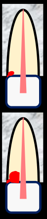
Introduction and Terminology345
 |  |  |
 |  |  |
 |  |
Classification of Tooth Resorption6
External
See - External root resorption types
The full external-resorption list is retained in L5 external classification. See the topic overview in Root Resorption Classification.
Lecturer — Resorption Classification
The lecture places external invasive resorption within a broader classification of resorptive conditions.
- Three types are internal in origin, while eight types are external in origin.
- Physiological resorption is a normal process; the other types discussed are pathological.
- Surface, inflammatory, and replacement resorptions share some similarities, but external invasive resorption has distinct clinical and histological features.
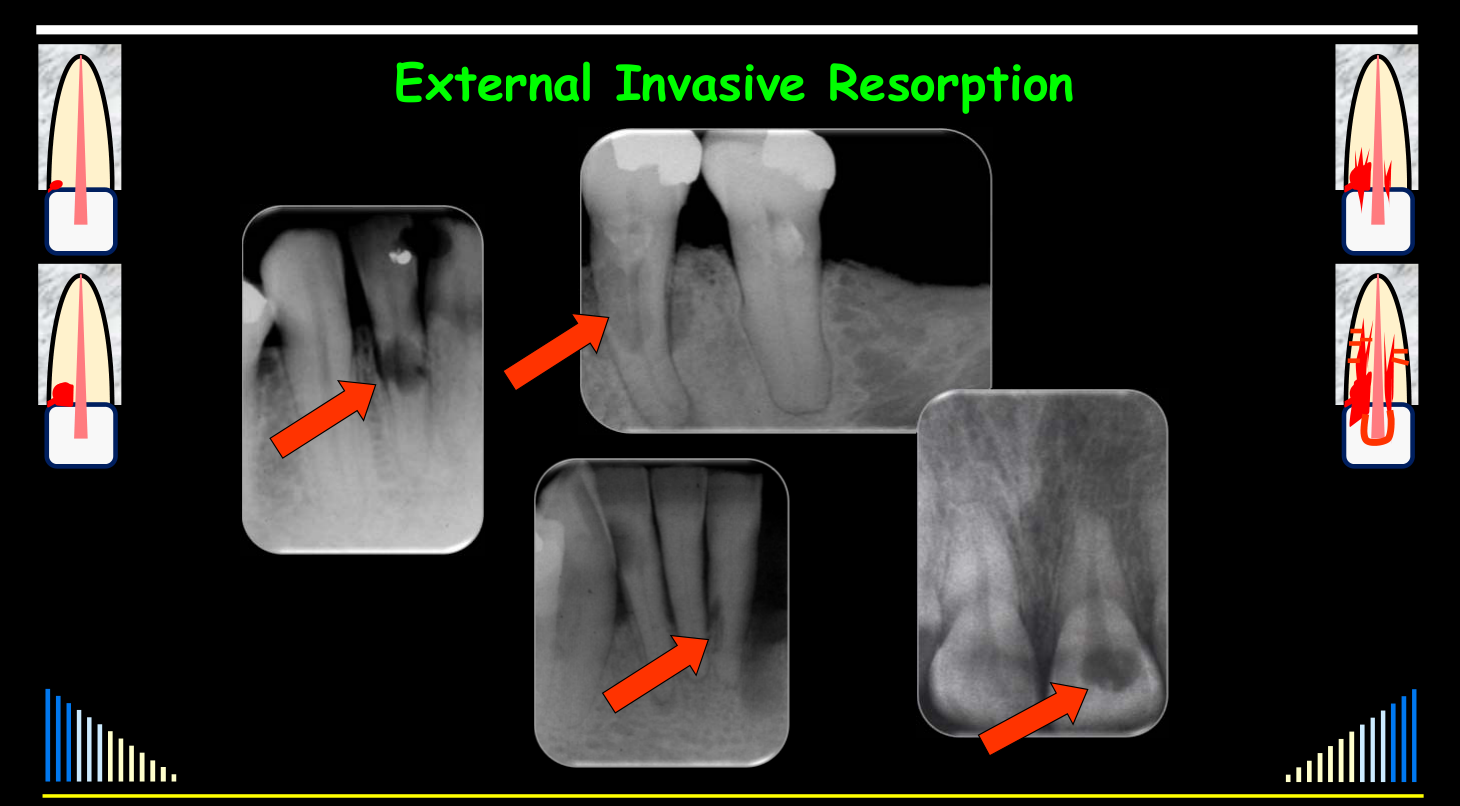
External Invasive Resorption
Other terminology that has been used:
- Idiopathic external resorption
- Late cervical resorption
- Cervical resorption
- External cervical invasive resorption
- External cervical inflammatory resorption
- Peripheral inflammatory resorption
- Extra-canal invasive resorption
- Fibrous dysplasia of teeth
Lecturer — Terminology
Several terms have been used, but each has limitations; the preferred term is external invasive resorption because the resorptive tissue invades the tooth from an external, subgingival location.
- Idiopathic external resorption does not indicate whether the process is replacement, inflammatory, pressure-related, or invasive.
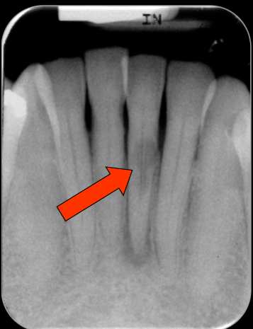
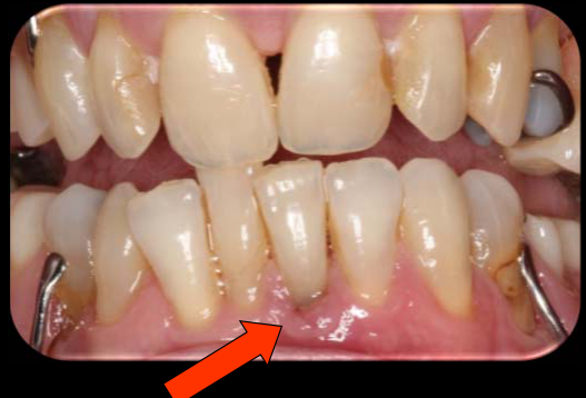
- “Late” cervical resorption is inappropriate because the condition is not restricted to teeth with a long interval after trauma, and “cervical” is incomplete because the lesion starts subgingivally and may occur below the cervical region when gingival recession is present.
- Inflammation is not part of the resorptive process; it may occur secondarily if plaque accumulates in a defect created by the resorption.
- Extra-canal resorption is inconsistent with the usual internal–external distinction: external resorption begins at the cementum and progresses through dentine, whereas internal resorption begins within the pulp or root canal and progresses outward.
- Although some radiographic similarities exist with fibrous dysplasia of bone, this condition is a resorptive process.
See - Terminology
Preferred terminology and its distinctions are in Introduction and Terminology.
Outline
- Definition and Terminology
- Histopathogenesis
- Potential Predisposing Factors
- Features / Classification
- Differential Diagnosis
- Management Strategies
- Outcomes
Topic map: Resorption Index.
Heithersay, G.S. — Quintessence International 1999; 30:
- A. Pages 27–37 — Clinical, radiologic and histopathologic features
- B. Pages 83–95 — Potential predisposing factors
- C. Pages 96–110 — Treatment with TCA, curettage and restoration
Also: Endodontic Topics 2004; 7: 73–92.
Lecturer — Heithersay Research
Much of the important research was conducted by Professor Geoffrey Heithersay, particularly during the 1980s and 1990s.
- His work culminated in three comprehensive publications in Quintessence International in 1990.
- A later paper in Endodontic Topics summarized this work.
- <mark>Orthodontic treatment and trauma</mark>
- <mark>Developmental defects at the cemento-enamel junction</mark>
Lecturer — Predisposing Factors
These factors should be regarded as potential predisposing factors rather than proven causes.
- Gingival inflammation alone cannot explain the condition, because many people have gingivitis without developing external invasive resorption.
- Trauma may be involved in some cases, but many cases have no history of trauma.
- Toxic or caustic materials may pass through cervical accessory canals, and bleaching has also been considered, but many treated or bleached teeth do not develop resorption.
- In one review of about 250 teeth, orthodontic treatment was the most frequently identified potential factor; the detailed chart and evidence limits are in Heithersay 1999 - Predisposing Factors Pages 83-95.
- Patients should not be told that orthodontic treatment, trauma, bleaching, or another individual factor necessarily caused the resorption.
- Enamel and cementum meet directly in approximately 30% of teeth
- In approximately 5–10% of teeth, enamel and cementum do not meet, leaving dentine exposed
- Exposed dentine has been reported in up to 30% of upper incisors
- The defect does not need to involve the entire circumference of the root; exposure in one area may be sufficient
Lecturer — Cemento-Enamel Junction
A developmental defect at the cemento-enamel junction may provide the underlying susceptibility, while another event may stimulate clastic cells to recognize exposed dentine as bone-like tissue.
- Upper incisors are frequently involved and may also be exposed to trauma, internal bleaching after discoloration, endodontic treatment, and orthodontic movement, including palatal crown movement.
- These events may be relevant, but none is necessarily sufficient by itself.
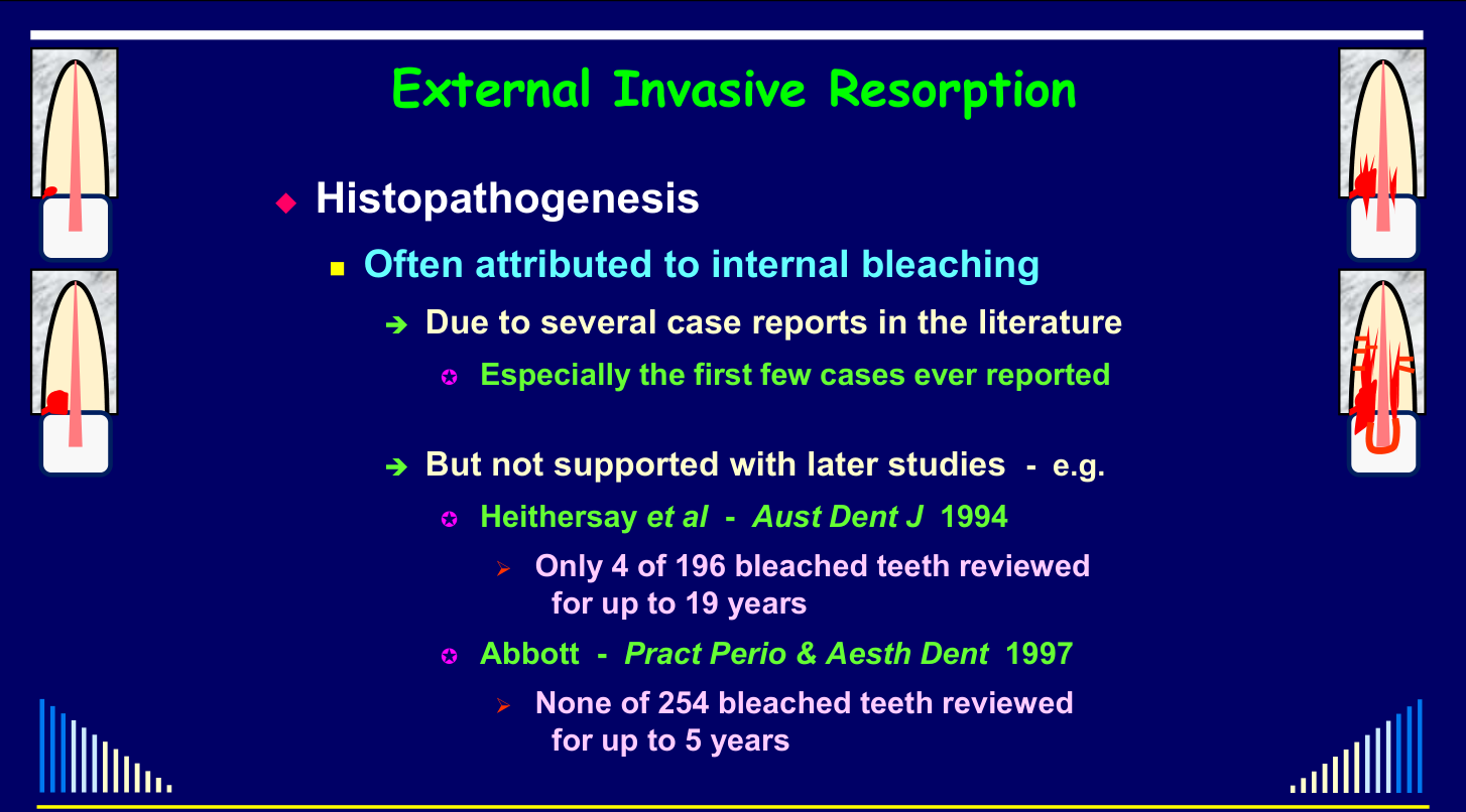
- Internal bleaching was initially overemphasized because the first reported cases occurred in internally bleached teeth, involved small numbers, and often also included trauma and root canal treatment.
- In a review of almost 200 bleached teeth followed for up to 19 years, only four developed external invasive resorption; all four had multiple potential factors.
- In another review of 254 bleached teeth, none developed external invasive resorption within five years, although later development could not be excluded.
Histopathogenesis and Predisposing Factors78
External Invasive Resorption9
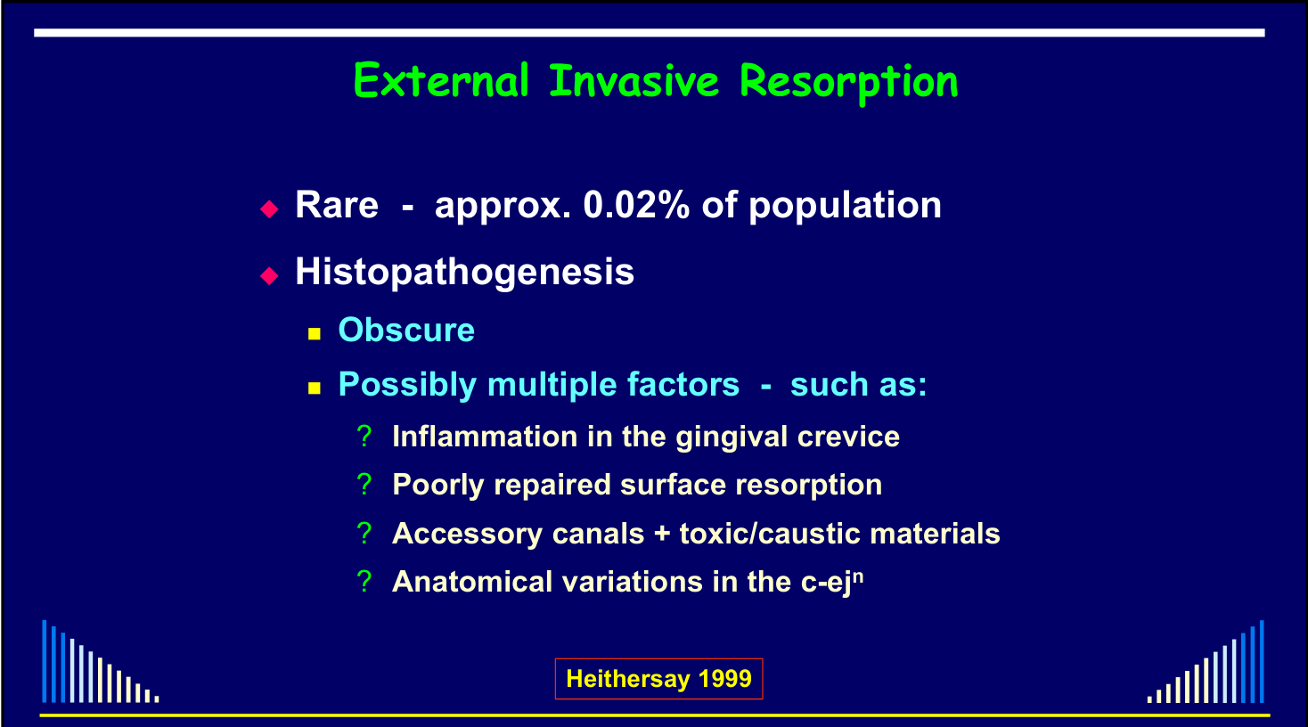
- Rare — approx. 0.02% of population
- Histopathogenesis
- Obscure
- Possibly multiple factors — such as:
- ? Inflammation in the gingival crevice
- ? Poorly repaired surface resorption
- ? Accessory canals + toxic/caustic materials
- ? Anatomical variations in the c-ejⁿ
Heithersay 1999
 | 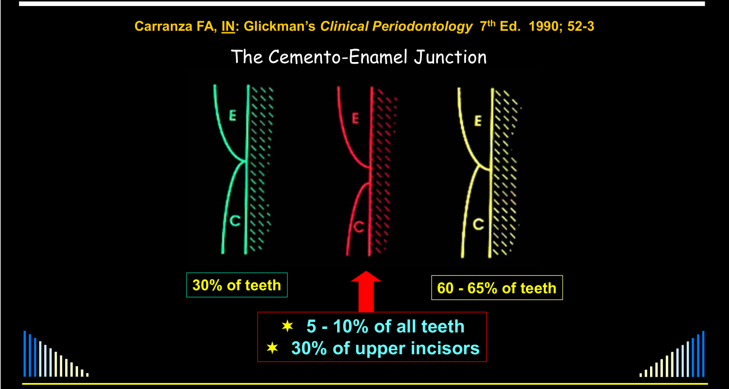 |
The Cemento-Enamel Junction
Carranza FA, IN: Glickman’s Clinical Periodontology 7th Ed. 1990; 52-3.
-
5–10% of all teeth
-
30% of upper incisors
-
60–65% of teeth
-
Histopathogenesis
- Often attributed to internal bleaching
- Due to several case reports in the literature
- Especially the first few cases ever reported
- Due to several case reports in the literature
- Often attributed to internal bleaching
| Case reports |
|---|
| Harrington & Natkin 1979 — 7 cases |
| Lado et al 1983 — 1 case |
| Montgomery 1984 — 1 case |
| Cvek & Lindvall 1985 — 11 cases |
| Friedman et al 1988 — 4 cases |
- Cvek & Lindvall 1985 — 11 cases
Heithersay 1999
Epidemiology and Prevalence
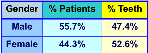
Analysis of a Referral-based Endodontic Practice. Part 1. Demographic Data and Reasons for Referral10
Abbott PV. J Endod 1994; 20: 93-96.
Lecturer — Incidence Trends
Heithersay estimated in 1999 that external invasive resorption affected approximately 0.02% of the population. The current incidence is thought to be higher, probably because of increased awareness, recognition, specialist referrals, and collaboration between endodontists and periodontists. The apparent increase may therefore reflect increased recognition rather than an actual increase in disease frequency.
Abbott 1994 referral finding
Of 2,000 patients referred, 57 had external cervical invasive resorption; 36% of those invasive cases were referred as internal resorption, not 36% of all 2,000 patients.
Full Table 5: Abbott 1994 - Referral Practice Table 5.
An Analysis of 219 Patients with External Invasive Resorption in 293 Teeth
56 Resorption Sites in One Patient
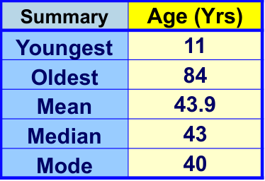
An unusual case involved a 27-year-old patient with 56 resorption sites in 32 teeth. The teeth and gingival tissues appeared attractive clinically, but extensive resorptive defects were present underneath, and the teeth ultimately required extraction.
2021 referral cohort
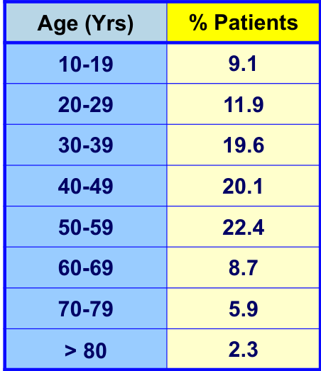
This unpublished series includes 219 patients and 293 teeth; class-specific treatments, follow-ups, media, and reported tally discrepancies are preserved in Abbott and Sunthoramoorthy 2021 - Invasive Resorption Cohort.
See - Lecture outline
Return to Introduction and Terminology.
Lecturer — Multiple-Tooth Involvement
Most people have only one tooth affected, but some patients develop multiple sites involving two or three teeth.
- Multiple-tooth involvement appears somewhat more common in patients with a history of orthodontic treatment.
- If orthodontic treatment is part of the history, obtain a panoramic radiograph to assess the other teeth.
- Further radiographs or a CT scan may be considered depending on the panoramic findings.
Lecturer — Clinical Variation
The condition varies considerably in its presentation and response.
- Location
- Radiographic appearance
- Extent
- Speed of progression
- Relationship to the pulp
- Relationship to the periodontal ligament
- Treatment response and prognosis
 |  |
 |
Clinical Features
Related diagnostic overview: Internal and External Resorption Diagnosis.
Features1112
-
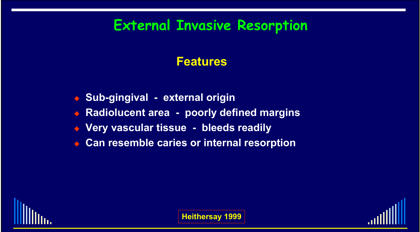
Sub-gingival – external origin
-
Radiolucent area – poorly defined margins
-
Very vascular tissue – bleeds readily
-
Can resemble caries or internal resorption
-
Considerable variation in radiographic appearance
-
Little or no pain, with many lesions discovered incidentally during routine radiography or dental hygiene treatment
-
A dental hygienist or dentist may catch an instrument in the resorptive area during scaling and cleaning Heithersay 1999. Publication details: Heithersay 1999 - Clinical Features Pages 27-37.
 |  |  |
 |  |  |
 |
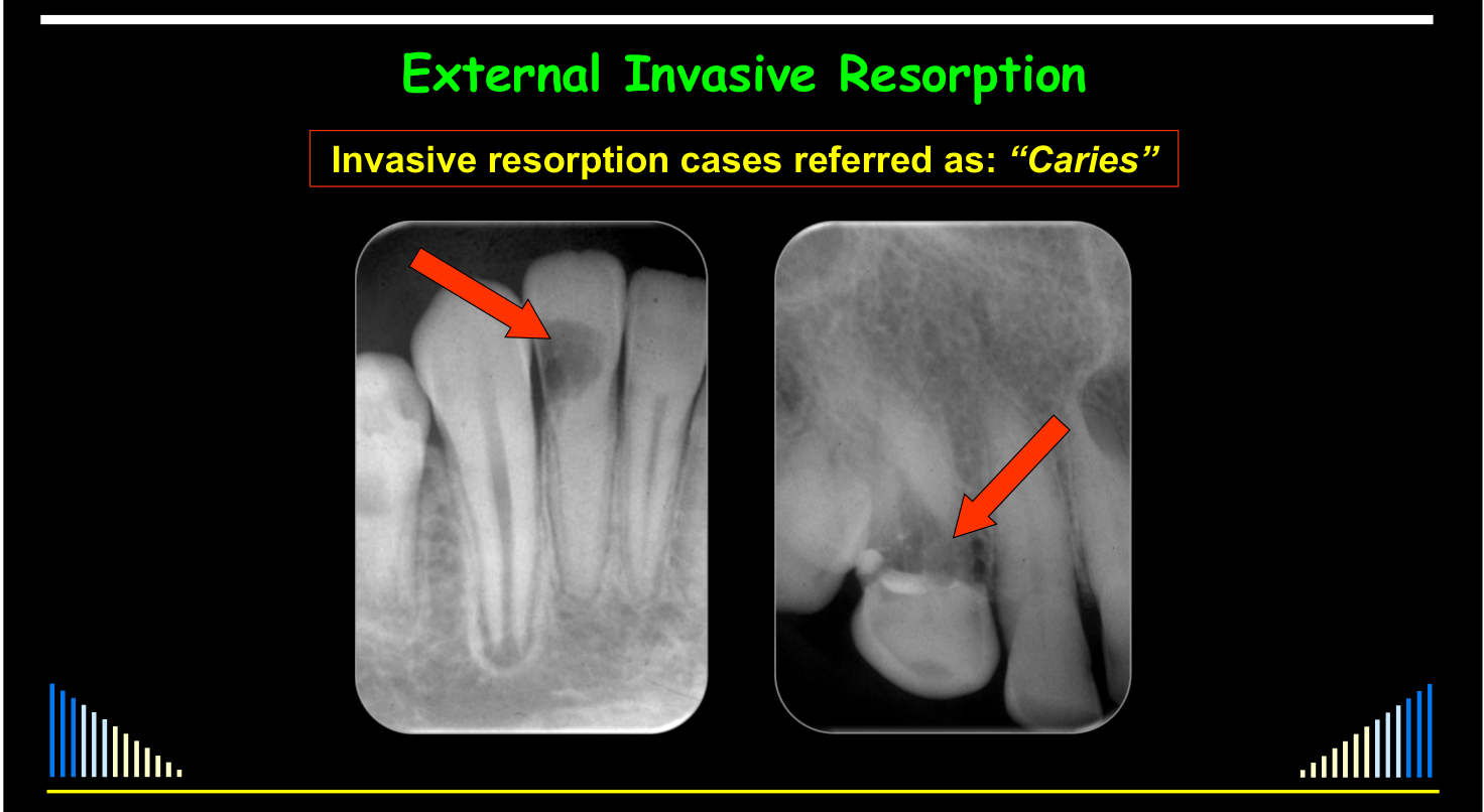
Referred as “Caries”
Invasive resorption cases referred as: “Caries”
Lecturer — Caries Misdiagnosis
External invasive resorption can be mistaken for caries because the defects do not develop in the usual caries pattern.
- The cemento-enamel junction, margins, and overall appearance may differ from caries.
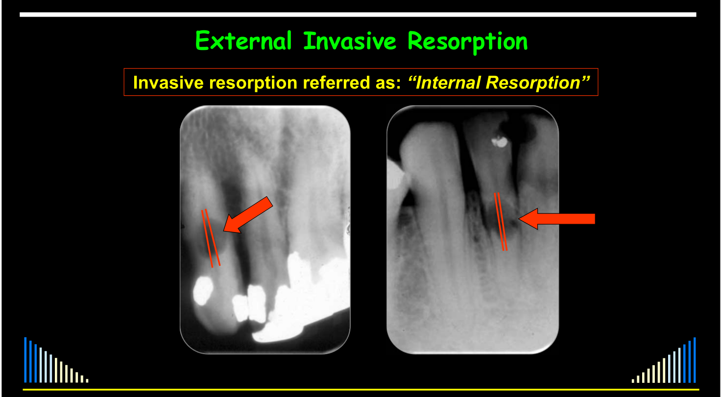
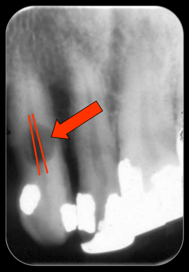
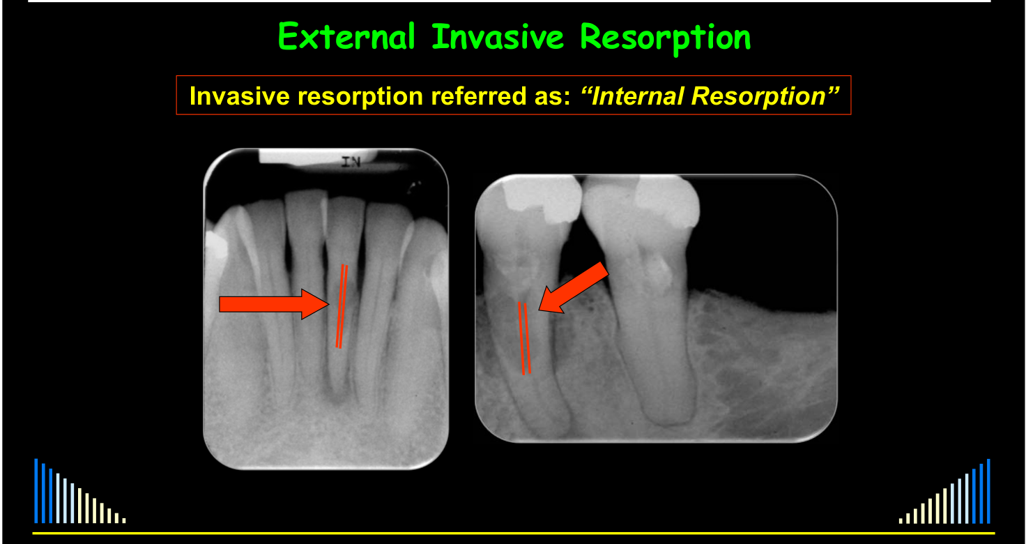
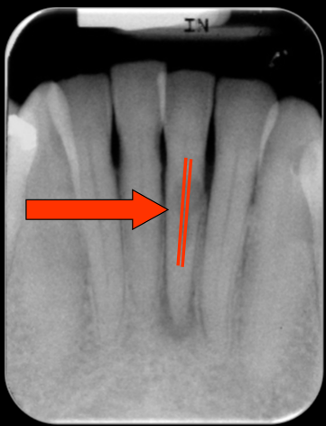
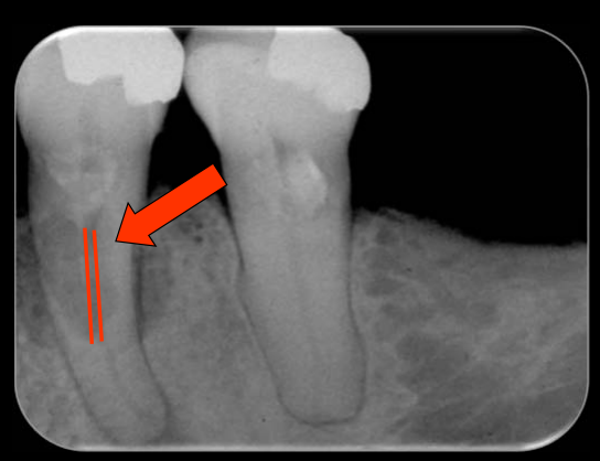
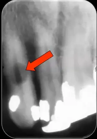
- The root canal outline may appear to pass through the defect because the defect is superimposed over the canal; tracing the canal walls helps distinguish external invasive resorption from internal resorption.
Invasive resorption referred as: “Internal Resorption”
Invasive resorption referred as: “Internal Resorption"
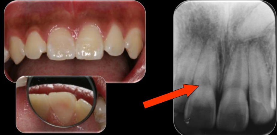
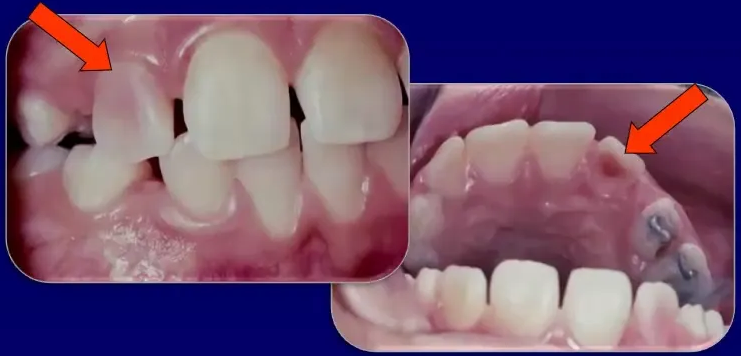
"Pink Teeth”13
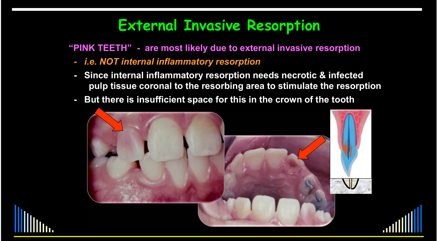
“PINK TEETH” – are most likely due to external invasive resorption:
-
i.e. NOT internal inflammatory resorption
-
Since internal inflammatory resorption needs necrotic & infected pulp tissue coronal to the resorbing area to stimulate the resorption
-
But there is insufficient space for this in the crown of the tooth
-
The pink colour results from highly vascular tissue in the early resorptive lesion and is not necessarily a sign of internal inflammatory resorption
-
A pink tooth should prompt consideration of external invasive resorption
Referral Data: Types and Incidence of Root Resorption
Abbott PV. Analysis of a Referral-based Endodontic Practice. Part 1. Demographic Data and Reasons for Referral. J Endod 1994; 20: 93-96.
See - Abbott 1994 referral table
The 36% refers to 57 patients with invasive resorption in a 2,000-patient referral cohort. Full Table 5 and denominator details: Abbott 1994 - Referral Practice Table 5.
An Analysis of 219 Patients with External Invasive Resorption in 293 Teeth14
2021 referral label
In the 2021 cohort, 48 of 293 teeth (16.4%) were referred as internal resorption; this tooth-level percentage is distinct from the 1994 patient-level 36% result. Full cohort data and source tallies: Abbott and Sunthoramoorthy 2021 - Invasive Resorption Cohort.
Lecturer — Multiple Sites
Most affected patients have one tooth involved, although multiple teeth can occur.
- Orthodontic treatment appears to be associated with a greater likelihood of multiple sites.
- The number and distribution of lesions may be extensive, and the reason for multiple-tooth involvement is not known.
- Resorption is by INVASIVE tissue
- NOT by inflammatory tissue
- But adjacent tissue may become inflamed
- As a secondary response to:
- The defect on the root surface
- Plaque retention in the defect
- As a secondary response to:
Heithersay 1999
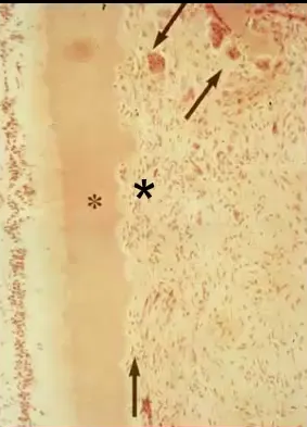
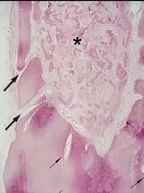
Nature of the Resorbing Tissue151617
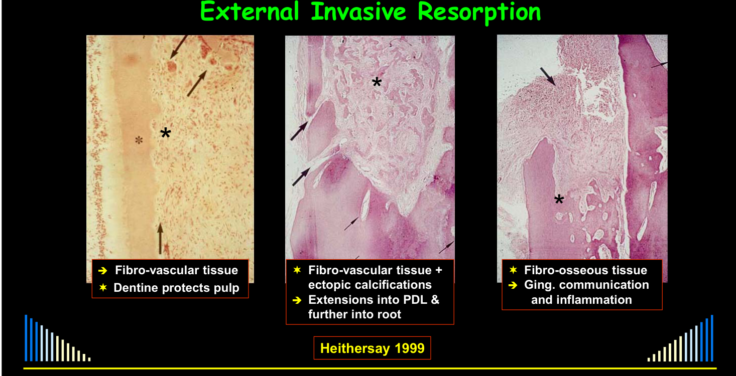
- Fibro-vascular tissue
- Dentine protects pulp
- Fibro-vascular tissue + ectopic calcifications
- Extensions into PDL & further into root
- Fibro-osseous tissue
- Gingival communication and inflammation
Lecturer — Resorbing Tissue
The invading tissue itself does not show inflammation, although gingival inflammation may occur secondarily when plaque accumulates in an irregular defect.
- Large blood vessels are present, and early lesions bleed readily when probed.
- Additional communication channels with the periodontal ligament can extend into the root, allowing blood vessels and potentially clastic-cell precursors to enter and maintain the lesion.
- Finger-like projections extend microscopically into the dentine and are difficult to identify clinically.
Heithersay 1999
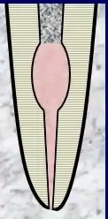
Pulp Space
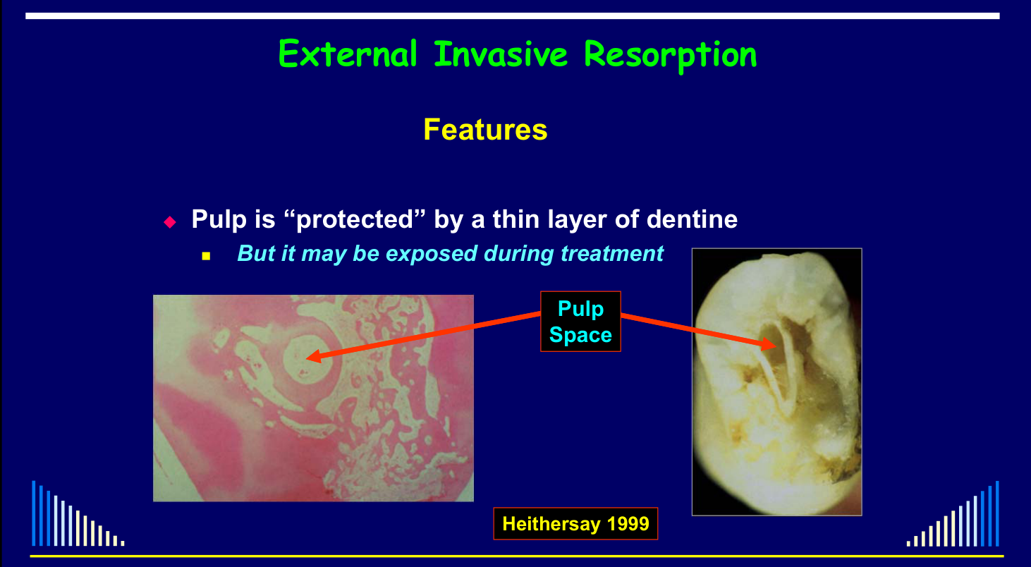
- Pulp is “protected” by a thin layer of dentine
- But it may be exposed during treatment
Lecturer — Pulp Protection
The invading tissue may encircle the pulp while the thin dentine layer remains intact, so external invasive resorption is not caused by pulp disease.
- The pulp is generally clinically normal unless bacteria enter secondarily through the defect.
- Curettage may remove the protective dentine layer and expose the pulp; root canal treatment may then be required, and in some situations exposure may make extraction necessary.
Heithersay 1999
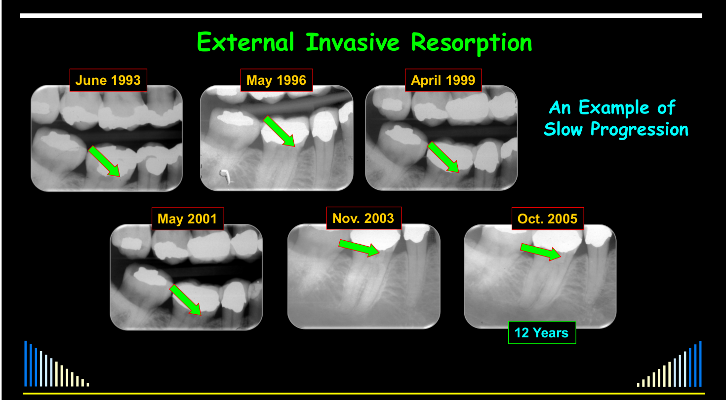
Speed of progression of invasive resorption
- Largely unknown, since time of initiation usually unknown
- Appears to be rapid initially
- Later very slow
- May have “bursts” of activity
Not investigated; clinical impression only
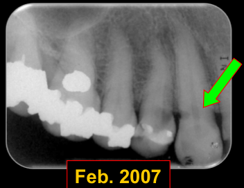
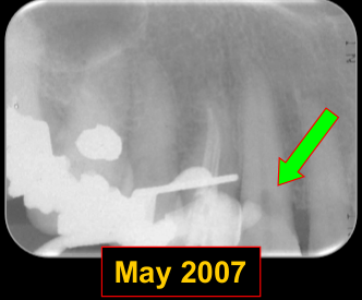
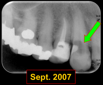
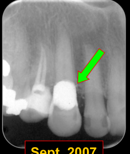
An Example of Rapid Progression — Feb. 2007, May 2007, Sept. 2007, Sept. 2007.
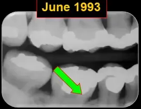
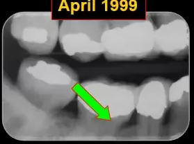
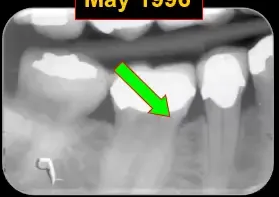
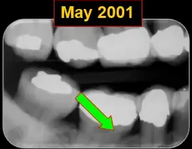
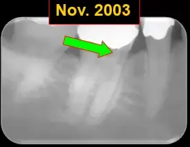
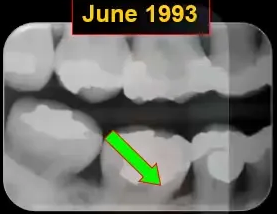
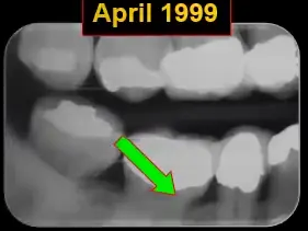
An Example of Slow Progression — June 1993, May 1996, April 1999, May 2001, Nov. 2003, Oct. 2005 (12 Years).
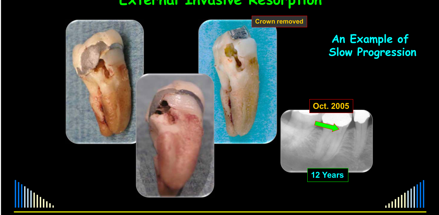
An Example of Slow Progression — Oct. 2005 (12 Years); crown removed.
Variable Progression
A first premolar was referred for routine endodontic treatment, but external invasive resorption was identified on the canine.
- The premolar received routine root canal treatment.
- The canine was treated by cauterization, curettage, and restoration in May 2007.
- At review approximately six months later, the resorption had progressed rapidly; it was treated again, but because of the defect size and recurrence, the prognosis was unpredictable.
A separate tooth showed very slow progression over approximately 12 years.
- A small defect was visible in 1993, with little change in 1996 and 1999, some progression by 2001, and a larger defect in 2003.
- The patient had vague symptoms related mainly to food packing and unsatisfactory contact points.
- Treatment was considered infeasible, so the tooth was monitored and extracted several years later.
Classification Systems18
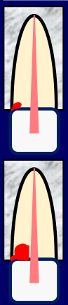
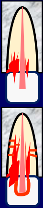
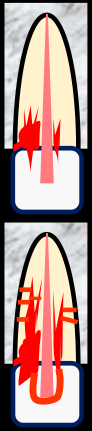
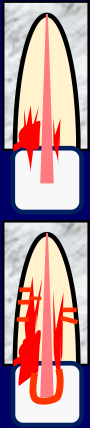
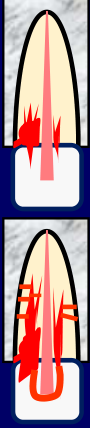
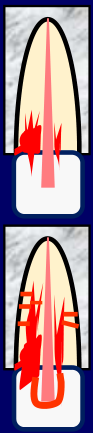
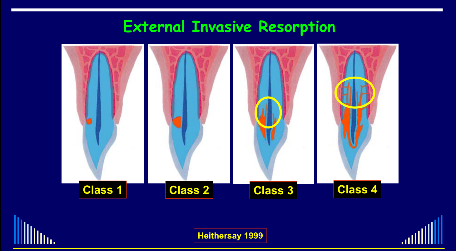
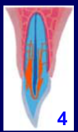
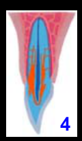
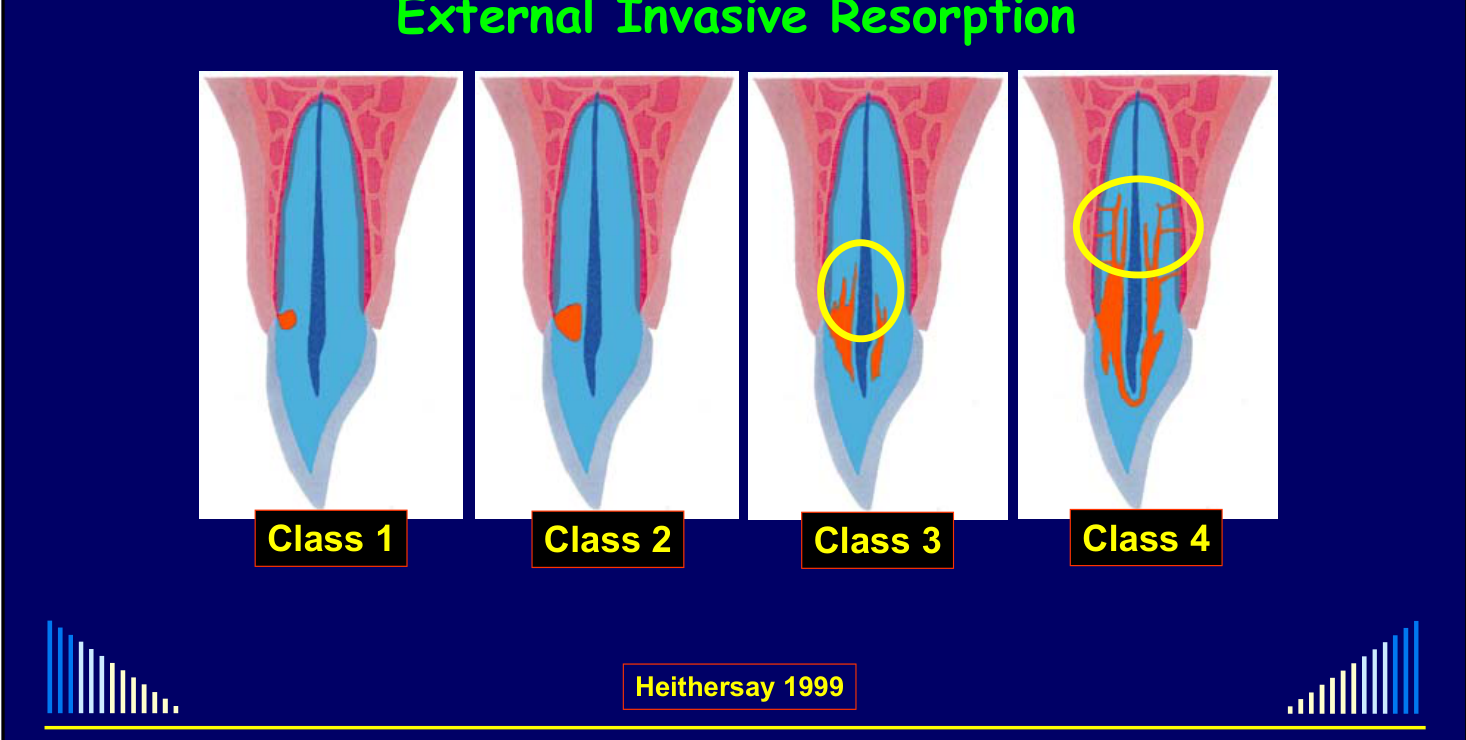
Heithersay Classification
- Class 1
- Class 2
- Class 3
- Class 4
Lecturer — Heithersay Classes
The classification is based mainly on the size and extent of the resorptive defect.
- Class 1 is a small, difficult-to-detect defect, usually not close to the pulp and often found incidentally.
- Class 2 is more extensive and extends closer to the pulp, although a thin layer of dentine usually remains.
- Class 3 may extend coronally or into the root, usually begins to wrap around the pulp, and may have finger-like projections into the dentine.
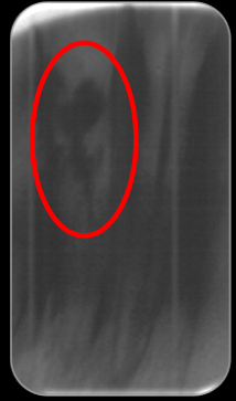
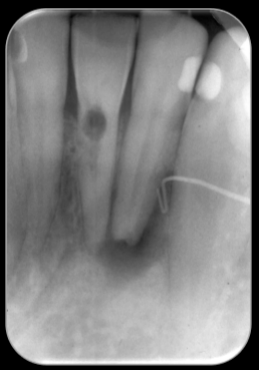
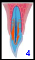
- Class 4 is very extensive, with finger-like projections far into the root and additional communications with the periodontal ligament; it may envelop the pulp and extend coronally, although this is not always the case.
- Classes 3 and 4 are more difficult to manage because of the microscopic projections and, in Class 4, the additional periodontal ligament communications.
Heithersay 1999.
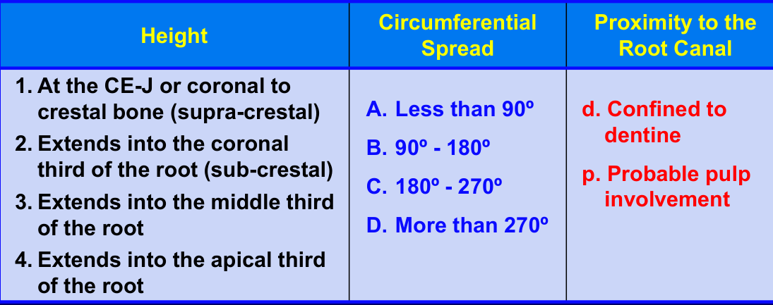
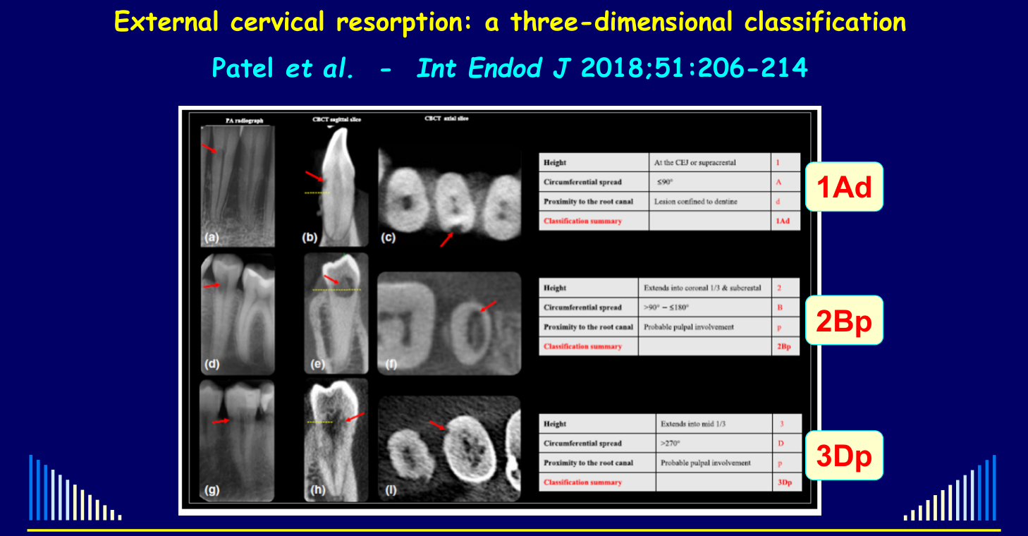
Three-Dimensional CBCT Classification1920
External cervical resorption: a three-dimensional classification — Patel et al., Int Endod J 2018;51:206-214.
For use with CBCT scans:
| Height | Circumferential Spread | Proximity to the Root Canal |
|---|---|---|
| 1. At the CEJ or coronal to crestal bone (supra-crestal) | A. Less than 90° | d. Confined to dentine |
| 2. Extends into the coronal third of the root (sub-crestal) | B. 90° – 180° | p. Probable pulp involvement |
| 3. Extends into the middle third of the root | C. 180° – 270° | |
| 4. Extends into the apical third of the root | D. More than 270° |
External cervical resorption: a three-dimensional classification — Patel et al., Int Endod J 2018;51:206-214.
Three-Dimensional Imaging Case
Three-dimensional imaging can demonstrate the extent and distribution of a lesion more clearly than conventional radiographs. In one case, CT images showed a large defect wrapping around the tooth, resorption at some levels of the root but not others, and the extent of the lesion in multiple directions.
- The lecture also described a case initially considered for extraction and implant replacement in which multi-slice CT provided additional information about the extent of the defect.
Example classifications: 1Ad, 2Bp, 3Dp.
2021 class distribution
Class 4 was the largest group (117 of 293 teeth; 39.9%); the full class and tooth-type distributions are in Abbott and Sunthoramoorthy 2021 - Invasive Resorption Cohort.
See - Lecture outline
Return to Introduction and Terminology.
Lecturer — Treatment Precautions
Treatment is performed under rubber dam isolation, with careful protection of adjacent tissues.
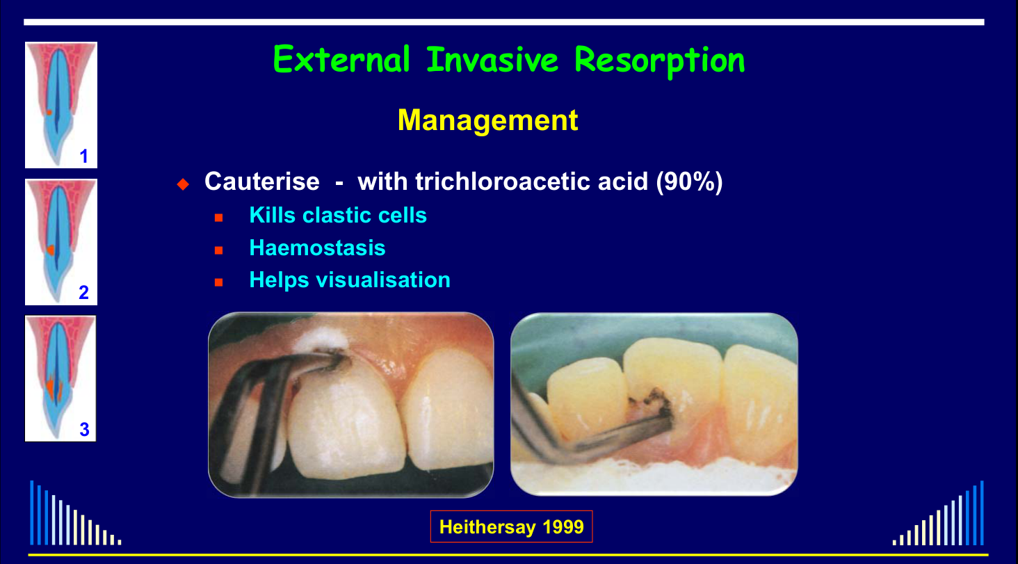
- Surrounding tissues may be painted with glycerin to counteract the acid.
- A very small, dry cotton pellet and only a small amount of trichloroacetic acid should be used, because excess liquid can run onto other tissues.
- The white necrotic tissue may slough off over several days, and referral to an experienced specialist endodontist is recommended.
Lecturer — Treatment Appointments
Treatment is usually carried out over two appointments approximately one to two weeks apart.
- At the first appointment, the lesion is cleaned and Cavit is placed as a temporary restoration, often slightly overcontoured to push tissue away from the defect.
- At the second appointment, the tissue is reassessed, further trichloroacetic acid is applied if necessary, and the dentine is inspected for residual projections before definitive restoration.
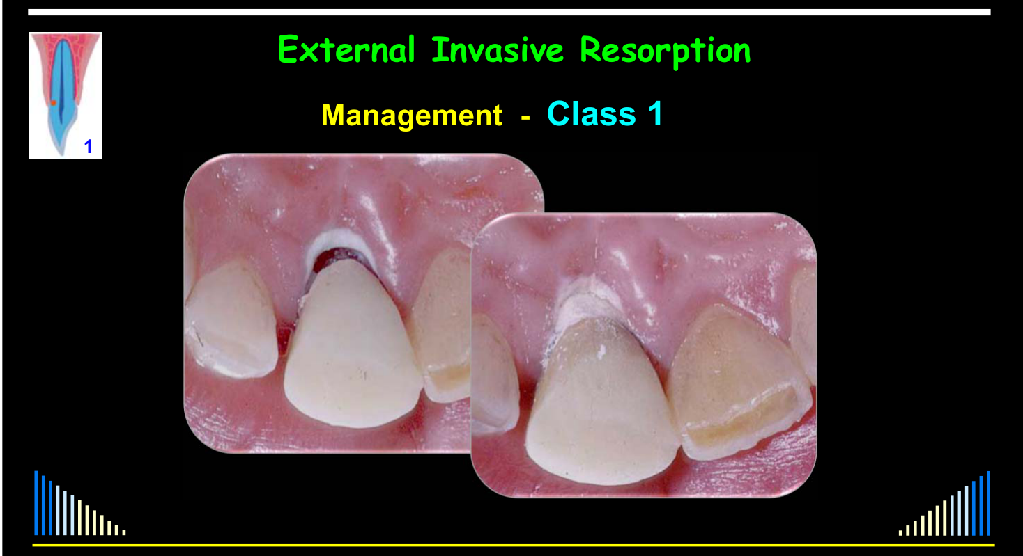
Lecturer — Class 1 and 2 Lesions
Class 1 and Class 2 lesions are generally manageable and relatively predictable.
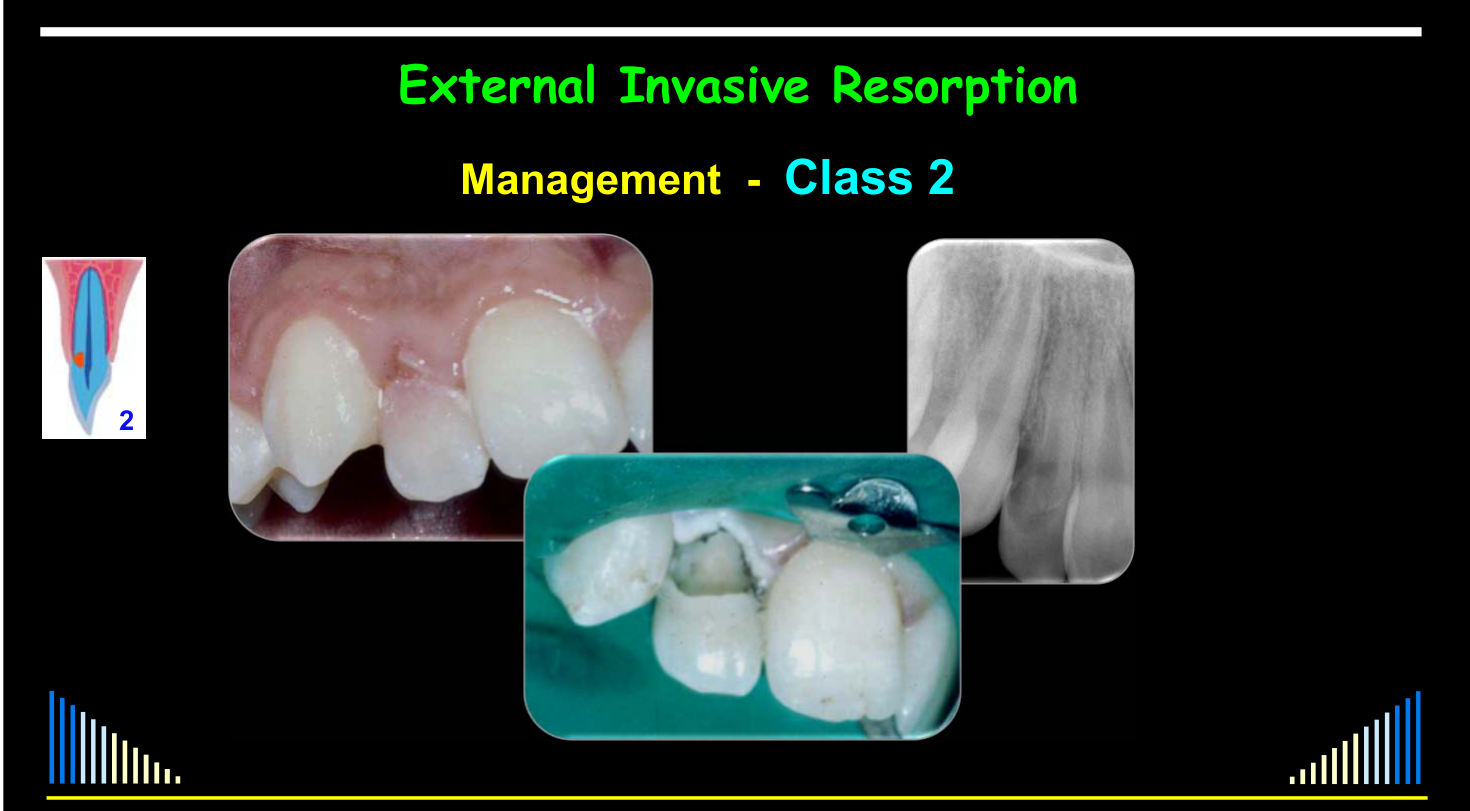
- Class 2 lesions are larger and carry a greater risk of pulp exposure, so patients should be warned that root canal treatment may become necessary if pulpitis develops or the pulp is exposed during curettage.
- A Class 2 lesion may initially receive a sedative lining over the pulp area followed by Cavit, with the pulp reassessed at the next appointment before definitive restoration.
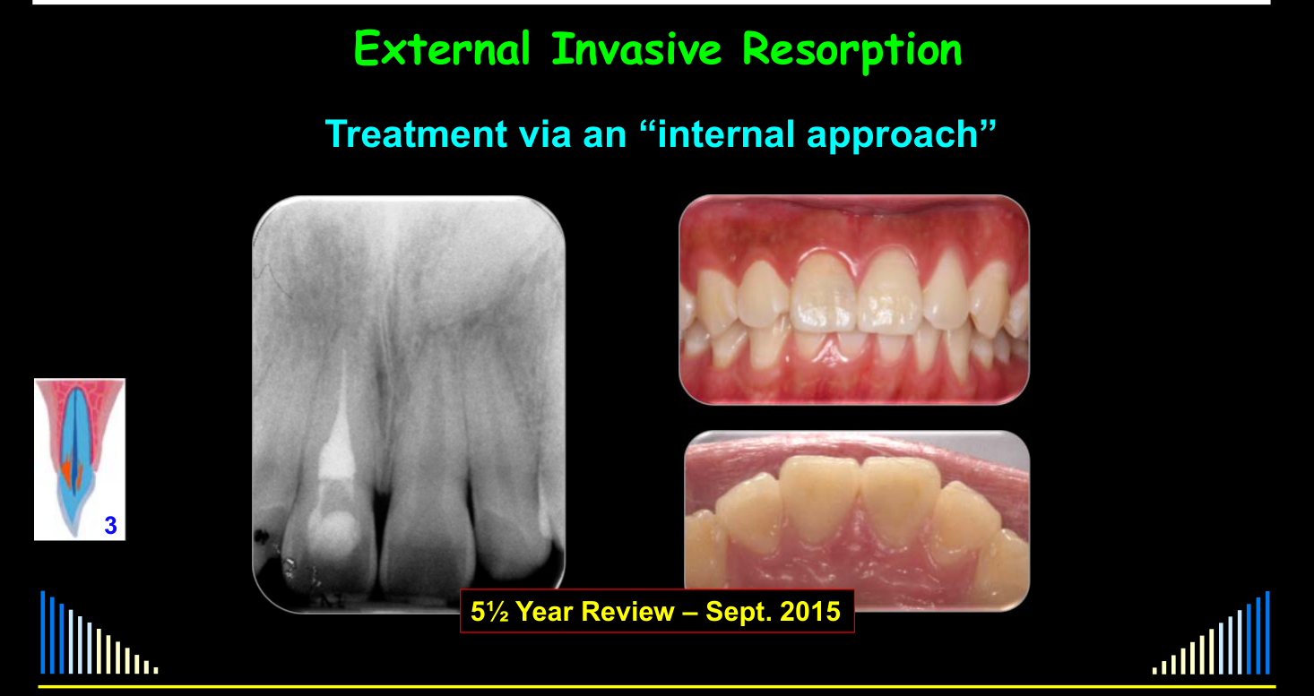
Lecturer — Internal Approach
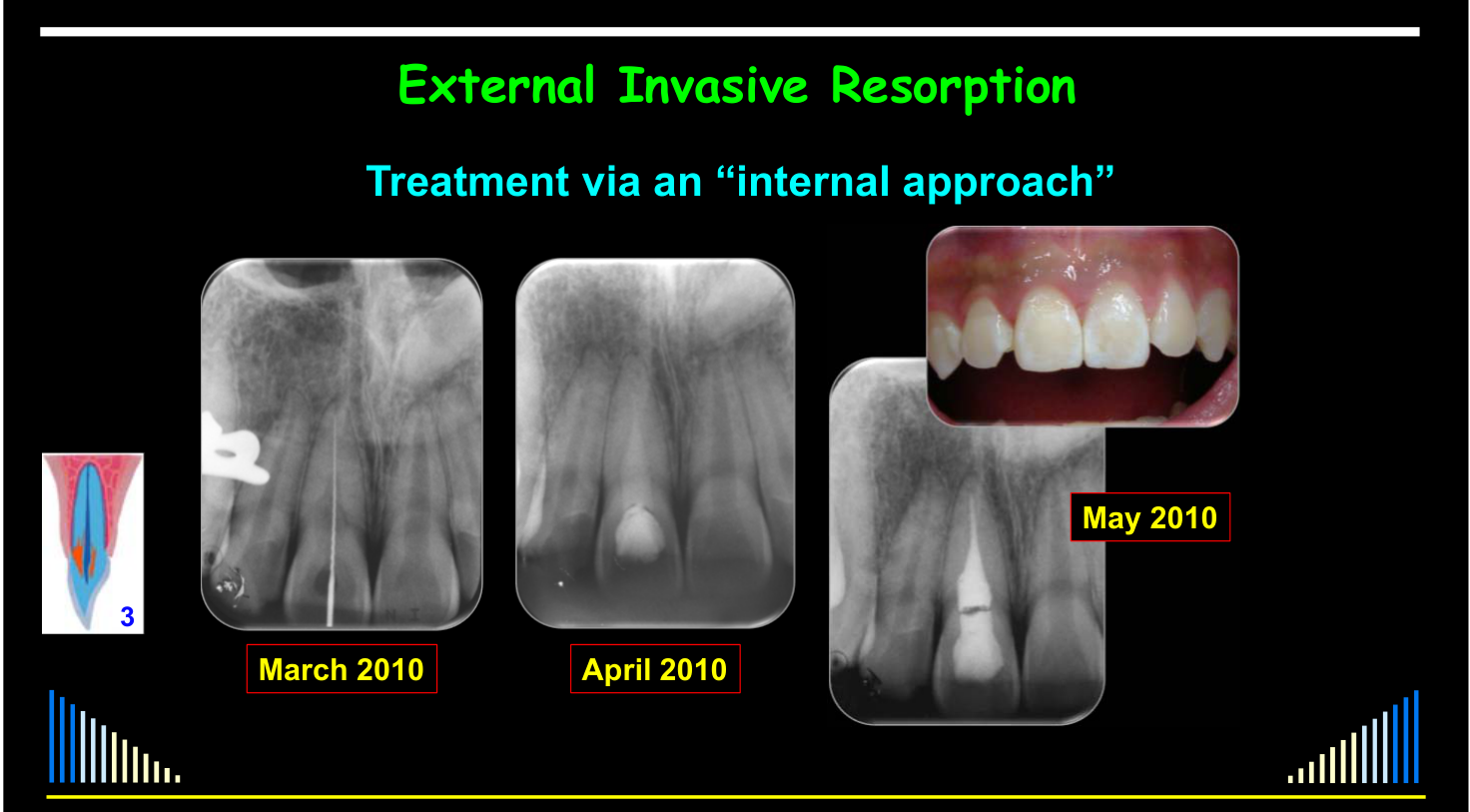
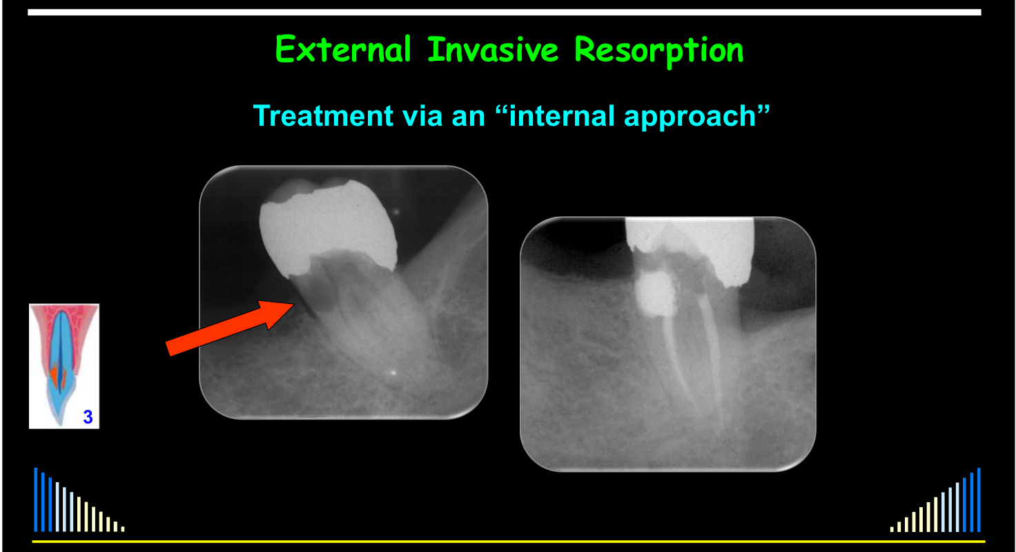
An internal approach may be selected when the external opening is small or cannot be located by probing, or when an external approach would cause excessive destruction of the tooth or surrounding tissues.
- The approach involves creating an access cavity, sacrificing the pulp, performing root canal treatment, applying trichloroacetic acid to the resorptive defect, and restoring the defect.
- It may also be used in posterior teeth when locating the external defect would require substantial bone removal.
Lecturer — Internal Treatment Outcome
An anterior case treated through an internal approach showed no further radiographic or clinical evidence of resorption at five and a half years.
- The interdental papilla was preserved.
- The soft tissues remained stable.
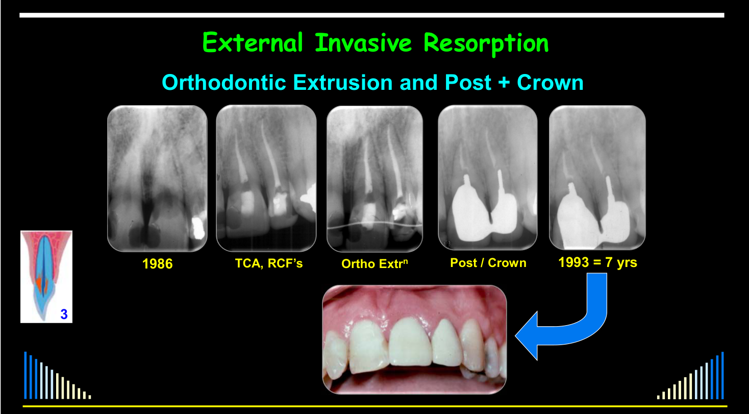
Lecturer — Orthodontic Extrusion
Orthodontic extrusion may be required when the lesion is extensive, restoration margins cannot otherwise be exposed adequately, or the tooth needs a post and core while maintaining normal oral hygiene.
- In the central incisor case, the tooth was treated by cauterization, curettage, and glass ionomer restoration before orthodontic extrusion.
- A prosthodontist subsequently placed a post, core, and crown, and the tooth was clinically stable with no ongoing resorption at seven years.
 |  |
 |
Management Strategies
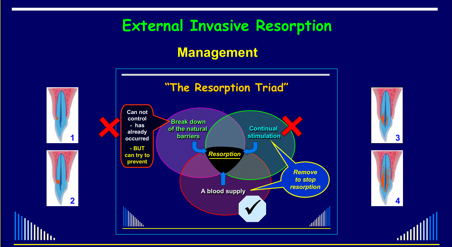
The Resorption Triad and Treatment Principles2122
See - Resorption triad foundations
The foundational requirements are explained in L2 foundational triad requirements and Resorption Triad.
The initial natural-barrier breakdown has already occurred and cannot be reversed; prevention aims to avoid that initial injury.
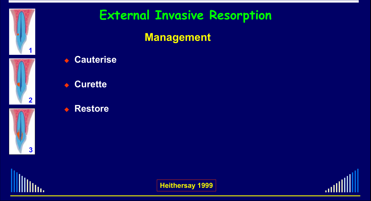
Remove to stop resorption
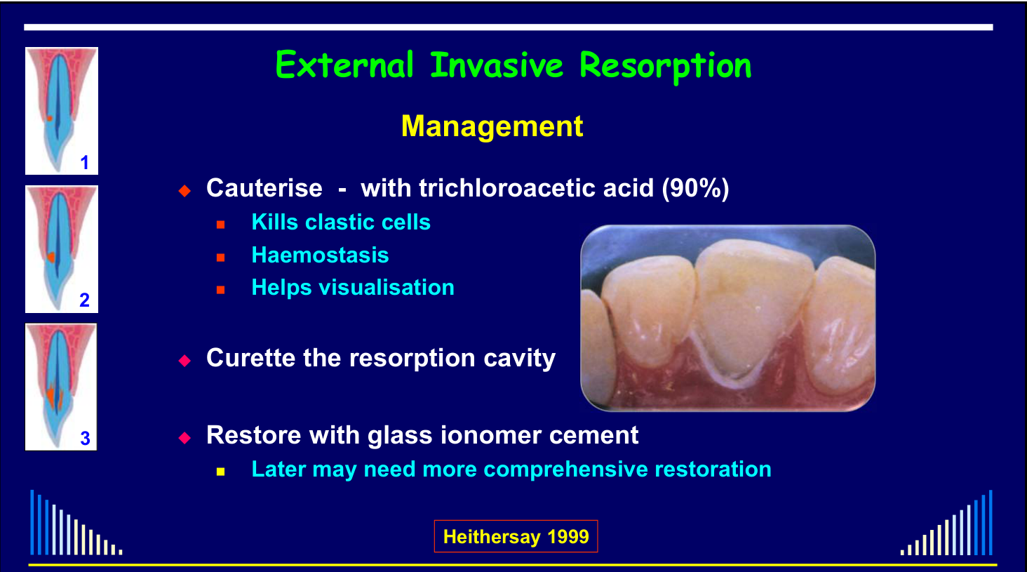
- Cauterise - with trichloroacetic acid (90%)
- Kills clastic cells
- Haemostasis
- Helps visualisation
- Curette the resorption cavity
- Restore with glass ionomer cement
- Later may need more comprehensive restoration
Heithersay 1999
 |  |
 |
Treatment by Class
Class 1 and Class 2 Management23
 |  |
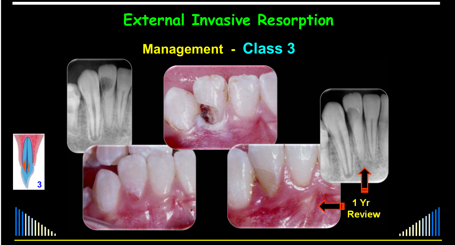
Class 3 Management2425262728
1 Yr Review
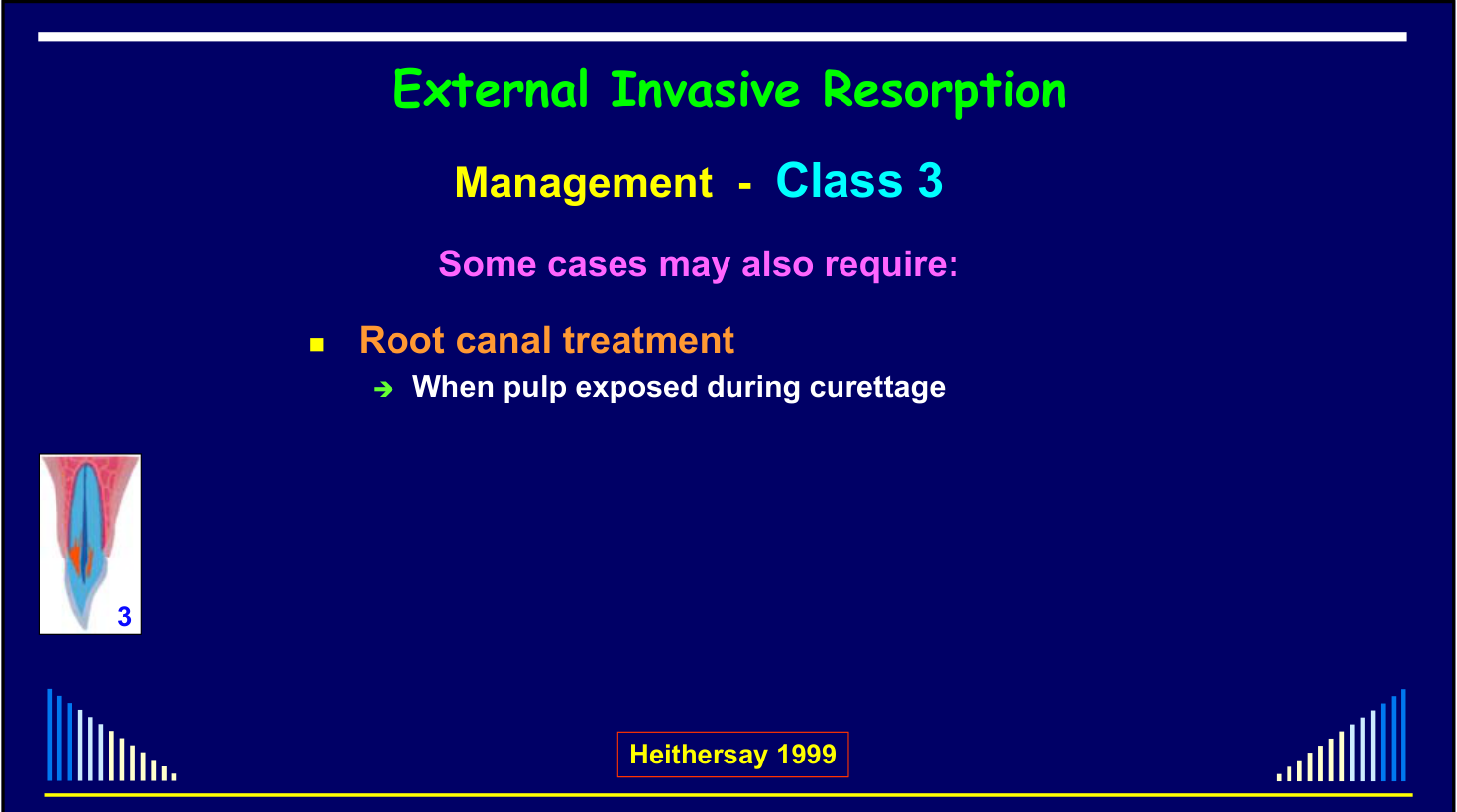
Some cases may also require:
- Root canal treatment
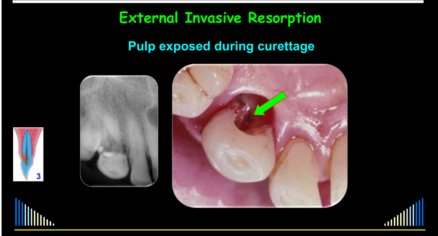
- When pulp exposed during curettage
Heithersay 1999
Some cases may also require:
- Root canal treatment
- When pulp exposed during curettage
- For an internal approach to the resorptive area
- May be advantageous and important to preserve the interdental papilla
- Especially with anterior teeth
- When external opening can not be detected
- May be advantageous and important to preserve the interdental papilla
Heithersay 1999
Treatment via an “internal approach”
- March 2010
- April 2010
- May 2010
5½ Year Review – Sept. 2015
Some cases may also require:
- Root canal treatment
- When pulp exposed during curettage
- For an internal approach to the resorptive area
- Advantageous to preserve the interdental papilla
- When needed to restore the tooth (e.g. with a post)
- Orthodontic extrusion
- When required to expose / restore margins
Heithersay 1999
Orthodontic Extrusion and Post + Crown
| 3 | 1986 | TCA, RCF’s | Ortho Extrⁿ | Post / Crown | 1993 = 7 yrs |
|---|

Class 4 Management293031323334
- Leave & observe
- → If not causing any symptoms
- → If no concurrent pulp disease
- → If no periodontal problems
- Extraction eventually required
- But may survive for a long time!
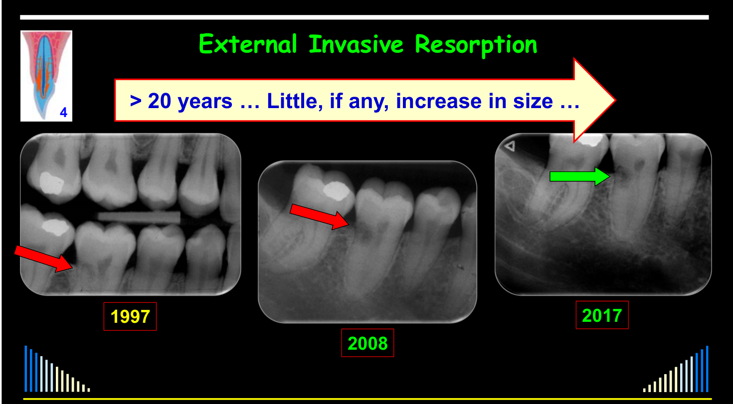
2008
20 years … Little, if any, increase in size …
Class 4 Lesion Monitored for 20 Years
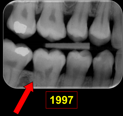
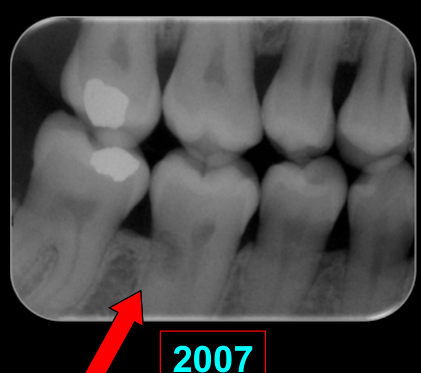
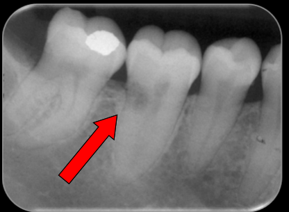
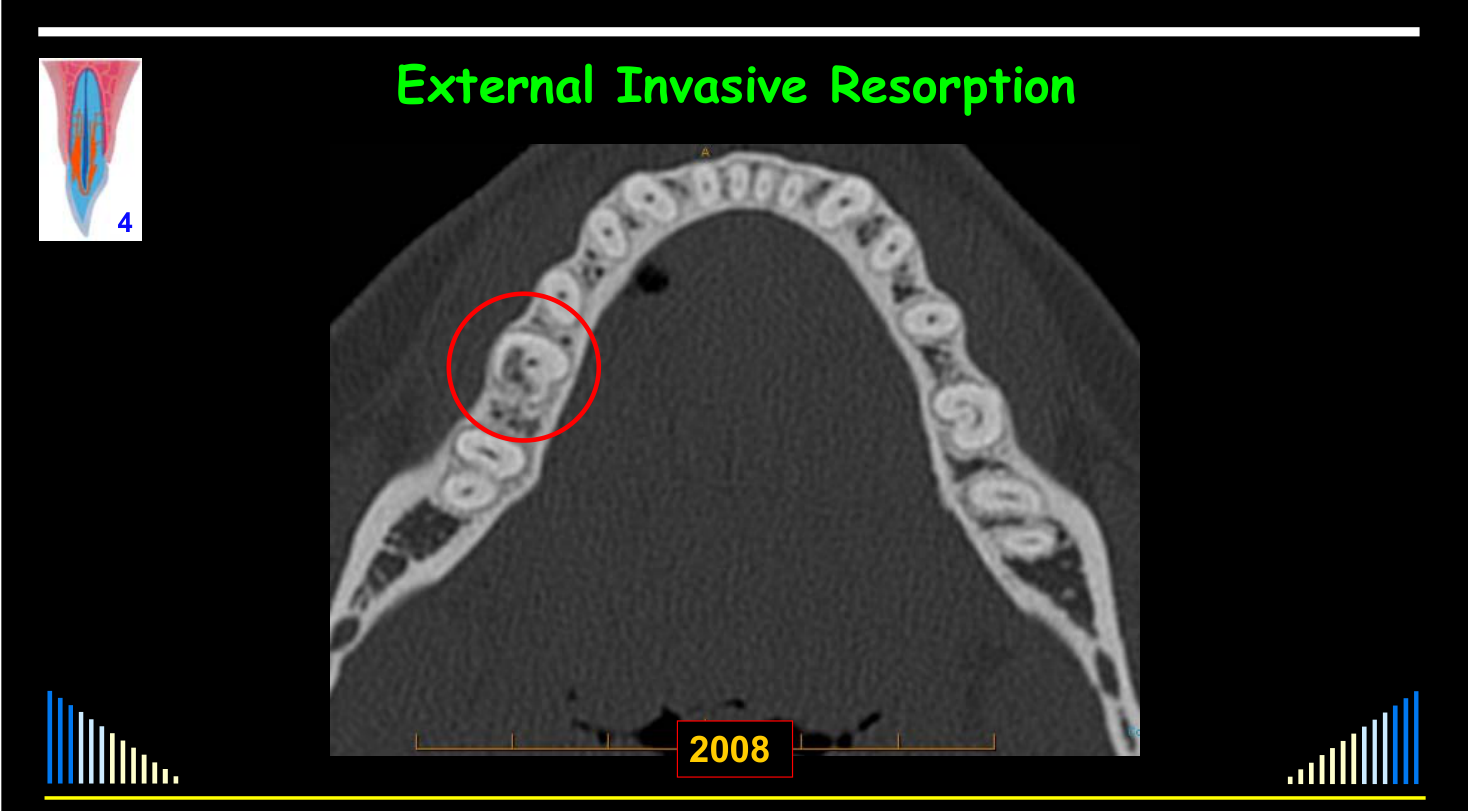
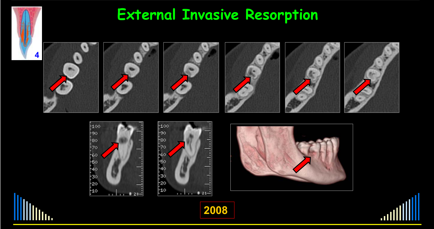
A bitewing from 1997 showed a resorptive defect on the distal aspect of a molar. The patient was later advised by another dentist to have the tooth extracted and replaced with an implant, but CT imaging was arranged by a periodontist and a second opinion recommended retention and monitoring because the tooth had never caused symptoms. A 2017 radiograph showed little or no change over approximately 20 years.
1997 2008 2017
-
Leave & observe
- If not causing any symptoms or other problems
-
Extraction will eventually be required
- But may survive for a long time!
-
But advise the patient of the potential problems that may develop
- Pulpitis / Pulp Necrosis / Apical Periodontitis
- Periodontal Pocketing / Abscess
- Root Fracture - transverse / horizontal
- Ankylosis
-
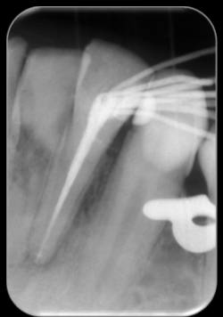
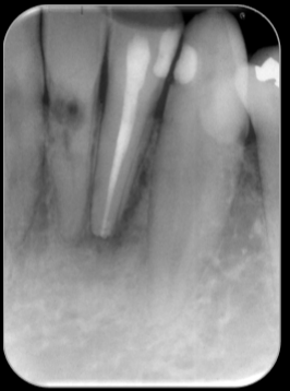
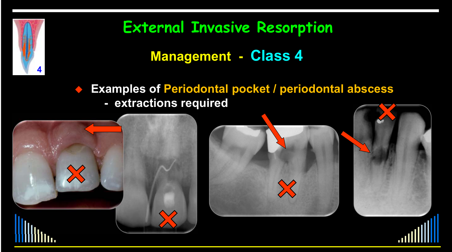
Examples of Periodontal pocket / periodontal abscess - extractions required
-
Example of Ankylosis - extraction required
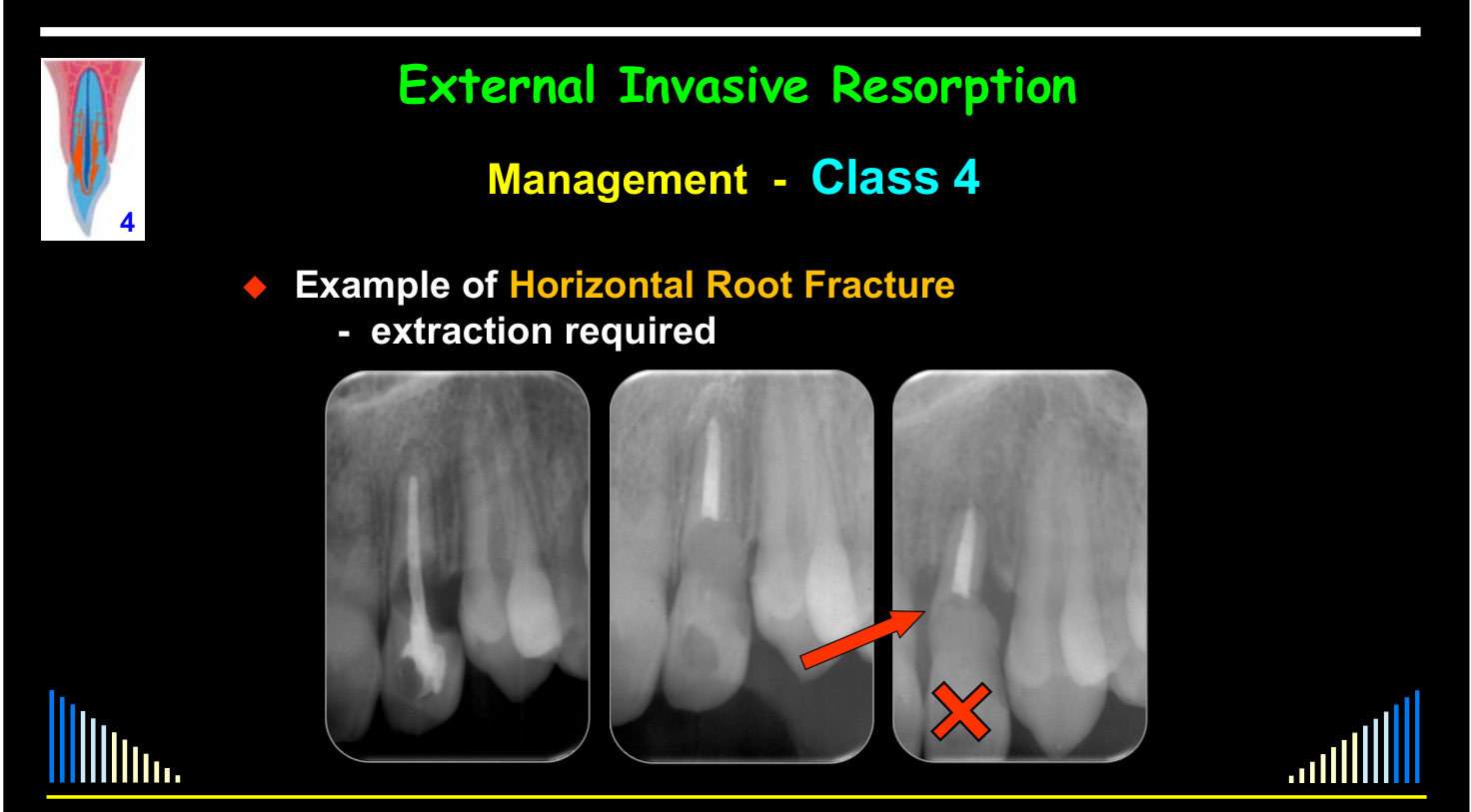
Lecturer — Class 4 Extraction Decisions
In advanced lesions, attempting treatment may be overly ambitious when the remaining tooth structure is compromised.
- Fracture can lead to bone loss and compromise later implant or prosthetic replacement.
- Earlier extraction may therefore be preferable to retaining a structurally compromised tooth.
Example of Horizontal Root Fracture - extraction required
Treatment Outcomes35
Heithersay Five-Year Outcomes36
In Heithersay’s five-year series, all three outcome measures were 100% for Classes 1 and 2 and lower for Classes 3 and 4, especially overall success; the extract gives no class denominators. Full outcome table and source context: Heithersay 1999 - TCA Treatment Pages 96-110.
Lecturer — Heithersay Treatment
Heithersay assessed treatment using trichloroacetic acid, curettage, and restoration.
- Class 3 failures were often related to periodontal or restorative problems.
- Class 4 results are too poor to justify routine attempts at treatment.
- Advanced Class 3 lesions may be better monitored when the tooth is asymptomatic and retaining it has no consequences.
Abbott and Sunthoramoorthy Case Series Outcomes
An Analysis of 219 patients with External Invasive Resorption in 293 Teeth — 2021, Abbott & Sunthoramoorthy (unpublished data).
Class 1 Cases — 12 Teeth (Follow-up: 6 mths – 6 yrs)37
See - 2021 Class 1 cohort records
Treatment and follow-up detail for the 12 Class 1 teeth is preserved in Abbott and Sunthoramoorthy 2021 - Invasive Resorption Cohort.
Class 2 Cases — 60 Teeth (Follow-up: 6 mths – 10 yrs)38
See - 2021 Class 2 cohort records
Treatment and follow-up detail for the 60 Class 2 teeth is preserved in Abbott and Sunthoramoorthy 2021 - Invasive Resorption Cohort.
Class 3 Cases — 104 Teeth (Follow-up: 6 mths – 6 yrs)
See - 2021 Class 3 cohort records
Treatment and follow-up detail for the 104 Class 3 teeth is preserved in Abbott and Sunthoramoorthy 2021 - Invasive Resorption Cohort.
Class 4 Cases — 117 Teeth (Follow-up: 3 mths – 21 yrs)
See - 2021 Class 4 cohort records
Treatment and follow-up detail for the 117 Class 4 teeth is preserved in Abbott and Sunthoramoorthy 2021 - Invasive Resorption Cohort.
Lecturer — Class 2 Management
Class 2 lesions are larger and closer to the pulp, although a thin dentine layer may still protect it. Patients should be warned about possible pulp exposure and the potential need for root canal treatment; glass ionomer is commonly used initially, with later restoration if the tooth remains stable.
Lecturer — Class 3 Management
Class 3 lesions may wrap around the pulp, and finger-like projections can complicate complete removal.
- Root canal treatment may be required because of pulp exposure or subsequent pulpitis.
- An internal approach may preserve the interdental papilla or avoid unnecessary external destruction.
- Orthodontic extrusion and a post-retained restoration may be needed in selected cases.
Lecturer — Class 4 Management
Class 4 lesions may include finger-like projections, periodontal ligament communications, blood vessels and nutrients entering the lesion, and envelopment of the pulp. Treatment is generally not recommended because control and survival rates are poor.
- Monitoring is appropriate when the tooth is asymptomatic, has no pulp disease or periodontal complications, and the patient understands that extraction may later be required.
- Long-term monitoring may show little or no progression over many years.
- Extraction may eventually be required because of periodontal abscess, ankylosis, replacement resorption, root fracture, or loss of tooth structure.
An Analysis of 219 patients with External Invasive Resorption in 293 Teeth — 2021, Abbott & Sunthoramoorthy (unpublished data).
Lecturer — Treatment Selection
Treatment selection is based primarily on lesion size and extent.
- Class 1 and Class 2 lesions are usually treated because treatment is comparatively predictable.
- Class 3 lesions may be treated selectively, while Class 4 lesions are usually monitored.
Lecturer — Class 1 Management
Class 1 lesions are small and often difficult to detect, but treatment is generally straightforward when they are identified. The lesion can usually be cauterized, curetted, and restored, with follow-up to confirm that resorption has not continued.
All Cases — 293 Teeth — No. Needing Root Canal Treatment39
See - Cohort root-canal data
Full class-specific root canal counts are preserved in Abbott and Sunthoramoorthy 2021 - Invasive Resorption Cohort.
Lecturer — Root Canal Indications
External invasive resorption itself is not caused by pulp disease. Root canal treatment is performed only when the pulp becomes involved, is intentionally sacrificed for access, or is needed for restoration.
- It may be required when the pulp is exposed during curettage or when pulpitis develops after treatment.
- It may also be required for an internal approach or to facilitate a post-retained restoration.
All Cases — 293 Teeth — Follow-up: 3 mths – 21 yrs
See - Cohort follow-up outcomes
Class-specific follow-up, treatment, and outcome tallies are preserved as reported in Abbott and Sunthoramoorthy 2021 - Invasive Resorption Cohort.
Lecturer — Long-Term Monitoring
Long-term outcomes depend strongly on lesion classification. Monitoring must include clinical examination and periodic radiography, and patients should understand that a tooth may eventually require extraction even if it remains symptom-free for many years.
See - Lecture outline
Return to Introduction and Terminology.
Clinical Cases
Case: Lower anterior teeth with recession and subcervical resorption
Question
Scenario: A patient presents with lower anterior teeth exhibiting gingival recession.
What’s shown: A periapical radiograph and a clinical intraoral photograph of the lower anterior teeth.
Consider: How the presence of gingival recession influences the starting location of the resorptive defect.
Answer
Observations:
- Gingival recession is present on the lower anterior teeth.
- The resorptive defect begins below the cervical region of the tooth.
Reasoning: External invasive resorption typically starts subgingivally. When gingival recession is present, the resorption can start below the cervical region and may extend anywhere along the tooth root, depending on the severity of the recession.
Takeaway: External invasive resorption can start below the cervical region of the tooth if there is significant gingival recession.
Case: Central incisors with resorptive defects following root canal treatment and bleaching
Question
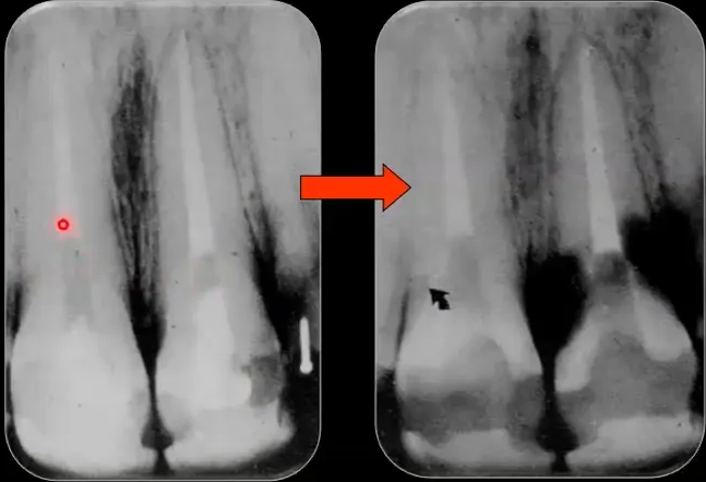
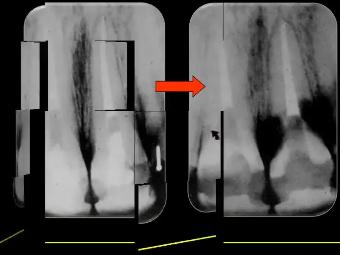
Scenario: A patient presents with a history of root canal treatment and internal bleaching of their central incisors.
What’s shown: Images displaying gutta percha at a deep level within the central incisors, alongside extensive resorptive defects on the left central incisor and early defects on the right central incisor.
Consider: What is the diagnosis for the defects, and how should the patient’s history of bleaching be interpreted in relation to this condition?
Answer
Observations:
- Gutta percha is present down to a deep level in the central incisors.
- Fairly extensive invasive resorptive defects are visible on the left central incisor.
- Early invasive resorptive defects are visible on the right central incisor.
Reasoning: The presence of these resorptive defects in teeth that have undergone root canal treatment and internal bleaching is characteristic of external invasive resorption. The lecturer uses this case to illustrate that while early case reports linked bleaching to resorption, it is likely just one of multiple potential predisposing factors rather than the sole cause.
Takeaway: External invasive resorption can develop in teeth with a history of root canal treatment and internal bleaching, but bleaching is likely only one of multiple potential predisposing factors rather than the sole etiology.
Case: Invasive resorption referred as caries, two radiographs
Question
Scenario: Two cases of teeth with lesions that were initially referred for treatment as caries.
What’s shown: Two dental X-ray images displayed side-by-side showing radiolucent lesions.
Consider: Based on the radiographic appearance and location, what is the actual diagnosis and how does it differ from caries?
Answer
Observations:
- The lesions are located at the cementoenamel junction (CEJ) level.
- The pattern of the radiolucency does not match typical caries.
Reasoning: Caries does not typically develop at the CEJ level or in this specific pattern. The appearance and location are characteristic of external invasive resorption, making it distinctly different from caries.
Takeaway: External invasive resorption can be misdiagnosed as caries, but its location at the CEJ and specific radiographic pattern help distinguish it.
Case: Radiographs demonstrating root canal superimposition over resorptive defect
Question
Scenario: Several radiographs of teeth with resorptive defects, presented to differentiate them from internal inflammatory resorption.
What’s shown: Multiple periapical dental X-rays showing radiolucent resorptive defects with the root canal outline visible through them.
Consider: How can you use the radiographic appearance of the root canal to distinguish external invasive resorption from internal inflammatory resorption?
Answer
Observations:
- The outline of the root canal can be traced going right through the radiolucent resorptive defect.
- The resorptive defect is superimposed over the root canal.
Reasoning: In internal inflammatory resorption, the defect originates from within the canal, altering its outline. In external invasive resorption, the root canal walls remain intact and can be seen superimposed over the defect, allowing careful examination of the radiographs to distinguish between the two.
Takeaway: Tracing the intact root canal outline through a radiolucent defect helps distinguish external invasive resorption from internal inflammatory resorption.
Case: Clinical photographs of a “pink tooth”
Question
Scenario: Two clinical photographs of a tooth exhibiting a pink coloration, a feature often misattributed to internal inflammatory resorption.
What’s shown: Clinical photographs of a tooth with a pink hue visible in the crown.
Consider: What is the actual pathological cause of the pink tooth appearance?
Answer
Observations:
- The tooth exhibits a pink coloration in the crown area.
Reasoning: The pink color is caused by highly vascular external invasive resorption tissue tracking down into the crown. Internal inflammatory resorption cannot cause this in the crown because it requires necrotic, infected pulp tissue coronal to the resorbing area, which would not produce a pink appearance.
Takeaway: A “pink tooth” is a clinical sign of external invasive resorption tracking into the crown, not internal inflammatory resorption.
Case: Histology and macro sections of external invasive resorption
Question
Scenario: Histological and macroscopic sections of teeth with external invasive resorption at various stages of development.
What’s shown: Micrographs and macro sections showing fibrovascular tissue, ectopic calcifications, bone growth, and a thin layer of dentine surrounding the pulp.
Consider: What are the histological and macroscopic features of external invasive resorption, and how does the pathological process affect the pulp?
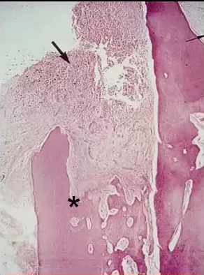
Answer
Observations:
- Early stages show very vascular, fibrous invading tissue with large blood vessels.
- Ectopic calcifications and fibro-osseous tissue (bone growing into the defect) can develop over time.
- Finger-like projections extend into the dentine, creating communications with the periodontal ligament.
- A thin layer of dentine protects the pulp, with the invading tissue encircling it.
- Inflammation is only a secondary response to plaque, not the primary pathological process.
Reasoning: The invading tissue is fibrovascular and invasive, not primarily inflammatory. The thin dentine layer protects the pulp, but curettage of the tissue may expose the pulp. The pulp itself is usually clinically normal unless secondarily infected.
Takeaway: External invasive resorption is driven by invasive fibrovascular tissue, not inflammation, and often leaves a thin protective layer of dentine around a clinically normal pulp.
Case: Canine tooth with invasive resorption, radiographic follow-up
Question
Scenario: A patient referred for endodontic treatment of a first premolar, where an incidental finding of invasive resorption on the adjacent canine was noted.
What’s shown: Periapical radiographs from May 2007 and September 2007 showing the canine tooth with a resorptive defect before and after treatment, and at a 6-month review.
Consider: What is the appropriate management and expected prognosis for this tooth?
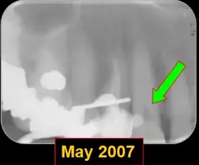
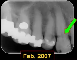
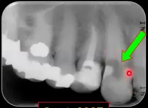
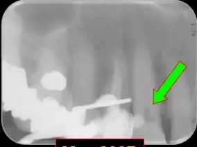
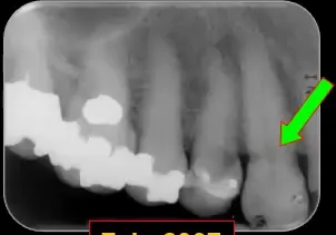
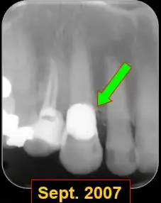
Answer
Observations:
- Invasive resorption defect on the canine tooth.
- Restoration placed after cauterization, curettage, and restoration of the defect.
- At 6-month review, the resorption has continued and progressed rapidly.
Reasoning: The defect was treated, but the resorption continued rapidly, indicating an unpredictable prognosis due to the fairly large defect.
Outcome: The first premolar received routine root canal treatment. The canine’s resorptive defect was treated by cauterization, curettage, and restoration. At a 6-month review, the resorption had continued rapidly, requiring further treatment. The prognosis for the canine is somewhat unpredictable.
Takeaway: External invasive resorption can progress rapidly after treatment, making the prognosis unpredictable, especially for larger defects.
Case: Canine with invasive resorption found during premolar endodontic referral
Question
Scenario: A patient was referred for endodontic treatment of a first premolar. During the examination, an incidental finding was noted on an adjacent tooth.
What’s shown: Radiographs from May 2007 and September 2007 showing a resorptive defect on a canine tooth before and after initial treatment, and at a six-month review.
Consider: What was the incidental finding, how was it initially managed, and what was the outcome at the six-month review?
Answer
Observations:
- An area of invasive resorption was noted on the canine tooth during the examination for the premolar; it was unknown to be present until a radiograph was taken.
- The first premolar was treated with routine root canal treatment, while the canine’s resorptive defect was treated with cauterization, curettage, and restoration, with a restoration placed in May 2007.
- At the six-month review in September 2007, the resorption had continued and progressed quite rapidly.
Reasoning: Despite the initial treatment of the fairly large defect, the resorption recurred and progressed rapidly within six months. This rapid recurrence after treatment indicates that the prognosis for a tooth with such a defect is somewhat unpredictable.
Outcome: The tooth was treated again due to the rapid recurrence of the resorption.
Takeaway: Invasive resorption can progress rapidly and recur after initial treatment, making the prognosis of teeth with large defects unpredictable.
Case: Slowly progressing external invasive resorption over 12 years
Question
Scenario: A patient presented with a history of radiographs spanning from 1993 to 2003, showing a defect on a tooth. The patient experienced vague symptoms related to food packing.
What’s shown: A series of radiographs from 1993, 1996, 1999, 2001, and 2003 showing the progression of a mesial defect, along with clinical photographs of the extracted tooth revealing the extent of the defect.
Consider: How did the defect progress over time, what was the clinical management, and what was found upon extraction?
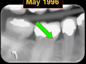
Answer
Observations:
- Radiographs from 1993 to 1999 showed a very tiny defect on the mesial with little to no change.
- By 2001, the defect had progressed a little, and by 2003, it was quite a bit more enlarged.
- The patient presented in 2003 with vague symptoms related to food packing, which was largely due to the angle of the tooth and unsatisfactory contact points.
- Upon extraction a couple of years after 2003, the tooth showed a large defect when the crown was removed.
Reasoning: The tooth was monitored for a while because it was not thought to be something that was going to be treatable at the time. The patient eventually decided to have the tooth extracted. The 12-year radiographic history demonstrated very slow progression, providing a stark contrast to the rapid progression seen in the previous case.
Outcome: The tooth was extracted a couple of years after the 2003 review. Examination of the extracted tooth confirmed a large internal defect.
Takeaway: The speed of progression of external invasive resorption can vary significantly, with some defects progressing very slowly over many years.
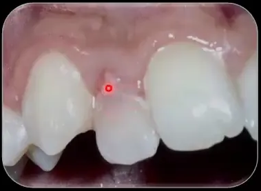
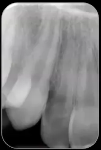
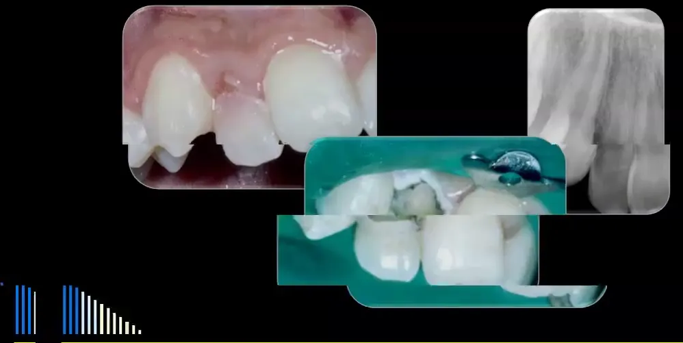
Case: Class 2 external invasive resorption with enamel breakdown
Question
Scenario: A patient presents with a tooth showing a pinkish hue and notes that enamel is breaking away from the labial surface.
What’s shown: Clinical photographs of an anterior tooth with a resorptive defect and broken enamel.
Consider: How to manage this defect, considering the risk of pulp involvement and the need for staged treatment.
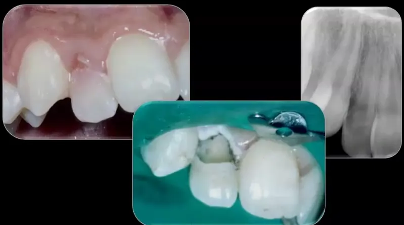
Answer
Observations:
- The tooth presents as a “pink tooth” with enamel breaking away from the labial surface.
- After cauterization and curettage, there is no pulp exposure.
Reasoning:
- The defect is classified as Class 2, meaning it is larger and carries a higher chance of pulp exposure or subsequent pulpitis compared to Class 1.
- Treatment is staged over two appointments to monitor pulp vitality and manage the resorptive tissue safely.
Outcome:
- The defect was cauterized with trichloroacetic acid (TCA) and the tissue was curetted.
- A sedative lining (Ledermix cement) was placed over the area where the pulp would be, followed by a Cavit temporary restoration.
- The patient was recalled in one to two weeks to reassess the pulp, remove the Cavit, and apply more TCA.
- The defect was then restored with glass ionomer cement.
- Ultimately, the tooth would receive a crown or composite restoration.
Takeaway:
- Class 2 external invasive resorption requires careful staging and monitoring for pulp vitality, often utilizing a sedative lining and temporary restoration between appointments before definitive restoration.
Case: External invasive resorption in a young boy with a history of trauma
Question
Scenario: A young boy presents with a tooth showing a pinkish hue and broken enamel. He has a history of a lateral luxation to the tooth seven or eight years prior.
What’s shown: Clinical photographs of the tooth showing the defect and broken enamel, as well as a one-year review image.
Consider: How the history of trauma and subsequent pulp changes influence the management and outcome of this condition.
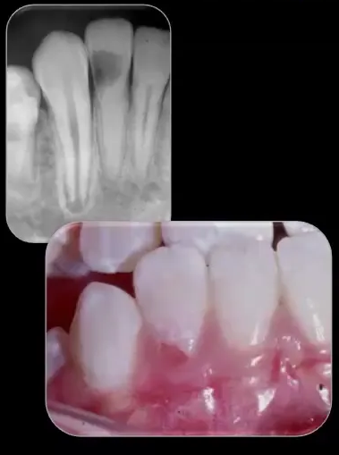
Answer
Observations:
- The tooth presents as a pink tooth with broken enamel.
- The tooth has pulp canal calcification resulting from a lateral luxation 7-8 years prior.
- During curettage, there is no pulp exposure.
Reasoning:
- The external invasive resorption may be related to the previous trauma or a developmental defect at the CEJ.
- The pulp canal calcification protected the pulp from exposure during the curettage of the extensive defect, which would have likely involved the pulp in a normal central incisor.
Outcome:
- The tissue was cauterized with TCA and curetted over a couple of appointments.
- The defect was restored with glass ionomer cement.
- At a one-year review, the glass ionomer had discolored slightly, but there was no further resorption.
- The patient was referred back to his general dentist to have the restoration covered with composite or entirely replaced.
Takeaway:
- Pulp canal calcification secondary to previous trauma can inadvertently protect the pulp from exposure during the surgical management of external invasive resorption, allowing for a more conservative endodontic approach.
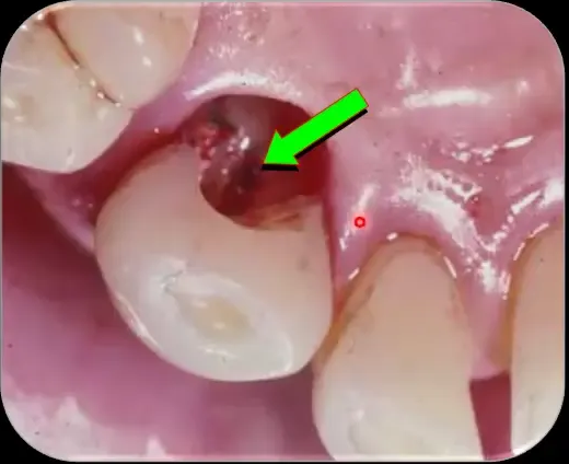
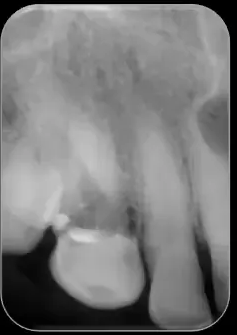
Case: Class 3 external invasive resorption with pulp exposure during curettage
Question
Scenario: A 60-year-old patient presents with a Class 3 external invasive resorption defect.
What’s shown: Clinical images showing the root canal and pulp exposure during the curettage process, as well as the tissue 10 days after trichloroacetic acid (TCA) application.
Consider: How to manage a Class 3 resorptive defect when the pulp is exposed during the curettage process.
Answer
Observations:
- The pulp is exposed during the curettage process.
- Tissue recovery is observed 10 days after TCA application, with the area free of inflammation.
- Lettermex cement and Cavit were placed as a temporary restoration.
Reasoning: Pulp exposure during curettage necessitates root canal treatment. Applying TCA and allowing the tissue to heal makes it easier to work on the tooth subsequently.
Outcome: Root canal treatment was started at the second appointment. The defect was initially restored with glass ionomer for follow-up.
Takeaway: Pulp exposure during the curettage of a Class 3 external invasive resorption defect requires root canal treatment.
Case: Upper anterior Class 3 external invasive resorption treated via an internal approach
Question
Scenario: An upper anterior tooth presents with a pinkish hue on the palatal aspect and a radiographic Class 3 defect.
What’s shown: A periapical radiograph showing the Class 3 defect, followed by images showing the access cavity, dressing placement, root filling, and final restoration.
Consider: The rationale and technique for managing this anterior defect using an internal approach.
Answer
Observations:
- Pinkish hue on the palatal aspect of the tooth.
- Radiographic evidence of a Class 3 defect.
- Internal approach taken, requiring an access cavity and sacrifice of the pulp.
Reasoning: An internal approach is advantageous for anterior teeth to preserve the interdental papilla and avoid the extensive tooth destruction and bone removal that an external approach would require. The access cavity allows for simultaneous root canal treatment and application of TCA to the defect.
Outcome: Root canal treatment was completed and the defect was restored. A 5.5-year review showed no further signs of resorption radiographically or clinically, and no loss of the interdental papilla.
Takeaway: An internal approach for anterior Class 3 defects preserves the interdental papilla and minimizes tooth destruction.
Case: Posterior Class 3 external invasive resorption treated via an internal approach
Question
Scenario: A posterior tooth has a Class 3 external invasive resorption defect where the external opening cannot be detected by probing.
What’s shown: A radiograph showing completed root canal treatment and the defect filled with Ketak silver, displaying an irregular margin.
Consider: The management strategy for a posterior defect when the external opening cannot be located.
Answer
Observations:
- The external defect could not be probed.
- Internal approach taken, sacrificing the pulp for access.
- Defect restored with Ketak silver, showing an irregular margin typical of resorptive defects.
Reasoning: Attempting an external approach when the defect cannot be probed risks destroying the tooth before finding the defect. An internal approach provides direct access to the defect without unnecessary bone removal or tooth destruction.
Outcome: Root canal treatment was completed and the defect was filled with Ketak silver.
Takeaway: When an external resorptive defect cannot be probed, an internal approach prevents unnecessary destruction of the tooth.
Case: Central incisor with Class 3 external invasive resorption requiring orthodontic extrusion
Question
Scenario: A central incisor has a Class 3 resorptive defect that ultimately requires a post and core restoration.
What’s shown: A horizontal series of radiographs showing the tooth before treatment, after root canal and resorptive treatment, after orthodontic extrusion, and after placement of a post, core, and crown, up to a 7-year review.
Consider: The sequence of treatment and the role of orthodontic extrusion in restoring this tooth.
Answer
Observations:
- Class 3 resorptive defect on the central incisor.
- Treated with cauterization, curettage, and glass ionomer restoration.
- Orthodontic extrusion performed to expose the margins.
- Restored with a post, core, and crown.
Reasoning: Orthodontic extrusion is used to bring the margins to a manageable level, allowing for adequate restoration and enabling the patient to maintain the tooth with normal oral hygiene measures.
Outcome: A 7-year review radiograph and clinical examination showed no signs of ongoing resorption and a stable result.
Takeaway: Orthodontic extrusion can be utilized in Class 3 cases to expose margins for adequate restoration and oral hygiene maintenance.
Case: Incidental finding of an asymptomatic Class 4 external invasive resorption defect
Question
Scenario: A patient is referred for treatment of tooth 42, which has a draining sinus. An extensive resorptive defect is incidentally noted on the adjacent tooth 41, which was not mentioned in the referral letter but was visible on a radiograph from 10 years prior.
What’s shown: A series of radiographs spanning from 1990 to 12 years later, showing the extent of the resorptive defect on tooth 41 and the treatment of tooth 42.
Consider: The management approach for an asymptomatic Class 4 external invasive resorption defect.
Answer
Observations:
- Tooth 42 has a draining sinus traced with a gutta percha point, indicating an infected root canal system and chronic apical abscess.
- Tooth 41 has an extensive Class 4 resorptive defect, visible on a radiograph from 10 years prior.
- Very little change in the defect on tooth 41 over a 12-year period.
Reasoning: Class 4 defects that are asymptomatic and lack concurrent pulp or periodontal disease can be managed by observation. The patient is warned that the tooth may eventually need extraction, but it can remain stable for many years.
Outcome: Tooth 42 was treated. Tooth 41 was observed, and a 12-year follow-up showed very little change in the resorptive defect.
Takeaway: Asymptomatic Class 4 external invasive resorption defects without concurrent disease can be managed by observation, as they may remain stable for many years.
Case: Resorptive defect on tooth 46 monitored over 20 years
Question
Scenario: A patient with a resorptive defect on the distal aspect of tooth 46, initially noticed on a bitewing in 1997, evaluated by multiple dentists, and followed up over a 20-year period.
What’s shown: A bitewing from 1997, a periapical radiograph from 2008, a multi-slice CT scan from 2007, and a follow-up radiograph from 2017.
Consider: How to manage an asymptomatic, extensive resorptive defect (Class 4) in a patient who is reluctant to undergo extraction and implant placement.
Answer
Observations:
- The 1997 bitewing shows a resorptive defect on the distal aspect of tooth 46.
- The 2008 periapical radiograph demonstrates the extensive nature of the resorption.
- The multi-slice CT scan reveals the defect wrapping around the tooth at various levels.
- The 2017 radiograph shows little to no change in the size of the defect over the 20-year period.
Reasoning: The patient was asymptomatic and reluctant to undergo extraction and implant placement due to the associated risks and costs. Class 4 resorptions can remain stable for many years without significant progression. Monitoring the tooth with occasional radiographs and clinical examinations is a reasonable approach, provided the patient understands the potential for future problems and the eventual need for extraction.
Outcome: The tooth was monitored and left in place. The patient returned in 2017, and the defect had not significantly changed over the 20-year period.
Takeaway: Asymptomatic Class 4 external invasive resorptions can be monitored over long periods with minimal change, avoiding unnecessary immediate extraction and implant placement.
Case: Resorptive defect misdiagnosed and treated as caries
Question
Scenario: A patient with a resorptive defect on a tooth that was mistakenly treated as if it were Class 5 caries.
What’s shown: An exhibit of a tooth with a resorptive defect and a subsequent periodontal abscess.
Consider: The potential complications of misdiagnosing and inadequately treating external invasive resorption.
Answer
Observations:
- The tooth had a resorptive defect.
- It was treated as if it were Class 5 caries.
- The treatment was inadequate for a Class 4 resorption.
- A periodontal abscess subsequently developed.
Reasoning: Treating a Class 4 resorption as caries is ineffective and does not address the underlying resorptive process. This mismanagement can lead to periodontal complications, such as a periodontal abscess, which may eventually necessitate extraction.
Outcome: The patient developed a periodontal abscess, highlighting the need for extraction or appropriate management of the resorption.
Takeaway: Misdiagnosing external invasive resorption as caries and treating it inadequately can lead to periodontal complications like abscesses, emphasizing the need for correct diagnosis and management.
Case: Ankylosis and external invasive resorption in a replanted tooth
Question
Scenario: A young patient with a history of tooth avulsion and replantation who developed ankylosis and external invasive resorption.
What’s shown: An exhibit of a tooth with infraocclusion, ankylosis, and external invasive resorption.
Consider: The implications of ankylosis developing alongside external invasive resorption in a replanted tooth.
Answer
Observations:
- The tooth is in infraocclusion.
- Ankylosis is present.
- External invasive resorption is also present.
Reasoning: Ankylosis leads to external replacement resorption. Once ankylosis starts, the tooth becomes increasingly difficult to extract later on due to the fusion with the bone. Therefore, early extraction is usually advised to prevent complications during future removal.
Outcome: The tooth is typically advised to be extracted once ankylosis develops to avoid a difficult extraction later.
Takeaway: When ankylosis develops alongside external invasive resorption, early extraction is recommended because the tooth will become increasingly difficult to remove as replacement resorption progresses.
Case: Root fracture following treatment of advanced resorption
Question
Scenario: A patient with an advanced Class 3 or 4 external invasive resorption where the orthodontist wanted to retain the tooth, leading to treatment and subsequent fracture.
What’s shown: A series of periapical radiographs showing a tooth with advanced resorption and a horizontal root fracture.
Consider: The risks of attempting to retain and treat a tooth with an extensive resorptive defect and reduced tooth structure.
Answer
Observations:
- The tooth had an advanced Class 3 or 4 resorptive defect.
- A significant amount of tooth structure was lost.
- The tooth fractured horizontally at the level of the defect.
- Bone loss occurred as a result of the fracture.
Reasoning: When a large amount of tooth structure is lost to resorption, the remaining tooth is structurally compromised and prone to fracture under normal function or trauma. Attempting to retain such a tooth can lead to fracture and subsequent bone loss, which complicates future implant placement or prosthetic rehabilitation.
Outcome: The tooth fractured, resulting in bone loss that created a non-ideal situation for implant replacement.
Takeaway: Attempting to retain teeth with extensive resorptive defects and compromised structure can lead to root fracture and bone loss; sometimes it is better to extract advanced cases earlier rather than later to preserve bone for future prosthetics.
Footnotes
-
Original PDF page 1: L9 Resorption 5 - External Invasive, p.1 ↩
-
Original PDF page 2: L9 Resorption 5 - External Invasive, p.2 ↩
-
Original PDF page 4: L9 Resorption 5 - External Invasive, p.4 ↩
-
Original PDF page 5: L9 Resorption 5 - External Invasive, p.5 ↩
-
Original PDF page 6: L9 Resorption 5 - External Invasive, p.6 ↩
-
Original PDF page 3: L9 Resorption 5 - External Invasive, p.3 ↩
-
Original PDF page 8: L9 Resorption 5 - External Invasive, p.8 ↩
-
Original PDF page 9: L9 Resorption 5 - External Invasive, p.9 ↩
-
Original PDF page 7: L9 Resorption 5 - External Invasive, p.7 ↩
-
Original PDF page 10: L9 Resorption 5 - External Invasive, p.10 ↩
-
Original PDF page 12: L9 Resorption 5 - External Invasive, p.12 ↩
-
Original PDF page 13: L9 Resorption 5 - External Invasive, p.13 ↩
-
Original PDF page 14: L9 Resorption 5 - External Invasive, p.14 ↩
-
Original PDF page 15: L9 Resorption 5 - External Invasive, p.15 ↩
-
Original PDF page 16: L9 Resorption 5 - External Invasive, p.16 ↩
-
Original PDF page 17: L9 Resorption 5 - External Invasive, p.17 ↩
-
Original PDF page 18: L9 Resorption 5 - External Invasive, p.18 ↩
-
Original PDF page 21: L9 Resorption 5 - External Invasive, p.21 ↩
-
Original PDF page 19: L9 Resorption 5 - External Invasive, p.19 ↩
-
Original PDF page 20: L9 Resorption 5 - External Invasive, p.20 ↩
-
Original PDF page 22: L9 Resorption 5 - External Invasive, p.22 ↩
-
Original PDF page 23: L9 Resorption 5 - External Invasive, p.23 ↩
-
Original PDF page 24: L9 Resorption 5 - External Invasive, p.24 ↩
-
Original PDF page 25: L9 Resorption 5 - External Invasive, p.25 ↩
-
Original PDF page 26: L9 Resorption 5 - External Invasive, p.26 ↩
-
Original PDF page 27: L9 Resorption 5 - External Invasive, p.27 ↩
-
Original PDF page 28: L9 Resorption 5 - External Invasive, p.28 ↩
-
Original PDF page 29: L9 Resorption 5 - External Invasive, p.29 ↩
-
Original PDF page 30: L9 Resorption 5 - External Invasive, p.30 ↩
-
Original PDF page 31: L9 Resorption 5 - External Invasive, p.31 ↩
-
Original PDF page 32: L9 Resorption 5 - External Invasive, p.32 ↩
-
Original PDF page 33: L9 Resorption 5 - External Invasive, p.33 ↩
-
Original PDF page 34: L9 Resorption 5 - External Invasive, p.34 ↩
-
Original PDF page 35: L9 Resorption 5 - External Invasive, p.35 ↩
-
Original PDF page 40: L9 Resorption 5 - External Invasive, p.40 ↩
-
Original PDF page 36: L9 Resorption 5 - External Invasive, p.36 ↩
-
Original PDF page 37: L9 Resorption 5 - External Invasive, p.37 ↩
-
Original PDF page 38: L9 Resorption 5 - External Invasive, p.38 ↩
-
Original PDF page 39: L9 Resorption 5 - External Invasive, p.39 ↩