L5 Resorption 3 - Ortho, Pressure, Physio, Idiopathic
External Resorption1
Lecturer — Orthodontic Mechanism
Orthodontic resorption is one of four types of external resorption discussed in the lecture, but its mechanism is not particularly well understood.
- It is not related to trauma, although trauma may have occurred concurrently in some patients.
Previous Trauma and Resorption
Previous trauma does not necessarily explain orthodontic resorption.
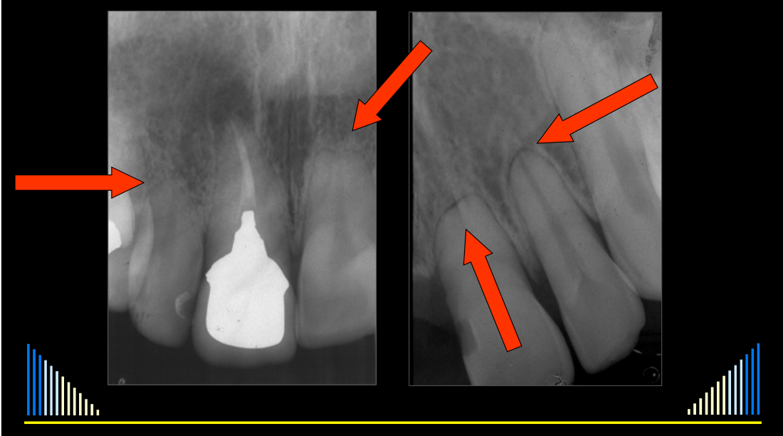
- In one example, the lateral incisor and central incisor had shortened roots.
- An adjacent tooth that had previously been root filled, suggesting previous trauma, had not developed orthodontic resorption.
- This supports the distinction between orthodontic resorption and resorption related to trauma or the condition of the root canal system.
Lecturer — Monitoring and Prevention
Orthodontic resorption should be assessed radiographically approximately six to nine months after treatment has commenced.
- Periapical radiographs would be ideal, while a panoramic radiograph can provide a useful screening assessment.
- If resorption is detected, the treatment plan may need to be modified by using lighter forces or adopting a different plan.
- If no resorption is visible at six to nine months, there is a good chance that resorption will not develop as treatment continues.
- Short roots combined with periodontal disease could result in the teeth being lost much more rapidly.
- Patients should be encouraged to wear a mouth guard during most sports, during training as well as competition, and during activities where the face could be struck; squash was given as an example because its hard racket is used in a confined space.
22/05/2020
Prof. Paul V. Abbott AO BDSc, MDS, FRACDS(Endo), FPFA, FADI, FICD, FACD, FIADT
- Specialist Endodontist
- Winthrop Professor of Clinical Dentistry
- UWA Dental School, The University of Western Australia
Oral Health Centre of Western Australia
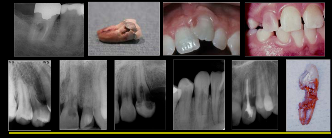
Orthodontic Resorption2
 |  |
Classification of Root Resorption — External3
- Surface
- Inflammatory
- Replacement
- Invasive
- Orthodontic
- Pressure
- Physiological
- Idiopathic
Related navigation: Root Resorption Classification; Resorption Index
Features
- Due to forces applied during orthodontic treatment
- Shortened tooth root with rounded or blunt apex
- More common and more severe in upper incisors
- PDL and pulp are normal — unless concurrent disease
Management
- Cease orthodontics — but this may not be possible!
- Monitor radiographically
- Ensure good oral hygiene to avoid periodontal disease
See - Full external root-resorption list
The full eight-type list remains under Classification of Root Resorption — External.
Pressure Resorption
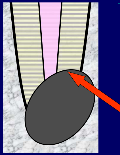
Features4
- Associated with impacted teeth or other pathosis
- e.g. cysts, tumours
- Resorption caused by pressure on the tooth root
- PDL and pulp are normal (unless concurrent disease)
Pressure Resorption Examples
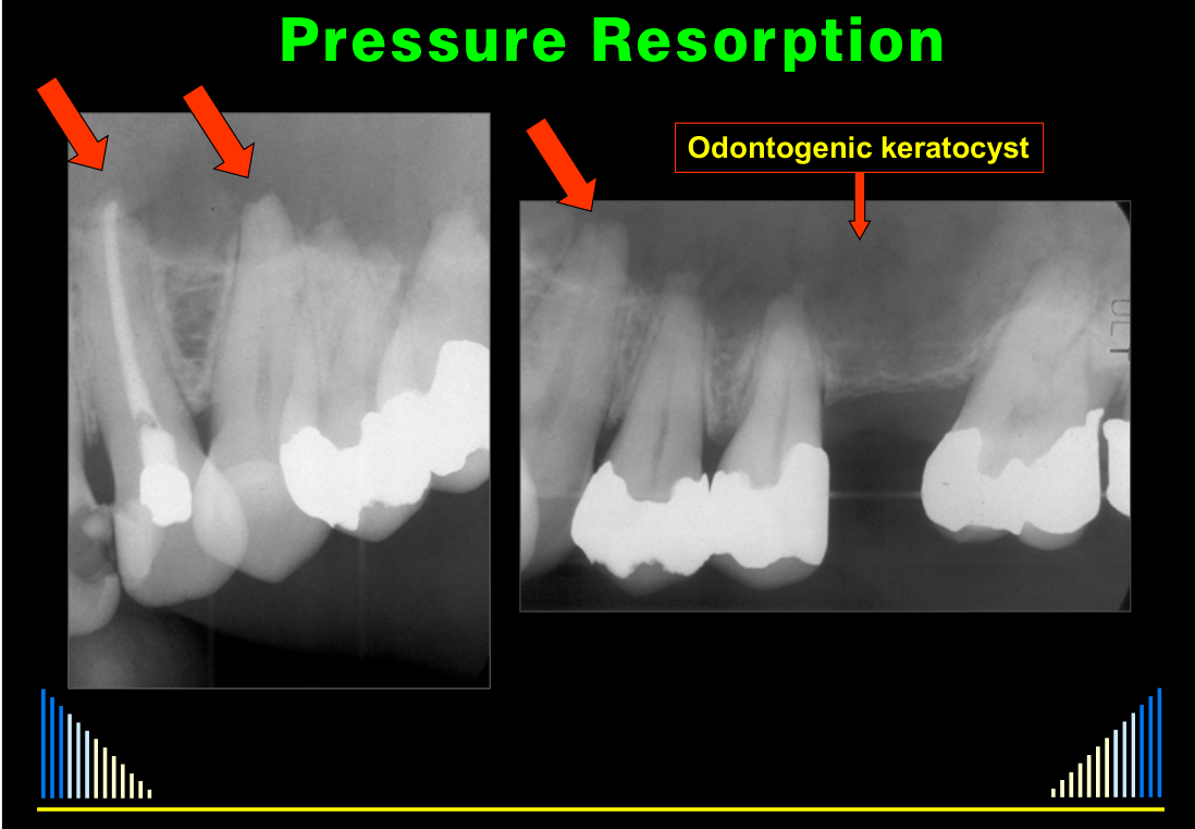
Several clinical examples illustrated the range of pressure resorption:
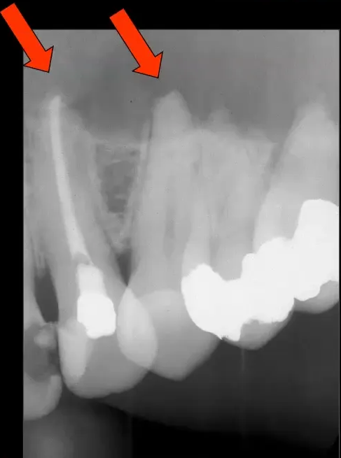
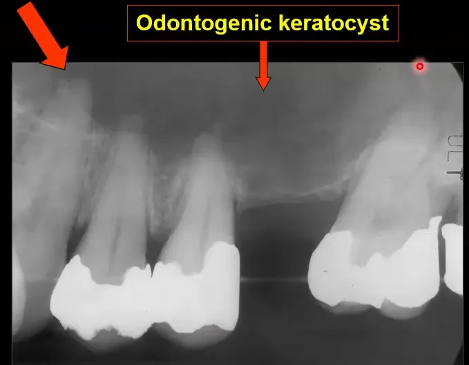
- An odontogenic keratocyst in the maxilla produced a radiolucency extending from the posterior to the anterior region; the canine root had an abnormal shape and the premolar apices were flattened.
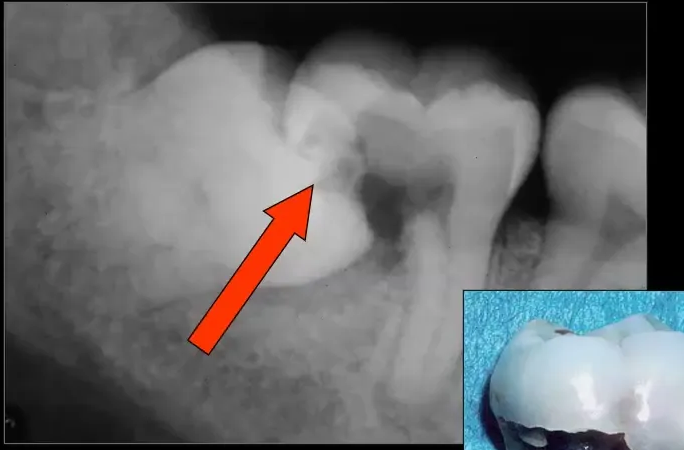
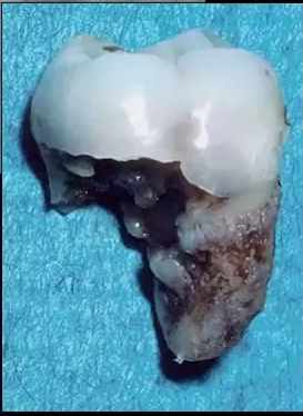
- An impacted lower third molar caused extensive resorption of the distal root of the lower second molar, which was extracted.
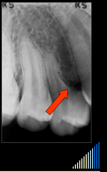
- An ectopically positioned canine caused resorption of the buccal root of the upper first premolar.
 |  |
Management5
Management will depend on the cause of the resorption:
- If caused by a cyst or tumour —
- Curette the lesion (and biopsy to confirm diagnosis!!)
- Watch & reassess the resorbing tooth — esp. pulp status
- If caused by an impacted tooth —
- Extract impacted tooth, then watch & reassess the resorbing tooth — esp. pulp
- OR: Extract both the resorbing & the impacted teeth
Lecturer — Surgical Pulp Risk
Removing the lesion should remove the pressure causing the resorption, but surgery may sever the pulp’s blood supply even though the resorption itself is not caused by pulp disease.
- The pulp must therefore be monitored, and root canal treatment may be required.
- In some situations, teeth may be treated endodontically before surgery.
Keratocyst Endodontic Treatment
In the patient with the odontogenic keratocyst, all teeth in the affected quadrant were root filled before removal of the keratocyst.
- The patient was able to retain the teeth, although extraction remained a possibility despite the prior root canal treatment.
Lecturer — Impacted Tooth Management
The choice between extracting the impacted tooth and monitoring the resorbing tooth, removing the resorbing tooth, or removing both teeth must be assessed on a case-by-case basis.
- Resorption should theoretically stop once pressure is removed, but the pulp must still be monitored because surgical removal may sever its blood supply.
Physiological Resorption6
 |  |
Features7
- Resorption of deciduous teeth during exfoliation
- Occurs with or without permanent successor present
- But rate may be very slow if no successor
Physiological Resorption Examples
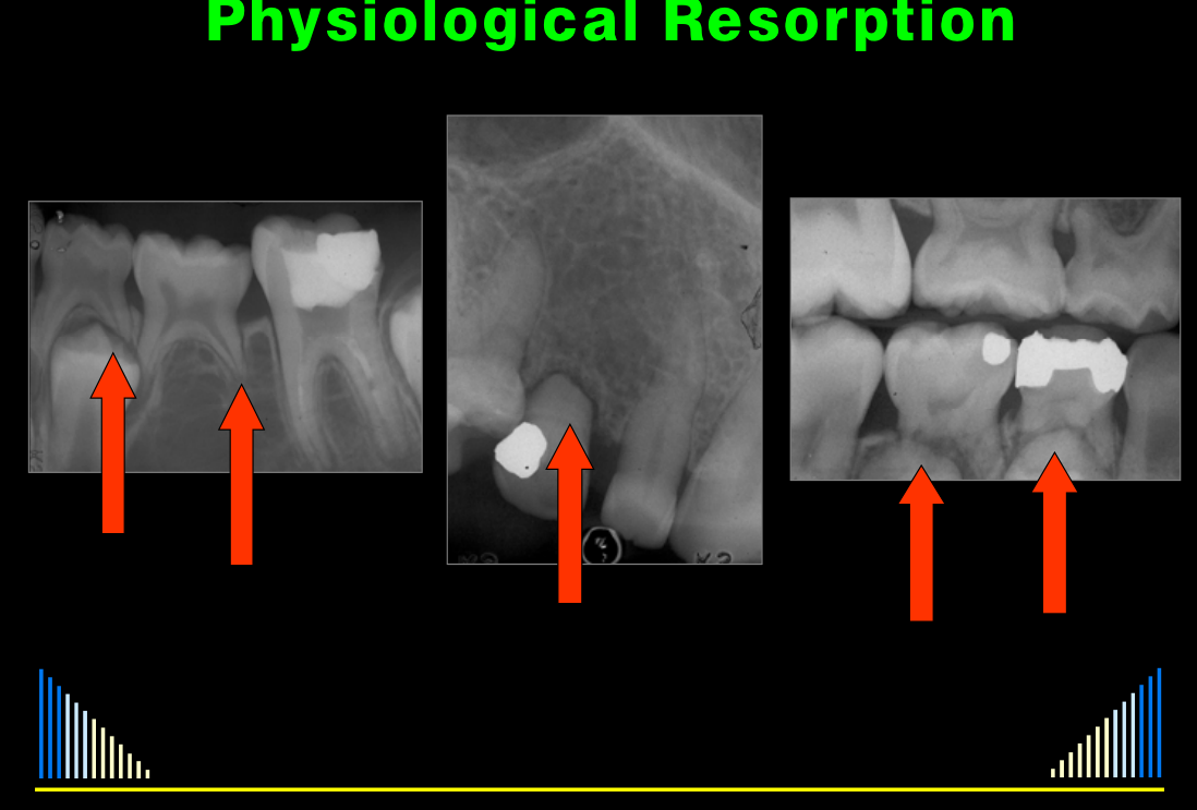
Physiological resorption is a normal process and is not generally a cause for alarm.
- Resorption of the roots of a first deciduous molar may occur as the premolar erupts.
- A second deciduous molar may have a somewhat shortened root even when there is no permanent successor tooth.
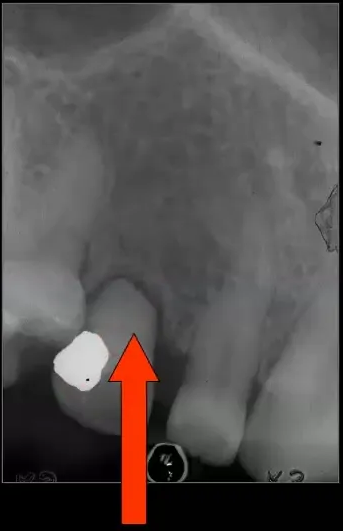
- A maxillary deciduous canine remained in place in a 93-year-old patient. It had undergone some physiological resorption but had not exfoliated, and its widened periodontal ligament space indicated increased mobility. The mobility was the reason the patient attended the dentist.
Management
-
Usually no treatment required
-
But: Extract if affecting eruption of permanent tooth
-
Earlier removal may be needed if the deciduous tooth has pulpitis or an infected root canal system, including when the permanent tooth is erupting.
Cheryl’s Initial Presentation
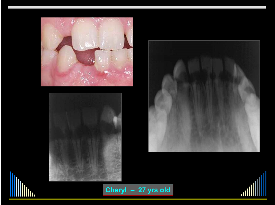
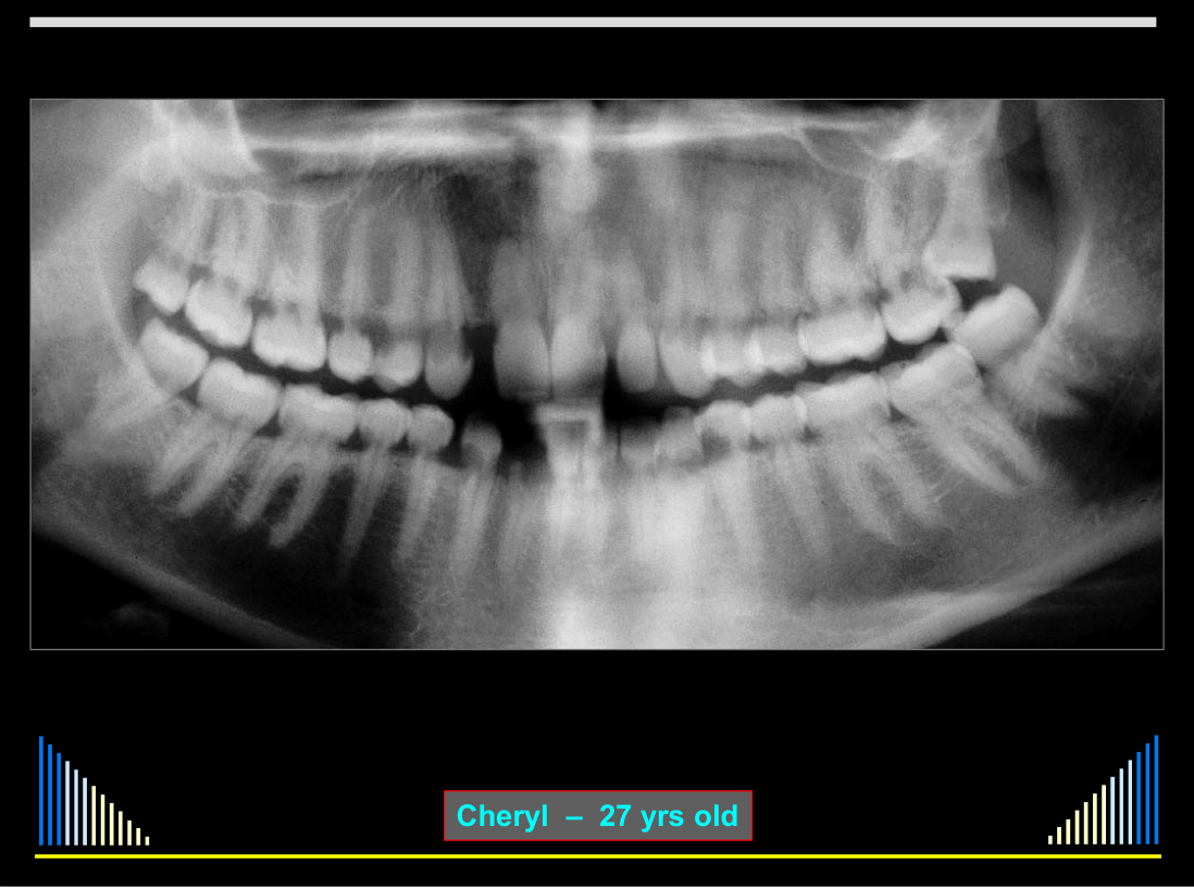
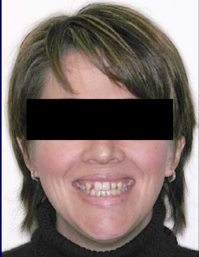
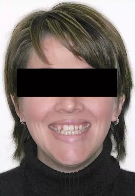
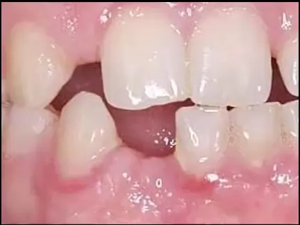
Cheryl was 27 years old and presented as an emergency after fracturing a lower lateral incisor while eating.
- The tooth fractured at the cervical level, was very mobile, and was painful on biting.
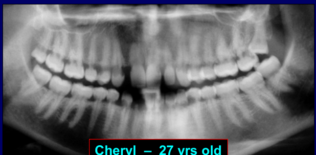
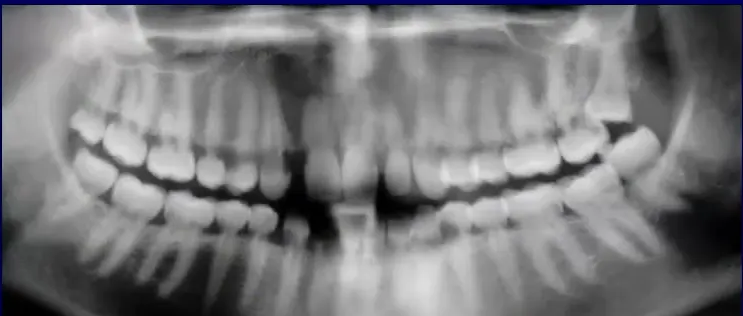
- Poor-quality initial radiographs prompted an occlusal radiograph and then a panoramic radiograph.
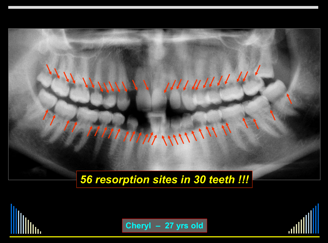
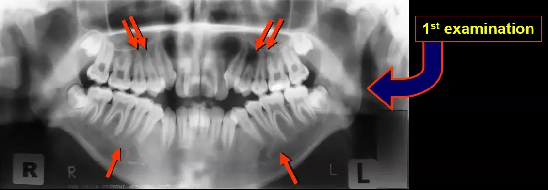
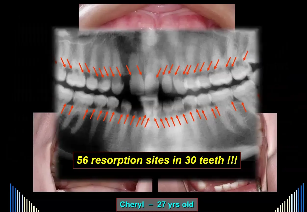
- The panoramic radiograph showed 56 sites of invasive resorption involving 32 teeth, with resorption in virtually every tooth and multiple sites in most affected teeth.
- Only the upper left third molar showed no visible resorption. The upper right lateral incisor was missing and may previously have fractured and been extracted for the same reason.
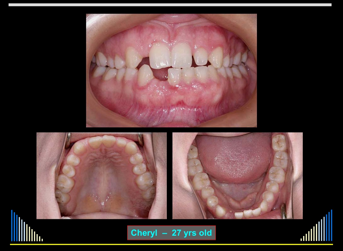
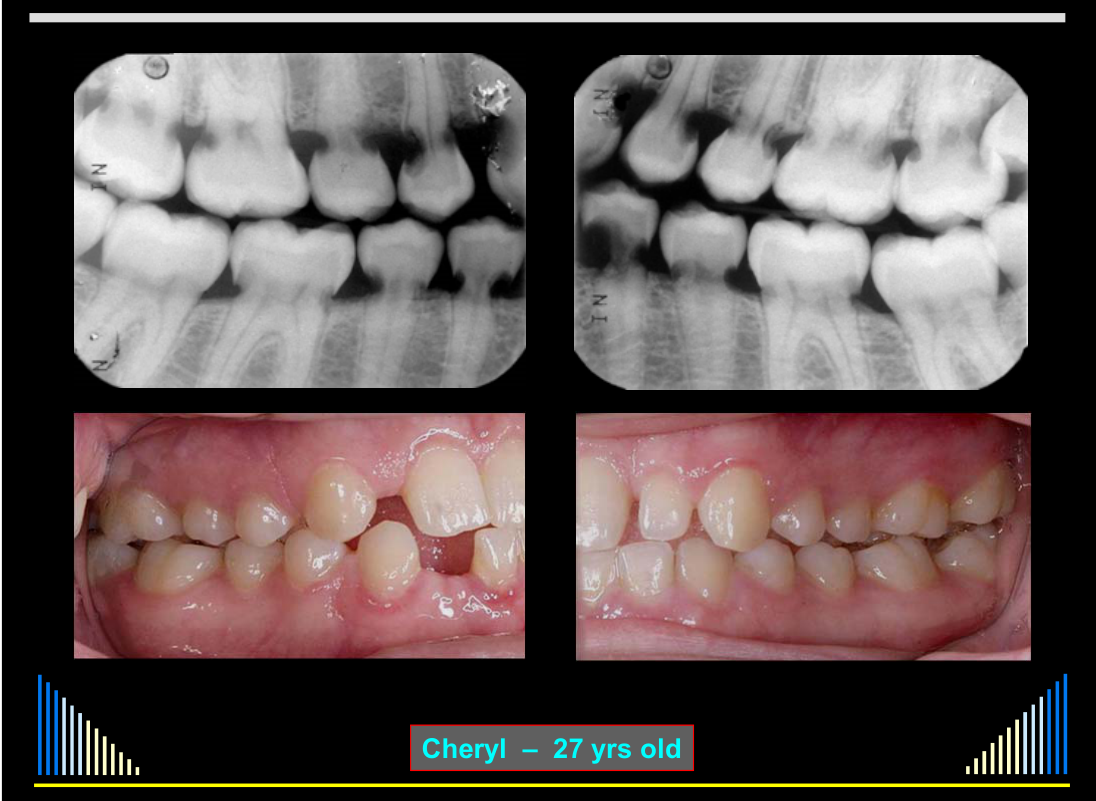
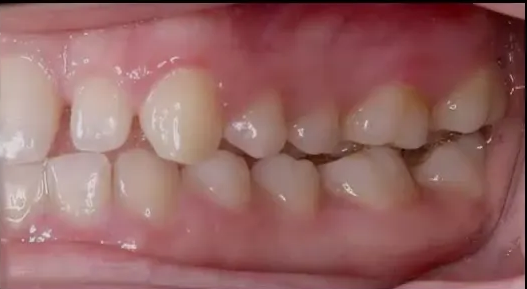
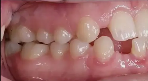
- Despite the extensive disease, the teeth appeared clinically normal, with no caries, restorations, periodontal pocketing, or bleeding on probing.
- The cause of the external invasive resorption could not be identified.
Source count discrepancy
The initial presentation reports 56 sites in 32 teeth, while later L5 case material reports 56 sites in 30 teeth; L9 Resorption 5 - External Invasive also reports 32. The counts remain unresolved, so both accounts are retained pending source verification.
Lecturer — Radiographic Progression
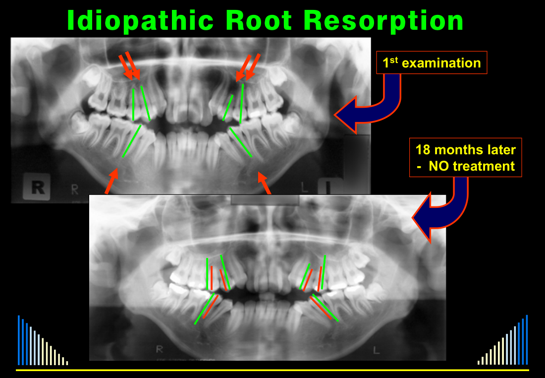
At the initial orthodontic assessment, treatment was deferred because the patient was considered too young, despite obvious crowding in the premolar, anterior, canine, lower premolar, and lower canine regions.
- Eighteen months later, no treatment had been carried out, but comparison of traced radiographs showed that the premolar teeth had become shorter.
- Although different radiographic angulation could have contributed to the apparent change, the other teeth did not show the same difference, supporting ongoing resorption for no known reason.
Idiopathic Resorption
Features8
- External resorption of numerous teeth
- Causes and mechanisms are unknown
- Can be apical resorption creating short roots
- OR: may be multiple sites of invasive resorption
- Causes and mechanisms are unknown
Management
- History & blood tests — to check for possible systemic causes
- Monitor rate of resorption — regular radiographs
- Probably extractions eventually + prostheses
Lecturer — Monitoring and Treatment
Idiopathic resorption is classified as idiopathic because no cause can be identified.
- Plain radiographs are preferred for regular monitoring because repeated CT scans expose the patient to more radiation.
- When orthodontic treatment is being considered, its safety cannot be assumed because the cause of the resorption is unknown.
- If extractions are needed during orthodontic treatment, teeth with the most severely shortened roots should be selected where possible.
Cheryl — 27 yrs old
Case Study: Cheryl910
 |  |
 |  |
First Examination11
- NO treatment
 |  |
18 Months Later — Medical History
- Adenoidectomy — age 8 y.o.
- Difficulty swallowing
- Microlaryngoscopy
- Mild reflux
- ? Raynaud’s Syndrome
- ? Autoimmune disease
- FBC: NAD
- No auto-antibodies
- Neck CT: NAD
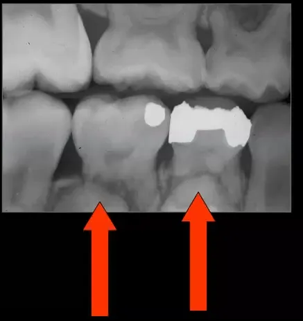
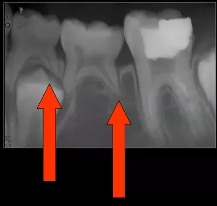
Radiographic examination images.
56 resorption sites in 30 teeth !!!
Diagnosis12
- External Invasive Resorption — 30 teeth
- Unknown / Idiopathic origin
Lecturer — Diagnostic Uncertainty
The orthodontic department identified the apical changes before orthodontic treatment had begun, but it was not possible to determine whether orthodontic treatment would be safe because the cause of the resorption was unknown.
 |  |
Diagnosis And Management Plan
Management Plan
- Full clearance + immediate dentures
- Implant-supported bridges
Cheryl’s Definitive Treatment
Individual treatment of isolated teeth was considered potentially possible, but the widespread resorption made conservation of the dentition impractical.
- Cheryl underwent full clearance of the teeth and received immediate dentures followed by implant treatment.
- An implant company provided the implants and surgical insertion as part of training procedures, while Cheryl paid for the implant-supported bridges.
- The lecturer described this as a particularly sad case because unexplained widespread resorption resulted in loss of the entire dentition.
Summary Of Root Resorption Types13
Internal
- Surface
- Inflammatory
- Replacement
External
See - Full external root-resorption list
The full eight-type list remains under Classification of Root Resorption — External.

Clinical Cases
Case: Orthodontic resorption, radiographic examples
Question
Scenario: Radiographic examples of teeth that have undergone orthodontic treatment.
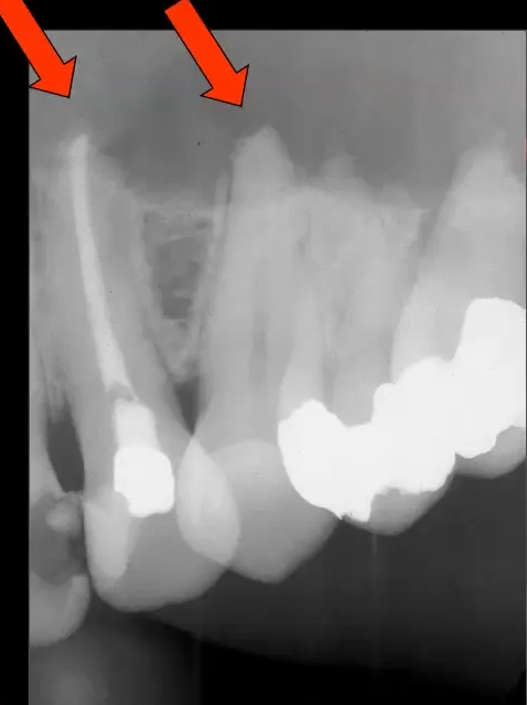
What’s shown: Radiographs showing teeth with shortened roots. One image shows a root-filled tooth with a crown alongside lateral and central incisors. Another image shows two teeth with very shortened roots.
Consider: Identify the features of orthodontic resorption and distinguish it from other causes of root shortening.
Answer
Observations:
- The lateral and left central incisors have shortened roots.
- A root-filled tooth with a crown (suggesting past trauma) shows no orthodontic resorption.
- Another example shows two teeth with very shortened roots.
Reasoning: Orthodontic resorption presents as shortened roots with rounded or blunt apices due to pressure during tooth movement. It is not related to trauma or the condition of the root canal system, as evidenced by the root-filled tooth showing no resorption despite a history of trauma.
Outcome: Management involves monitoring radiographically, ensuring good oral hygiene to prevent periodontal disease, and using mouthguards during sports to prevent trauma. If resorption occurs, orthodontic treatment may need to be modified with lighter forces or a different plan.
Takeaway: Orthodontic resorption is characterized by shortened, blunted roots due to orthodontic forces, is distinct from trauma-related resorption, and requires monitoring and protective measures.
Case: Maxillary odontogenic keratocyst causing pressure resorption
Question
Scenario: A patient with an odontogenic keratocyst in the maxilla.
What’s shown: A radiograph showing a radiolucency involving the entire maxilla on one side, extending from the posterior to the anterior region. The canine tooth has an abnormal root shape, and the premolars have flattened apices.
Consider: Identify the cause of the root resorption and the appropriate management for this patient.
Answer
Observations:
- Extensive radiolucency in the maxilla from the posterior to the anterior region.
- The canine tooth has an abnormal root shape.
- The premolars have flattened apices.
Reasoning: The extensive radiolucency is an odontogenic keratocyst. The abnormal root shape of the canine and flattened apices of the premolars are examples of external pressure resorption caused by the pressure from the cyst.
Outcome: All of the teeth in the affected quadrant were electively endodontically treated (root canal treatment) prior to the surgical removal of the keratocyst to prevent pulp necrosis from the surgery.
Takeaway: Pressure resorption from cysts or tumors causes abnormal root shapes and flattened apices; management involves removing the lesion and may require elective root canal treatment prior to surgery to protect the pulp.
Case: Impacted third molar causing resorption of lower second molar
Question
Scenario: A patient presenting with an impacted third molar.
What’s shown: A radiograph showing the impacted third molar and the adjacent lower second molar.
Consider: What is the effect of the impacted tooth on the adjacent tooth, and what was the management?
Answer
Observations:
- There is a very extensive area of resorption on the distal root of the lower second molar.
Reasoning: The impacted third molar is causing pressure resorption on the adjacent tooth root. While it is uncommon to see resorption this extensive, it can occur due to the pressure from the impacted tooth.
Outcome: The lower second molar was extracted.
Takeaway: Pressure resorption from an impacted tooth can cause extensive root resorption of an adjacent tooth, which may necessitate the extraction of the resorbing tooth.
Case: Ectopic canine causing resorption of upper first premolar
Question
Scenario: A patient presenting with an ectopic canine.
What’s shown: A radiograph showing the ectopic canine and the adjacent upper first premolar.
Consider: What is the effect of the ectopic tooth on the adjacent tooth?
Answer
Observations:
- There is resorption of the buccal root of the upper first premolar.
Reasoning: The ectopic canine erupted and caused pressure resorption on the root of the adjacent premolar.
Outcome: Not stated.
Takeaway: An ectopic erupting tooth can cause pressure resorption on the root of an adjacent tooth.
Case: Retained deciduous canine in a 93-year-old patient
Question
Scenario: A 93-year-old patient who presented to the dentist due to increased mobility of a retained deciduous tooth.
What’s shown: A radiograph of a maxillary deciduous canine.
Consider: What are the radiographic findings and the cause of the patient’s symptoms?
Answer
Observations:
- The deciduous canine has undergone some physiological resorption but has not exfoliated, despite the patient having had the tooth for a long time.
- A widened periodontal ligament space is visible around the tooth.
Reasoning: The widened periodontal ligament space indicates increased mobility, which explains why the patient visited the dentist. The tooth has undergone physiological resorption over a long period without exfoliating.
Outcome: The patient sought dental care due to the mobility of the tooth.
Takeaway: Retained deciduous teeth can undergo physiological resorption over a long period without exfoliating, and increased mobility may present radiographically as a widened periodontal ligament space.
Case: Idiopathic root resorption in a patient considered for orthodontic treatment
Question
Scenario: A patient presenting to the orthodontic department for an examination, found to have crowding and disappearing tooth apices.
What’s shown: Two panoramic radiographs taken 18 months apart, with traced lines indicating the initial and subsequent tooth lengths.
Consider: What is happening to the teeth over the 18-month period, and how should it influence the orthodontic treatment plan?
Answer
Observations:
- The initial radiograph shows obvious crowding, particularly in the premolar and anterior areas, and disappearing apices.
- The radiograph taken 18 months later shows that the premolar teeth have become shorter (traced with red lines compared to the green lines from the first radiograph).
Reasoning: The shortening of the teeth over 18 months without any treatment indicates the teeth are resorbing for no known reason (idiopathic root resorption). While radiographic angles might account for some difference in length, other teeth do not show the same effect, confirming active resorption.
Outcome: The orthodontist was advised that if extractions are part of the treatment plan, they should extract the teeth that have the most severe shortening of the roots.
Takeaway: In cases of idiopathic root resorption where the cause is unknown, orthodontic treatment plans involving extractions should prioritize extracting the most severely resorbed teeth.
Case: Extensive radiolucent lesions in a 27-year-old woman
Question
Scenario: A 27-year-old woman presented as an emergency after fracturing her lower lateral incisor while eating on Christmas Day. She had a vague medical history of an autoimmune disease during her youth, but specific details were unknown. An emergency dentist took radiographs on Boxing Day and was shocked by the findings, subsequently referring her for further evaluation.
What’s shown:
- A clinical photograph showing the fractured lower lateral incisor and a temporary suck-down denture.
- Initial panoramic and occlusal radiographs showing multiple radiolucent areas, particularly in the lower anterior teeth.
- A subsequent panoramic radiograph revealing widespread radiolucent defects across almost the entire dentition.
- Intraoral photographs showing clinically healthy teeth and soft tissues with no caries, restorations, or periodontal pocketing.
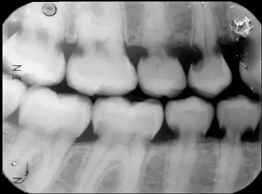
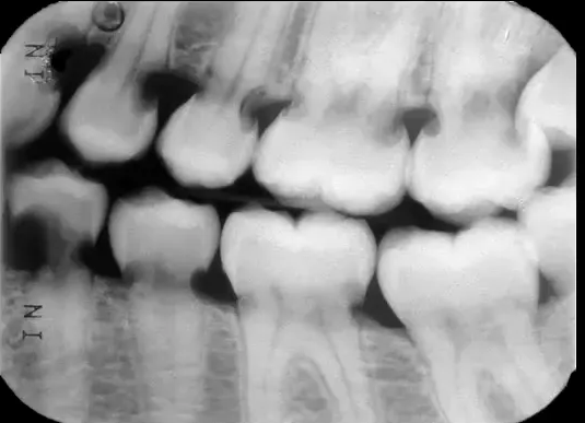
- Bitewing radiographs confirming varying extents of radiolucent areas on the teeth.
Consider: Identify the extent of the radiographic findings, contrast them with the clinical presentation, and determine the appropriate management for this condition.
Answer
Observations:
- The panoramic radiograph revealed 56 sites of invasive resorption across 32 teeth; only the upper left third molar was completely unaffected.
- The upper right lateral incisor was missing, and the patient noted it had broken in a similar manner to the lower lateral incisor.
- Clinically, the teeth and soft tissues appeared healthy, with no caries, restorations, or periodontal disease (minor gingival inflammation was present only due to irritation from the temporary denture).
- Bitewing radiographs confirmed both extensive and small areas of resorption on the teeth.
Reasoning:
- The widespread radiolucent areas on the radiographs indicate severe, multiple external invasive resorption affecting virtually the entire dentition.
- The stark contrast between the severe radiographic destruction and the normal clinical appearance highlights the hidden, destructive nature of this condition.
- The exact cause of the resorption remained unknown (idiopathic), despite the queried history of an autoimmune disease.
- Because almost every tooth had multiple extensive sites of resorption, treating and preserving individual teeth was not feasible.
Outcome:
- The patient underwent a full clearance of all teeth and was fitted with immediate dentures.
- She was later accepted into a training program by an implant company, which supplied the implants and surgical insertion for free; she only had to pay for the final implant-supported bridges.
Takeaway:
- Multiple invasive resorption can be extremely extensive and destructive across the entire dentition while remaining clinically asymptomatic and hidden, often necessitating full clearance and implant rehabilitation when individual tooth preservation is unfeasible and the cause is unknown.
Footnotes
-
Original PDF page 1: L5 Resorption 3 - Ortho, Pressure, Physio, Idiopathic, p.1 ↩
-
Original PDF page 3: L5 Resorption 3 - Ortho, Pressure, Physio, Idiopathic, p.3 ↩
-
Original PDF page 2: L5 Resorption 3 - Ortho, Pressure, Physio, Idiopathic, p.2 ↩
-
Original PDF page 4: L5 Resorption 3 - Ortho, Pressure, Physio, Idiopathic, p.4 ↩
-
Original PDF page 5: L5 Resorption 3 - Ortho, Pressure, Physio, Idiopathic, p.5 ↩
-
Original PDF page 7: L5 Resorption 3 - Ortho, Pressure, Physio, Idiopathic, p.7 ↩
-
Original PDF page 6: L5 Resorption 3 - Ortho, Pressure, Physio, Idiopathic, p.6 ↩
-
Original PDF page 8: L5 Resorption 3 - Ortho, Pressure, Physio, Idiopathic, p.8 ↩
-
Original PDF page 10: L5 Resorption 3 - Ortho, Pressure, Physio, Idiopathic, p.10 ↩
-
Original PDF page 11: L5 Resorption 3 - Ortho, Pressure, Physio, Idiopathic, p.11 ↩
-
Original PDF page 9: L5 Resorption 3 - Ortho, Pressure, Physio, Idiopathic, p.9 ↩
-
Original PDF page 12: L5 Resorption 3 - Ortho, Pressure, Physio, Idiopathic, p.12 ↩
-
Original PDF page 13: L5 Resorption 3 - Ortho, Pressure, Physio, Idiopathic, p.13 ↩