L4 Resorption 2 - External Surface, Replacement
External Resorption1
22/05/2020
Prof. Paul V. Abbott AO BDSc, MDS, FRACDS(Endo), FPFA, FADI, FICD, FACD, FIADT
Lecturer — Lecture Scope
This lecture continues the discussion of external resorption. It covers external surface, replacement, orthodontic pressure, physiological, and idiopathic resorption; external inflammatory and invasive resorption are reserved for separate lectures.
Related resorption topics
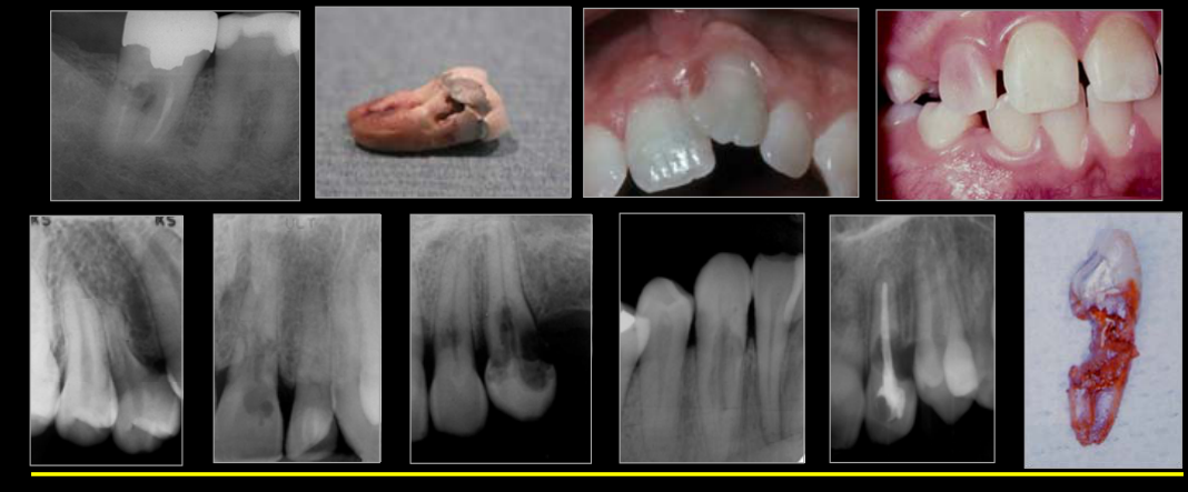
Classification of Root Resorption
Root Resorption2
See - Root-resorption definition and external types
Lecturer — Root Resorption
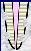
- In external replacement resorption, lost tooth structure is replaced by bone.
- The periodontal ligament separates the tooth from the alveolar bone, while cementum also helps protect the tooth.
- If the periodontal ligament and cementum are lost, bone may contact dentine, and osteoclasts involved in normal bone turnover may treat dentine as though it were bone.
- These cells can become dentinoclasts and remove tooth structure, after which osteoblasts form new bone where the tooth structure was removed.
Lecturer — Surface Resorption
External surface resorption may follow concussion, subluxation, extrusion, or lateral luxation when the root has not sustained extensive damage.
- Loss of a small amount of cementum may require an appropriate radiographic angle for detection; lesions on non-proximal surfaces are unlikely to be visible on radiographs or CT scans.
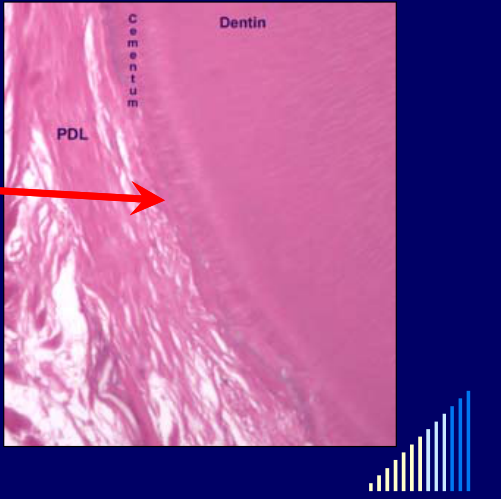
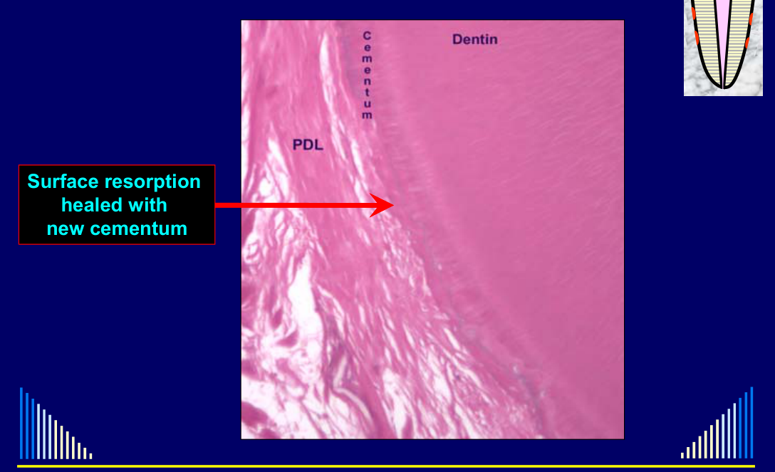
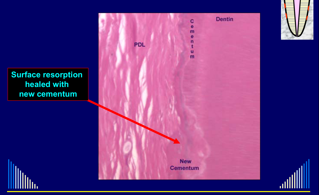
- Histologically, a line may separate the original cementum from the newly formed cementum.
- If the lesion extends into dentine, external inflammatory resorption may develop, but the root canal system would also need to be infected.
Lecturer — Replacement Resorption
External replacement resorption has also been called ankylosis-related resorption, but replacement resorption more accurately describes the tissue changes.
- Tooth structure is lost and replaced by bone; alveolar bone grows into the tooth, with no periodontal ligament between bone and dentine and no cementum in the affected area.
- Ankylosis is loss of the periodontal ligament and may occur without resorption, after other forms of external resorption, or before replacement resorption is radiographically evident.
- Prevention is the main management approach: replant an avulsed tooth immediately when possible, or store it in milk or saliva, and use a simple, passive, correctly positioned, preferably flexible splint.
- Once ankylosis and replacement resorption have developed, treatment cannot stop the process; endodontic treatment does not resolve it and may worsen the situation if it damages the periodontal ligament or uses toxic materials at an inappropriate time.
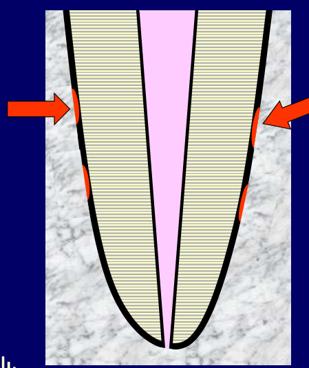
External Surface Resorption
Features3
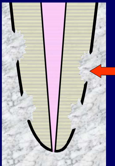
- Small resorption areas
- Usually only in cementum
- But may extend into dentine to a shallow depth
- Due to a localised injury to the cementum and the periodontal ligament
- Unlikely to be seen on radiographs
- Because too small and only proximal is visible
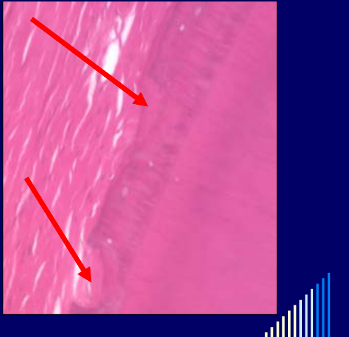 |  |
Management45
- A self-limiting process
- Usually heals with new cementum
- No treatment required
Surface resorption healed with new cementum
Ankylosis Before Replacement Resorption
An avulsed right central incisor showed loss of the periodontal ligament space in one area but a normal periodontal ligament space around the rest of the root.
- The affected area was ankylosed, but replacement resorption was not yet present.
- Because the protective separation between bone and tooth had been lost, replacement resorption was expected to follow over time as bone-remodelling cells resorbed dentine and replaced it with bone.
 |  |
 |  |
External Replacement Resorption
“Ankylosis-related resorption”
- Alternate terminology suggested by some authors
- BUT: The name of any disease/condition should reflect what is happening in the tissues — not what it is related to
- AND: ankylosis can also be related to other types of external resorption
Hence this term is confusing and inappropriate
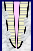
Features67
- Loss of tooth structure
- Root is replaced by bone
- Also have ankylosis — since there is:
- Loss of periodontal ligament
- AND
- Bone “fuses” to tooth root
 |  |
Ankylosis8
- “Fusion” of bone & root
- No periodontal ligament
- Can occur WITHOUT any resorption
- Can occur after ALL forms of resorption
Replacement resorption usually follows — eventually
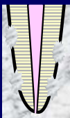
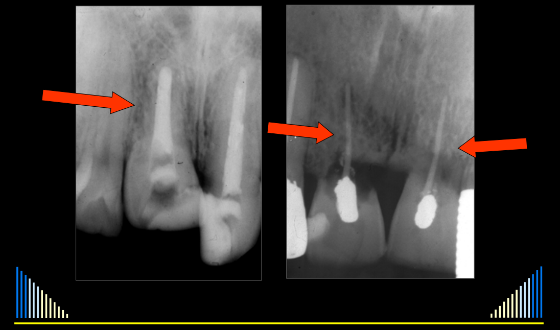
Types9
- Progressive
- Associated with drying or removal of PDL
- Gradually involves the whole root
- Typically in avulsion, intrusion, lateral luxation
Lecturer — Lateral Luxation
With lateral luxation, the damage may be limited to particular regions of the root.
- The palatal cervical region may be damaged when the root is pushed against the palatal bone.
- The apical region may be damaged when the root scrapes across the lip of bone after the labial socket wall breaks.
- Transient
- Minor areas of damage
- Later disappear — normal PDL forms
- Transient replacement resorption is not commonly seen and may be considered similar to surface resorption, depending partly on how deeply the resorption extends.
 |  |
Contributing Factors
- Extra-alveolar time
- Storage conditions
- Damage during injury and repositioning
- Splinting
- Endodontic treatment
Extra-Alveolar Time1011
- As short as possible
- To keep periodontal ligament cells on the tooth root alive
- 12–15 minutes critical
- If dry storage
Lecturer — Avulsion Timing
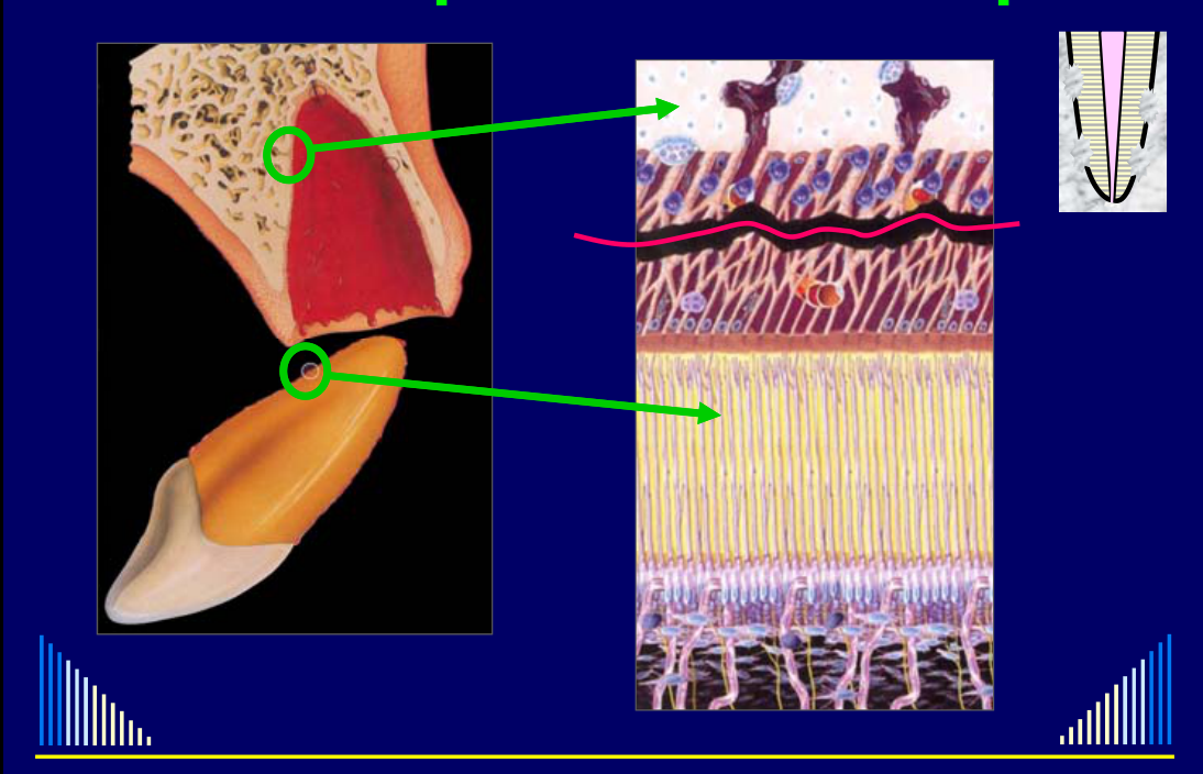
If an avulsed tooth remains dry outside the mouth for more than approximately 12–15 minutes, periodontal ligament cells begin to die.
- After approximately 20–30 minutes, about half of the periodontal ligament cells on the root may have died.
- After approximately one hour, almost all periodontal ligament cells are likely to have died.
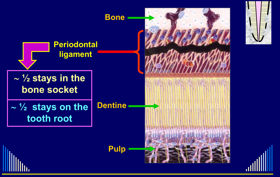
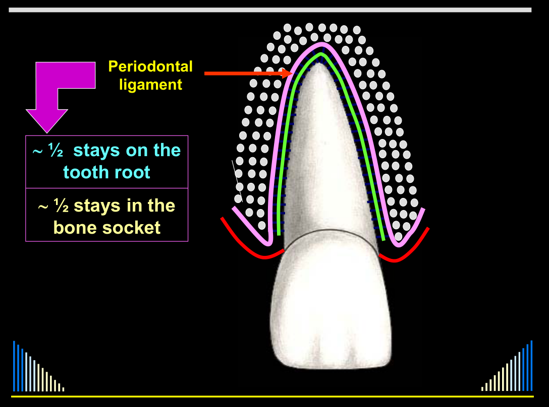
Periodontal ligament: ~½ stays in the bone socket, ~½ stays on the tooth root. Labels: Dentine, Pulp, Bone.
- ~ ½ stays on the tooth root
- ~ ½ stays in the bone socket
Lecturer — Immediate Replantation
The periodontal ligament remaining in the socket still has its normal blood supply, although it has been damaged by the injury. The portion attached to the root is particularly vulnerable because it can dry out and sustain further damage outside the mouth.
- Immediate replantation is encouraged even if the tooth cannot initially be positioned perfectly.
- Returning the tooth to its socket places it in the most appropriate environment, and the dentist can reposition it correctly later.
 |  |
 |
Storage Conditions12
- Storage media
- Factors associated with maintaining the viability of periodontal cells
- Osmolarity
- pH
- Temperature
- Factors associated with maintaining the viability of periodontal cells
Tap water
- → Rapid cell lysis
- → Only quick rinse if nothing else available
- ☼ But saliva is usually available !!!
Saline
- → Max. 1 hour
- → No nutrients
Saliva
- → Max. 2 hours
- ☼ PDL cells have low resistance to salivary bacteria
Plastic wrap
- → Max. 1 hour — prevents dehydration
- → Combine with saliva, if possible
Milk
- Max. 6 hours
- pH and osmolarity suitable
- Temperature not important — if < 37°C
- NOT yoghurt or sour milk
- pH too low
- Milk is practical because it is often available in homes, workplaces, schools, sporting venues, and nearby shops; low-fat, skim, long-life, powdered, goat, and sheep milk were all described as generally effective.
- pH too low
Tissue culture media
- Ideal
- But rarely available
Lecturer — Commercial Storage Media
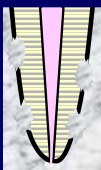
Commercial products such as Saver Tooth contain tissue culture solution and may be effective, but they are expensive and have a relatively short shelf life.
- The tooth is held in a soft net inside a plastic cage placed in the medium.
- The net prevents the tooth from rubbing against the container, while the cage allows insertion and removal without physically handling the tooth.
 |  |
Damage During Injury and Repositioning
- Damage during the injury
- Mechanical damage
- Can not be controlled
- Mechanical damage
- During repositioning/replant procedure
- Minimise trauma
- If replanted by the dentist
- BUT — can not always be controlled
- e.g. if replanted by patient, parent or other person at the accident scene
- Minimise trauma
Splinting13141516
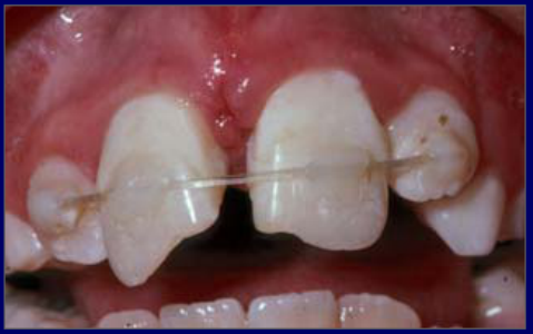
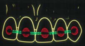
Two types of splints:
- Flexible
- Rigid
Flexible splint
- If NO root or bone fractures
- Allows “functional” healing of PDL
- Reduces ankylosis & replacement resorption
- 7–10 days only
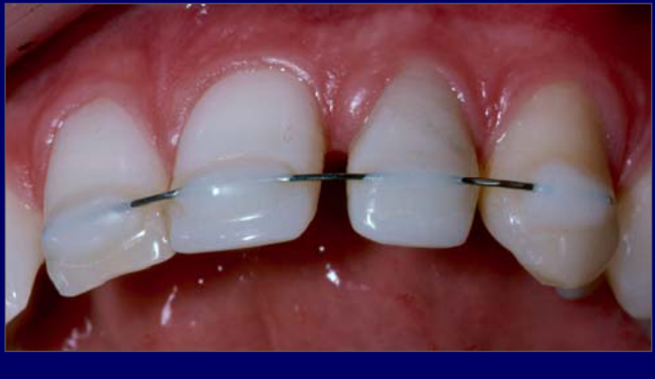
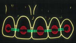
Rigid splint
ONLY use if:
- A bone fracture → 6 weeks
- OR if: a root fracture → 3 months
Requirements of a splint
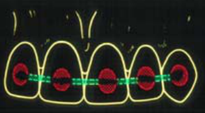
- Keep it simple
- Avoid gingival tissues
- Allow interproximal cleaning
- Instruct the patient how to clean
- Check tooth position radiographically
- Relieve from occlusion
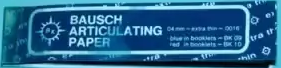
- Check with articulating paper
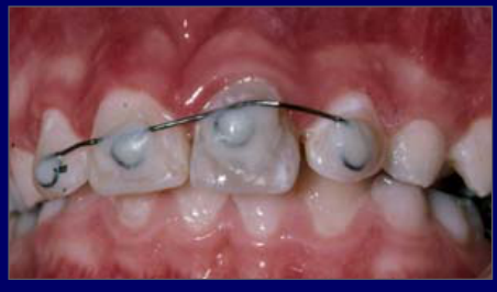
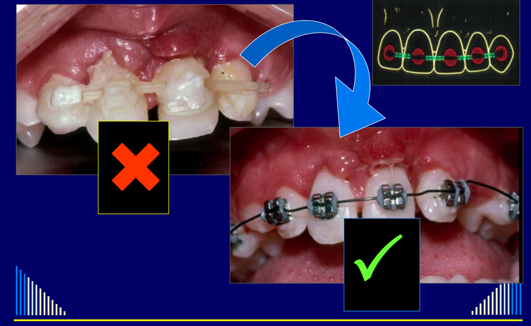
Unsatisfactory Splint
Several avulsed teeth and a tooth with a horizontal root fracture were splinted together without adequate anchorage. The composite extended around the gingival margins, the patient developed substantial gingival inflammation, and the splint remained for approximately one month, longer than appropriate for a flexible splint.
- A later, more rigid splint was extended to the premolar teeth to obtain anchorage.
DeTrey / RAUSCH articulating paper; radiograph to check if splint is right position
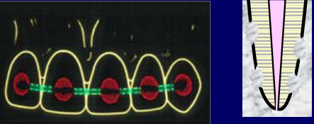
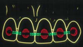
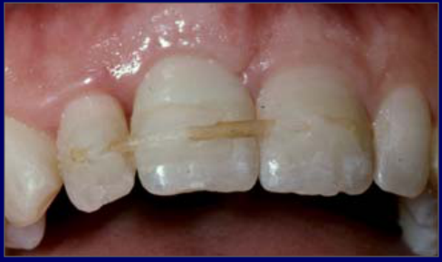
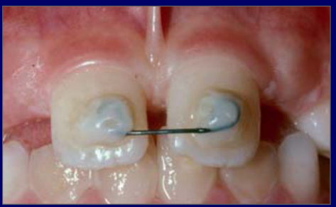
Flexible splint
Fishing line + composite resin
Rigid splint
SS wire + composite resin
SS wire + composite resin
Can put loops to make it more rigid
Lecturer — Splint Rigidity
A splint can be made more rigid by using thicker wire, adding loops, extending the composite slightly across the labial surface without involving the proximal surfaces, or increasing the amount of composite when using nylon fishing line.
Unsatisfactory splint
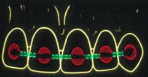
Endodontic Treatment
- Use of toxic materials may destroy more PDL cells — hence more replacement resorption occurs
- ∴ AVOID:
- early use of Ca(OH)₂ → (early moves within 3 months)
- immediate root filling
- extra-oral endodontic treatment
- ✓ Instead USE: a corticosteroid-antibiotic mixture
- e.g. Ledermix paste
Management
- Endodontic treatment can NOT arrest or treat external replacement resorption
- Since this resorption is due to damage to the PDL and root surface
- ⇒ i.e. it is NOT due to pulp disease
- Only do endodontics to prevent or treat external inflammatory resorption
- Some added benefits from using intracanal medicaments that inhibit clastic cells
- Corticosteroid / tetracycline paste
- Some added benefits from using intracanal medicaments that inhibit clastic cells
Lecturer — Endodontic Indications
Immediate endodontic treatment after replantation may be recommended for avulsion or intrusion of fully developed teeth, and for avulsion or intrusion of immature teeth when there is also a crown fracture. The purpose is to prevent or treat external inflammatory resorption, not external replacement resorption.
Factors Summary
- Extra-alveolar time
- Keep as short as possible (ideally < 12 mins)
- Storage conditions
- Milk > saliva = plastic wrap ≠ water = dry
- Damage during injury and repositioning
- Avoid mechanical damage
- Splinting
- Flexible > Rigid
- Endodontic treatment
- Avoid toxic materials; use CS-AB’s
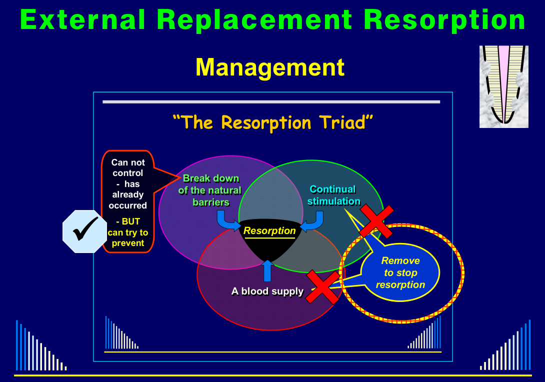
Management17
See - Resorption triad foundation
L2 foundational triad; navigation: Resorption Triad.
- Can not control — has already occurred — BUT can try to prevent
- Remove to stop resorption
 |  |
Clinical Cases
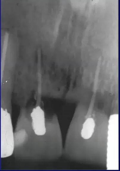
Case: Avulsed teeth with external replacement resorption
Question
Scenario: Two upper teeth that were avulsed some years ago are presented for evaluation.
What’s shown: A periapical radiograph showing the two teeth with extensive external replacement resorption.
Consider: What would you expect regarding the clinical mobility of these teeth, and why?
Answer
Observations:
- The radiograph shows two upper teeth with extensive external replacement resorption.
- The affected areas lack a periodontal ligament and cementum, with alveolar bone growing directly into the tooth.
Reasoning:
- The loss of the periodontal ligament and the fusion of bone to the tooth root result in ankylosis.
- Due to this ankylosis and the absence of a periodontal ligament, the teeth do not have increased mobility; instead, they exhibit decreased mobility.
Outcome: None stated.
Takeaway: External replacement resorption leads to ankylosis, which clinically presents as decreased tooth mobility due to the loss of the periodontal ligament.
Case: Right central incisor with ankylosis post-avulsion
Question
Scenario: A right central incisor that was avulsed approximately three years ago is evaluated.
What’s shown: A radiograph with an arrow pointing to a specific area on the root of the right central incisor, while the rest of the root shows a normal periodontal ligament space.
Consider: What does the area indicated by the arrow represent, and what is the status of replacement resorption in this tooth?
Answer
Observations:
- The area indicated by the arrow shows an absence of the periodontal ligament space.
- The rest of the tooth root displays a normal periodontal ligament space.
Reasoning:
- The loss of the periodontal ligament space indicates ankylosis (fusion of bone to the root).
- Despite the presence of ankylosis, there is no evidence of replacement resorption at this stage.
Outcome: None stated.
Takeaway: Ankylosis can occur without replacement resorption, though replacement resorption typically follows ankylosis over time due to the loss of the periodontal ligament’s protective function.
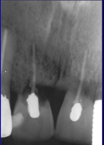
Case: Periapical radiographs showing root resorption
Question
Scenario: Radiographs of maxillary anterior teeth are presented for evaluation.
What’s shown: Two side-by-side periapical radiographs.
Consider: What type of resorption is demonstrated, and what is the typical clinical history associated with this finding?
Answer
Observations:
- Radiographs show root resorption.
Reasoning: The findings represent progressive external replacement resorption, which will gradually involve the entire tooth root and lead to tooth loss. This condition is typically associated with traumatic injuries causing significant displacement (such as avulsion, intrusion, or lateral luxation) that result in drying, removal, or damage to the periodontal ligament. The specific example shown follows an avulsion injury.
Takeaway: Progressive external replacement resorption is a severe consequence of traumatic dental injuries that damage the periodontal ligament, leading to gradual root loss and eventual tooth loss.
Case: Rigid splint, radiographic check
Question
Scenario: A patient with a rigid splint is being reviewed to verify the position of the injured tooth.
What’s shown: A radiograph displaying a rigid wire splint in place on the anterior teeth.
Consider: What is the primary radiographic check required after placing a rigid splint?
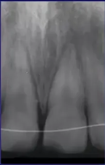
Answer
Observations:
- A rigid splint made of wire is visible on the teeth.
- The injured tooth is positioned back within its socket.
Reasoning: After placing a rigid splint (e.g., for a bone or root fracture), it is essential to verify radiographically that the tooth has been correctly repositioned into its socket.
Outcome: The radiograph confirms the tooth is correctly seated in its socket.
Takeaway: Always check radiographically that a splinted tooth is correctly positioned in its socket.
Case: Splint using a local anesthetic needle
Question
Scenario: A dentist needs to place a rigid splint for a patient over the weekend but does not have wire available.
What’s shown: A clinical photograph of a splint where a local anesthetic needle is used as the connector.
Consider: What alternative material can be used as a connector for a rigid splint if standard wire is unavailable?
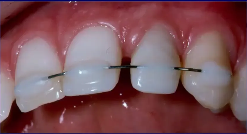
Answer
Observations:
- A long local anesthetic needle is utilized as the wire or connector for the splint.
Reasoning: The needle provides a suitable size and rigidity for a splint connector. It serves as a practical improvised alternative because local anesthetic needles are readily available in every dental surgery.
Takeaway: A local anesthetic needle can be used as an improvised connector for a rigid splint when wire is unavailable.
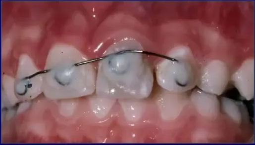
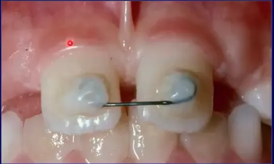
Case: Horizontal root fracture with missing teeth, splinted with wire loops
Question
Scenario: A patient presents with a horizontal root fracture in a right anterior tooth, complicated by missing deciduous and unerupted permanent lateral incisors.
What’s shown: A clinical photograph of a rigid splint constructed using wire with loops, applied to the limited available teeth.
Consider: How can an effective rigid splint be constructed when there are missing teeth and limited anchorage?
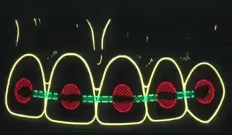
Answer
Observations:
- The patient is missing deciduous lateral incisors, and the permanent lateral incisors have not yet erupted.
- The right tooth has a horizontal root fracture at the mid-root level.
- A rigid splint is constructed using only one adjacent tooth, incorporating small loops in the wire.
Reasoning: When anchorage is limited due to missing teeth, wire loops can be incorporated into the splint to increase rigidity and hold the wire against the tooth more securely. This allows an effective splint to be placed using minimal teeth.
Outcome: An effective rigid splint was successfully placed using one tooth and wire loops to provide the necessary rigidity.
Takeaway: Wire loops can be added to a splint to increase rigidity when anchorage is limited, such as in cases with missing teeth.
Case: Unsatisfactory splint with multiple fishing lines and composite
Question
Scenario: A patient presents for review of a dental splint placed after trauma.
What’s shown: A clinical photograph of the splinted teeth showing multiple types of fishing line and composite resin.
Consider: What are the clinical findings regarding the splint design and the patient’s oral hygiene status?
Answer
Observations:
- The splint incorporates at least three different types or thicknesses of fishing line.
- Composite resin is placed between the teeth and extends right up to the gingival margin.
- There is significant plaque accumulation around the teeth.
- The gingiva shows extensive inflammation.
Reasoning: The use of multiple fishing lines suggests a lack of a standardized approach, while the composite extending to the gingival margin and between the teeth makes it impossible for the patient to clean effectively. This poor design directly leads to plaque retention and gingival inflammation.
Outcome: The splint is deemed highly unsatisfactory. The lecturer emphasizes that splints should be simple and allow for adequate oral hygiene, rather than using composite-only or poorly designed splints that hinder cleaning.
Takeaway: Splints must be designed to allow the patient to clean between the teeth; extending composite to the gingival margin and interproximal spaces causes plaque accumulation and gingival inflammation.
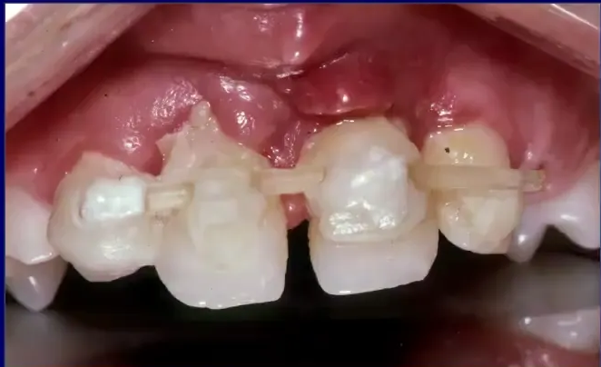
Case: Inadequately splinted avulsed and root-fractured incisors
Question
Scenario: A patient who suffered trauma resulting in the avulsion of multiple incisors and a horizontal root fracture of a lateral incisor, initially treated by another dentist.
What’s shown: Clinical photographs showing the initial inadequate splint with severe gingival inflammation, followed by images after splint removal and subsequent re-splinting.
Consider: What were the errors in the initial splinting management, and how was the situation corrected?
Answer
Observations:
- The initial splint used fishing line but lacked anchorage, as it only splinted the injured teeth together.
- Composite resin extended to the gingival margin, preventing the patient from cleaning.
- Severe gingival reaction and inflammation were present one month after placement.
- The flexible splint had been left on for far too long (flexible splints should only be left for 7 to 10 days).
- The teeth were very mobile upon removal of the initial splint.
Reasoning: The initial splint lacked adequate anchorage and prevented oral hygiene, leading to inflammation and a failure to heal. Because the teeth were very mobile and there was a root fracture requiring rigid stabilization, a new splint needed to provide more rigidity and proper anchorage.
Outcome: The initial splint was removed. A new, more rigid splint was placed using orthodontic brackets, extending back to the premolar teeth on each side to provide adequate anchorage.
Takeaway: Splints must have adequate anchorage, allow for oral hygiene, and be removed in a timely manner. Root fractures or severe mobility may require a rigid splint with extended anchorage.
Case: Broken composite resin splint
Question
Scenario: A patient presents with a failed composite resin splint.
What’s shown: A clinical photograph showing a composite splint that has broken, with different colored materials visible, covering the interproximal regions and extending to the gingival margin.
Consider: Why did this splint fail, and what are the general drawbacks of this type of splint design?
Answer
Observations:
- The composite splint originally extended to the lateral incisor but has broken.
- Different colored material is visible, indicating a break or previous repair.
- The composite covers the interproximal regions and extends right up to the gingival margin.
Reasoning: The splint broke because teeth naturally move, especially after injury, and if the occlusion is incorrect, this movement places stress on the composite. The material eventually gives way, typically by debonding from one or more teeth. Additionally, this design makes it very difficult for the patient to clean and for the dentist to remove.
Outcome: The splint became ineffective due to the breakage.
Takeaway: Composite-only splints are unsatisfactory because they hinder oral hygiene, are difficult to remove, and tend to break under the stress of natural tooth movement and occlusal forces.
Footnotes
-
Original PDF page 1: L4 Resorption 2 - External Surface, Replacement, p.1 ↩
-
Original PDF page 2: L4 Resorption 2 - External Surface, Replacement, p.2 ↩
-
Original PDF page 3: L4 Resorption 2 - External Surface, Replacement, p.3 ↩
-
Original PDF page 4: L4 Resorption 2 - External Surface, Replacement, p.4 ↩
-
Original PDF page 5: L4 Resorption 2 - External Surface, Replacement, p.5 ↩
-
Original PDF page 6: L4 Resorption 2 - External Surface, Replacement, p.6 ↩
-
Original PDF page 7: L4 Resorption 2 - External Surface, Replacement, p.7 ↩
-
Original PDF page 8: L4 Resorption 2 - External Surface, Replacement, p.8 ↩
-
Original PDF page 9: L4 Resorption 2 - External Surface, Replacement, p.9 ↩
-
Original PDF page 10: L4 Resorption 2 - External Surface, Replacement, p.10 ↩
-
Original PDF page 11: L4 Resorption 2 - External Surface, Replacement, p.11 ↩
-
Original PDF page 12: L4 Resorption 2 - External Surface, Replacement, p.12 ↩
-
Original PDF page 16: L4 Resorption 2 - External Surface, Replacement, p.16 ↩
-
Original PDF page 19: L4 Resorption 2 - External Surface, Replacement, p.19 ↩
-
Original PDF page 20: L4 Resorption 2 - External Surface, Replacement, p.20 ↩
-
Original PDF page 21: L4 Resorption 2 - External Surface, Replacement, p.21 ↩
-
Original PDF page 23: L4 Resorption 2 - External Surface, Replacement, p.23 ↩