Case 11
R
Worksheet prompts (Suggested Approach, applies to every case)
- Describe the image/radiograph.
- Identify relevant soft tissue findings.
- Identify relevant hard tissue findings.
- Estimate the patient’s dental age.
- Develop a problem list.
- Formulate differential diagnoses.
- Consider additional investigations.
- Develop a management plan.
Clinical Case
Answered from the lecturer’s comments in the recording only. The student presentations are left out.
1. Image / radiograph
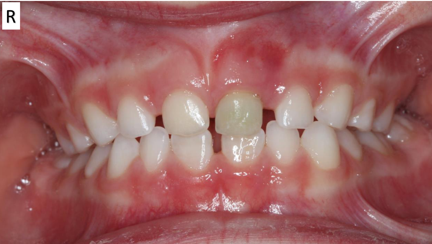
- Frontal intraoral photograph with the teeth in occlusion.
2. Soft tissue findings
- Redness and inflammation at the mucogingival junction / sulcus above 61. This is most likely the site of a draining sinus.
- The abscess (a collection of pus) builds up until the pressure makes it burst. The infection then drains, and the child often has no more pain because the pressure is gone and the tooth is necrotic.
- Parents often report “a big pimple on the gum that burst, so everything’s fine”. It is still a sign of infection, just past its peak collection.
- If unsure, compare with the contralateral side. This matters especially with darker, pigmented mucosa.
3. Hard tissue findings
- 61: darker (grey) discolouration.
- 51: also discoloured, with a slight yellow hue compared with the adjacent tooth.
- Lower central incisors: permanent.
- In this child the colour difference from the primary teeth is small, and width alone isn’t decisive.
- The tell is that the incisal edges are not quite in occlusion: the teeth are only partially erupted, which matches the age group.
- The lecturer said this distinction is hard and wouldn’t be examined.
- Faint intrinsic discolouration is hard to assess from the front. It is easiest to see from the palatal with a mirror and light shining through the teeth. Transillumination can also help.
4. Dental age
- About 6 years (“six is very good”).
- The mandibular central incisors erupt at about 5½–7 (6–7) years, which firms up 6 rather than earlier.
- No maxillary permanent incisors are in yet (they erupt at 7–8), so the child is not in the 7–8 bracket.
5. Problem list
- 61: discoloured, with a draining sinus, so infection is present.
- 51: intrinsic yellow discolouration.
- Likely past trauma, even if the parents deny it (“there’s been a trauma at some point”).
6. Differential diagnoses
- The lecturer said the group’s differentials were “perfect”: pulp canal obliteration, necrosis, or recent trauma (haemorrhage inside the tooth causing temporary discolouration).
- 61: necrotic, infected pulp space with a periapical abscess.
- 51: pulp canal obliteration (calcification), suggested by the yellow colour. Necrosis is possible, but the colour leans towards obliteration. Cause: probably the same trauma, or a separate episode.
7. Additional investigations
- History: trauma (it may have been years ago and forgotten), pain, and whether there was a lump on the gum that has since gone.
- Radiograph: for the primary dentition some time after trauma, usually just a maxillary occlusal, because 6-year-olds often struggle with periapical films.
- Look at the periapical area and the pulp space: is there pulp canal obliteration, and is there infection?
- Expected result here: pulp canal obliteration on 51 and a periapical radiolucency on 61.
8. Management plan
- Extract 61 because it is infected.
- It would normally exfoliate around 7–8, but necrotic teeth don’t always exfoliate normally.
- Over-retained teeth can cause crowding or ectopic eruption of the successors.
- This can be a difficult conversation with the family.
- Monitor 51.
- Pulp canal obliteration isn’t tied to necrosis; the pulp must be vital for it to occur.
- Act only on pain, abnormal mobility or radiographic signs of infection.
- If 51 is over-retained (for example, the contralateral tooth exfoliated 6 months earlier), extract it for that reason.
- Advise the family: if they see a pimple or redness on the gum when lifting the lip, or the child has pain, they should bring the child back.
Footnotes
-
Original PDF page 1: T1 Case 1, p.1 ↩