Case 31
R
Worksheet prompts (Suggested Approach, applies to every case)
- Describe the image/radiograph.
- Identify relevant soft tissue findings.
- Identify relevant hard tissue findings.
- Estimate the patient’s dental age.
- Develop a problem list.
- Formulate differential diagnoses.
- Consider additional investigations.
- Develop a management plan.
Clinical Case
Answered from the lecturer’s comments in the recording only. The student presentations are left out.
1. Image / radiograph
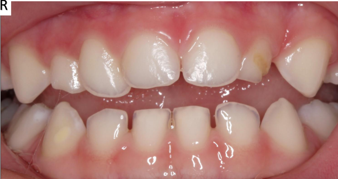
- Frontal intraoral photograph.
2. Soft tissue findings
- Pretty healthy gingiva, without excessive plaque.
3. Hard tissue findings
- Supernumerary tooth in the upper anterior region.
- Which one is extra? Compare with the contralateral side. The one that is much larger than its contralateral (about twice the size) is most likely the supernumerary, even though the other looks the more “perfect” incisor shape.
- Midline shift: there is a primate space mesial to the primary canine on one side but not on the other.
- 83: well-demarcated, creamy-yellow opacity on the buccal surface.
- 84: well-demarcated, white opacity on the mesiobuccal surface.
- How to describe a lesion: is it well demarcated or not, what colour is it, and is it an opacity.
- 63: buccal defect. Most likely a hypoplastic defect (a developmental defect of enamel) with secondary caries, not primary caries.
- The gingiva is healthy, other developmental defects are present, and the buccal surface isn’t in occlusion.
- There is also a wear facet, because the tooth sits further back in the arch than normal.
- Incisal edges: translucent, which is normal for this child (the upper arch is the same).
- Hypoplasia vs hypomineralisation: hypoplasia means tooth structure is missing from development. Hypomineralised enamel is the same amount of enamel with a different mineral content.
4. Dental age
- 3–5 years (“very reasonable”).
5. Problem list
- Supernumerary tooth: midline shift and a risk to the eruption of the permanent dentition.
- 63: hypoplastic defect with secondary caries.
- 83 and 84: developmental defects of enamel.
- Caries risk: this child is not considered high caries risk; the caries is due to a local factor.
6. Differential diagnoses
- 63: hypoplastic defect with secondary caries (favoured), versus post-eruptive breakdown, versus primary caries.
7. Additional investigations
- Maxillary occlusal radiograph: checks that all the permanent teeth are present and looks for other supernumeraries (“if you see one, always go looking for more”).
- Why not an OPG: the anterior region is outside the focal trough, the field there is narrow, and the central region often has ghosting. A slightly anterior or posterior supernumerary may not be visible.
- Timing: there is no need to take it at age 2. Ask whether it will change management. Waiting until 4–5 is fine, because there will be no intervention before then.
8. Management plan
- 63, option 1: composite (strip) crown.
- 63, option 2: conservative remineralisation with fluoride varnish. The area is open and cleansable, and with committed parents it may hold until exfoliation.
- Supernumerary: monitor the eruption of the permanent teeth.
- If the permanent lateral won’t resorb the primary roots and erupt normally, the supernumerary has to go.
- Often both the supernumerary and 62 are removed, because 62 has been pushed distally and is probably out of line with the permanent incisor.
- Timing of extraction: ideally about 6–7 years.
- Too early means a likely GA commitment and a higher risk of damaging the developing, not-yet-calcified permanent teeth.
- Go earlier if the child is already under GA, or if there is a severe impact on the erupting dentition.
- Technique: in a cooperative child these are easy extractions under local anaesthetic. Release the PDL with a luxator, then use upper straight forceps with a twist. The local anaesthetic takes longer than the extraction.
Footnotes
-
Original PDF page 1: T1 Case 3, p.1 ↩