Case 51
Worksheet prompts (Suggested Approach, applies to every case)
- Describe the image/radiograph.
- Identify relevant soft tissue findings.
- Identify relevant hard tissue findings.
- Estimate the patient’s dental age.
- Develop a problem list.
- Formulate differential diagnoses.
- Consider additional investigations.
- Develop a management plan.
Clinical Case
Answered from the lecturer’s comments in the recording only. The student presentations are left out.
1. Image / radiograph
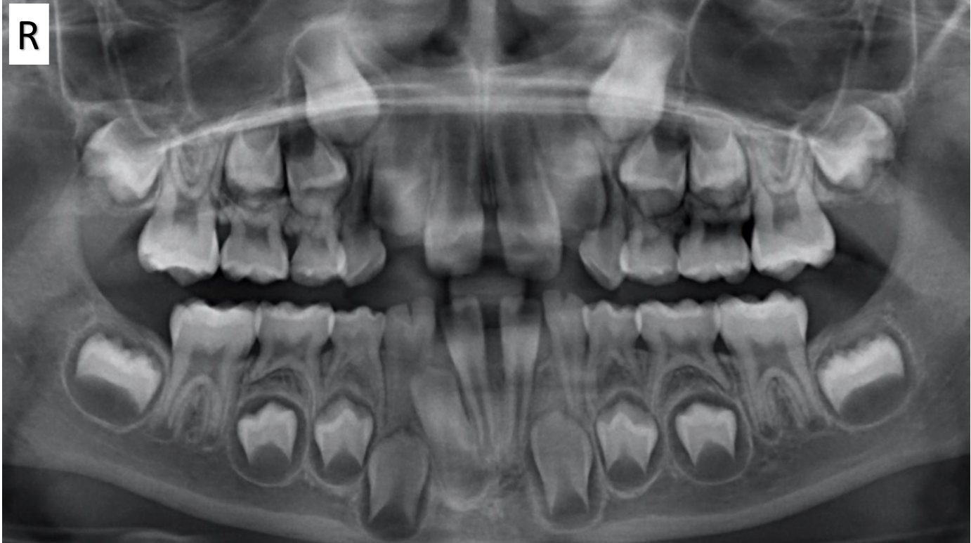
- OPG.
2. Soft tissue findings
- Not discussed (radiograph only).
3. Hard tissue findings
- Upper laterals: they look large, but partly because of positioning. On an OPG, objects closer to the source look larger.
- The anterior maxilla is likely crowded, with the laterals sitting palatally (a Div 1-type situation, with the centrals proclined).
- The laterals look potentially impacted.
- Primary canines: the laterals appear to have resorbed the primary canines to some extent. One canine may be lost earlier than the other, which risks a midline shift (consider a balancing extraction).
- Lower double teeth (primary): fusion versus gemination can’t really be told apart.
- A missing tooth suggests fusion. But permanent teeth have erupted, so it’s unknown whether primary laterals were there.
- There are two pulp chambers, which makes fusion more likely.
- Clinically it doesn’t matter. “Double tooth” is used for this reason.
- Incisal notches: pronounced on the double teeth, where the two crowns partially fused. This is a very plaque-retentive area, and the pulp is close.
- 32 is missing: a midline shift is possible as 42 erupts. Count every tooth, because a missing lower incisor is easy to miss.
- Lower canines: positioned nicely.
4. Dental age
- Not explicitly estimated by the lecturer. The referral advice was framed around a presentation like this at about 7–8 years.
5. Problem list
- Anterior maxillary crowding, with palatally positioned, possibly impacted upper laterals.
- Resorption of the primary canines by the laterals, with a risk of early unilateral loss and a midline shift.
- Lower primary double teeth: plaque-retentive notches close to the pulp.
- 32 missing: a potential lower midline shift.
6. Differential diagnoses
- Double teeth: fusion versus gemination (fusion more likely, given the two pulp chambers).
- Apparent macrodontia of the laterals: largely a positioning or magnification effect.
7. Additional investigations
- Not discussed beyond the OPG.
8. Management plan
- Seal the notch of the double teeth, even in a cooperative, low caries risk child, to make cleaning easier. Caries there could reach the pulp quickly.
- Don’t immediately extract the lower double teeth. The canines are well positioned, so monitor how they exfoliate.
- Don’t immediately extract the upper primary canines either.
- Refer to an orthodontist for a consultation at about 7–8 years with a presentation like this. The child may not need treatment: spacing and the “ugly duckling” stage are normal. But you don’t want to be the dentist who didn’t refer.
- If the parents decline, document it.
- The orthodontist may ask for the canines to be extracted to help the laterals, or may prefer to monitor.
- Missing 32: in this crowded child it is probably not the end of the world, and may even help.
Footnotes
-
Original PDF page 1: T1 Case 5, p.1 ↩