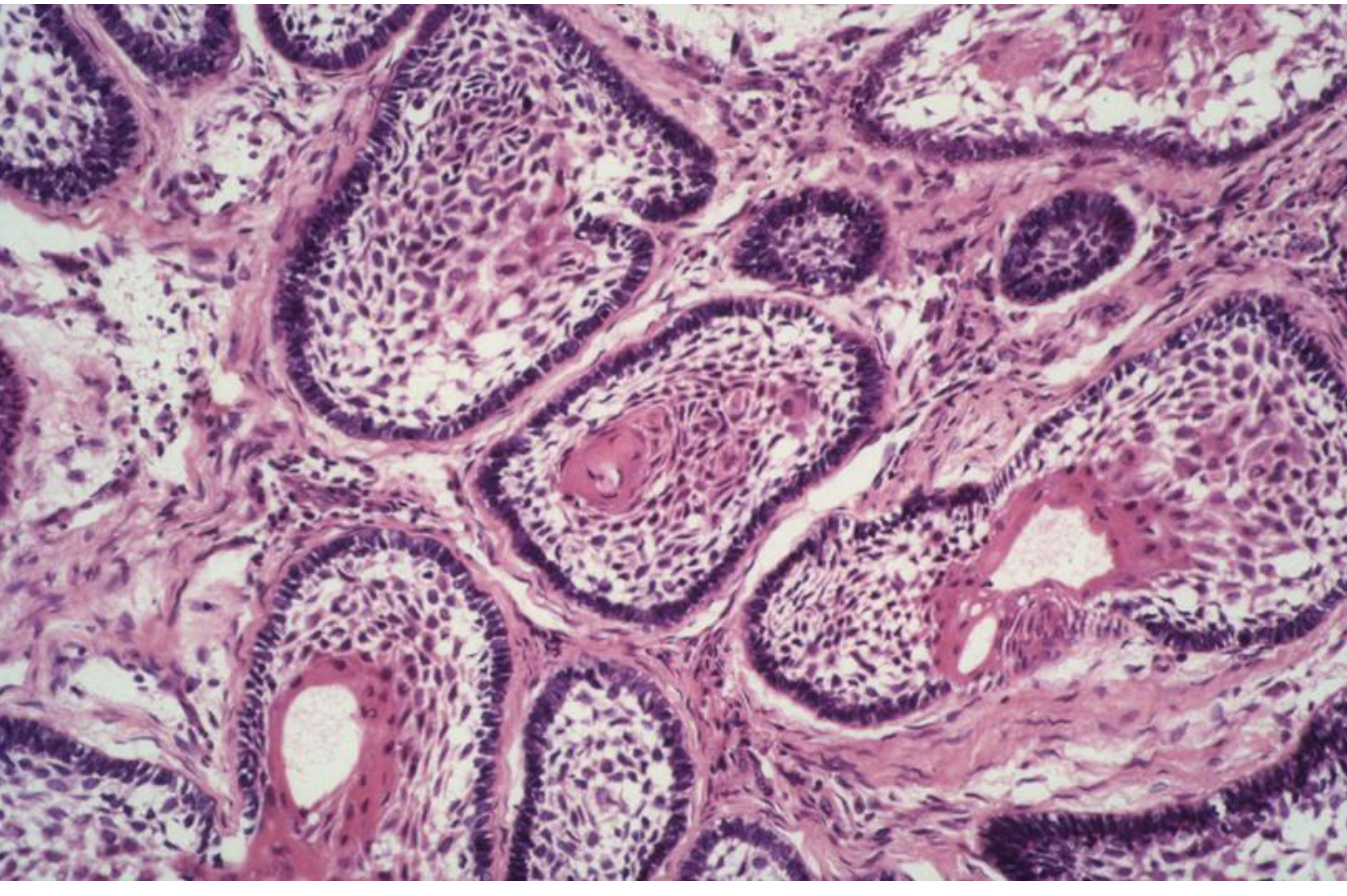
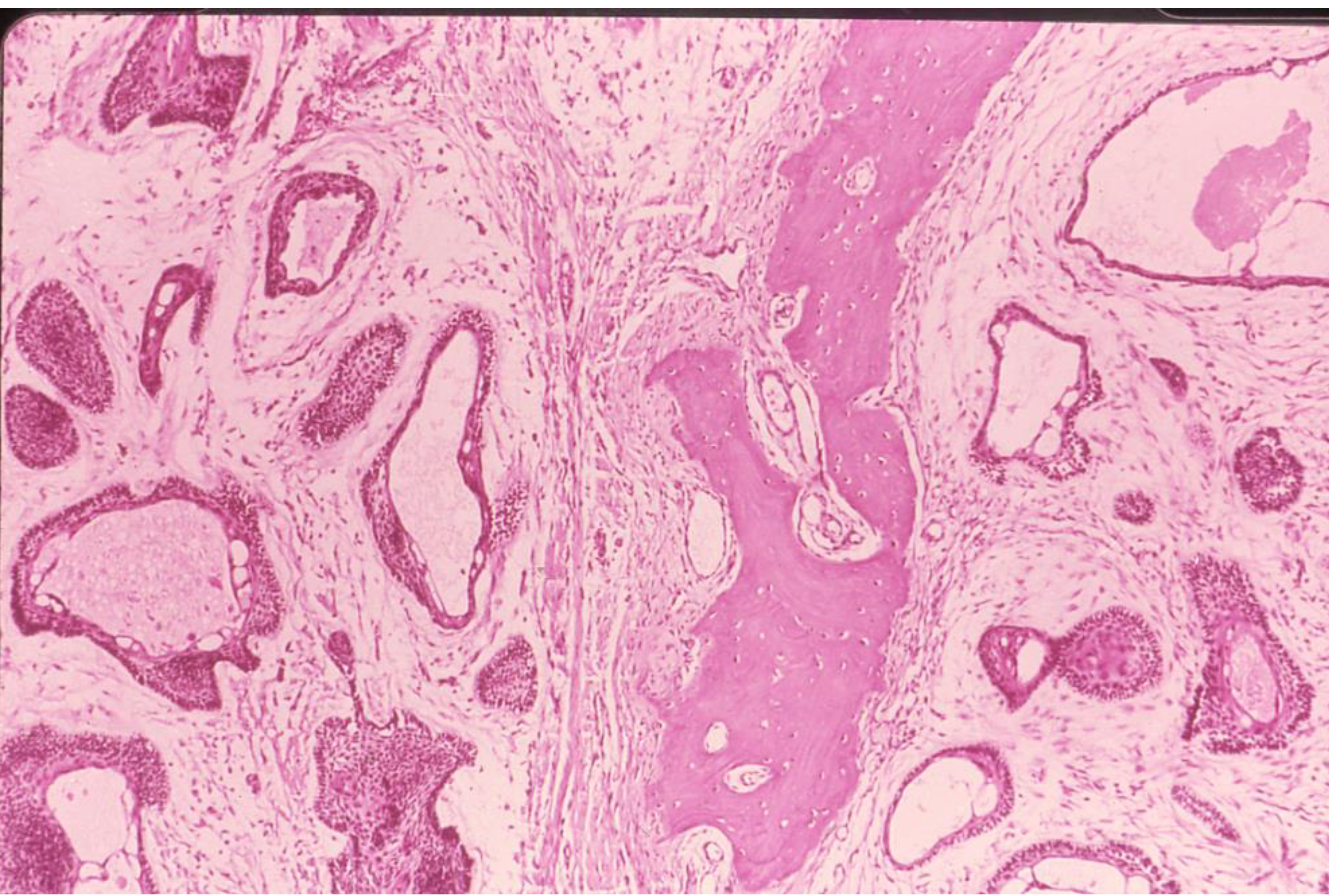
Ameloblastoma - plexiform patternAmeloblastoma - follicular patternAmeloblastoma - plexiform pattern6. Odontogenic tumours August 1, 2025 8:19 AM
Oral Pathology module
WHO Classification of Odontogenic Tumours (Kramer et al 1991)
- based on the similarity between tumours and various stages of tooth development
Classification of Benign Odontogenic Tumours
- Odontogenic epithelium without odontogenic ectomesenchyme
- Odontogenic epithelium with odontogenic ectomesenchyme, with or without dental hard tissue formation
- Odontogenic ectomesenchyme with or without included odontogenic epithelium
Odontogenic Tumour Classification Details
1. Odontogenic epithelium without odontogenic ectomesenchyme
- Ameloblastoma
- Squamous odontogenic tumour
- Adenomatoid odontogenic tumour
- Calcifying epithelial odontogenic tumour (Pindborg Tumour)
2. Odontogenic epithelium with odontogenic ectomesenchyme with or without dental hard tissue
- Ameloblastic fibroma
- Ameloblastic fibro-dentinoma
- Ameloblastic fibro-odontome
- Calcifying odontogenic cyst
- Complex odontome
- Compound odontome
3. Odontogenic ectomesenchyme with or without included odontogenic epithelium
- Odontogenic fibroma
- Myxoma [myxofibroma]
- Benign cementoblastoma (‘true’ cementoma)
Malignant Odontogenic Tumours
- Clear cell odontogenic carcinoma
- Malignant ameloblastoma
- Primary intra-osseous carcinoma
- Malignant variants of other odontogenic epithelial tumours
- Malignant changes in odontogenic cysts
Benign Odontogenic Tumours: Examples
**Ameloblastoma
Ameloblastoma - follicular pattern
Ameloblastoma - plexiform pattern
Unicystic ameloblastoma
.
- You should remember that ameloblastoma causes bone resorption
- You should also remembmer that you HAVE to see the columanr cells with rever polarity, this is the reason we even clal it unicystic ameloblastoma Unicystic ameloblastoma Sections
- Ameloblastomas also have “baloon” or parachute cells which is another feature, although the main feature remains the columnar cells with reverse polarization
Differential diagnosis
- Dentigerous cyst
- Squamous odontogenic tumour
- Ameloblastic carcinoma
**Ameloblastic fibroma
-
We have fibrous tissue which is a the main part of the tumor, we also expect to see epithelial components
- Usually they appear in younger patients and can effect any site in the oral cavity
-
You can see fibroblasts and spindle ells
-
These cells have a mixoid stroma
- Mixoid = less collagen fibers and more ecm (i.e. Glycogen)
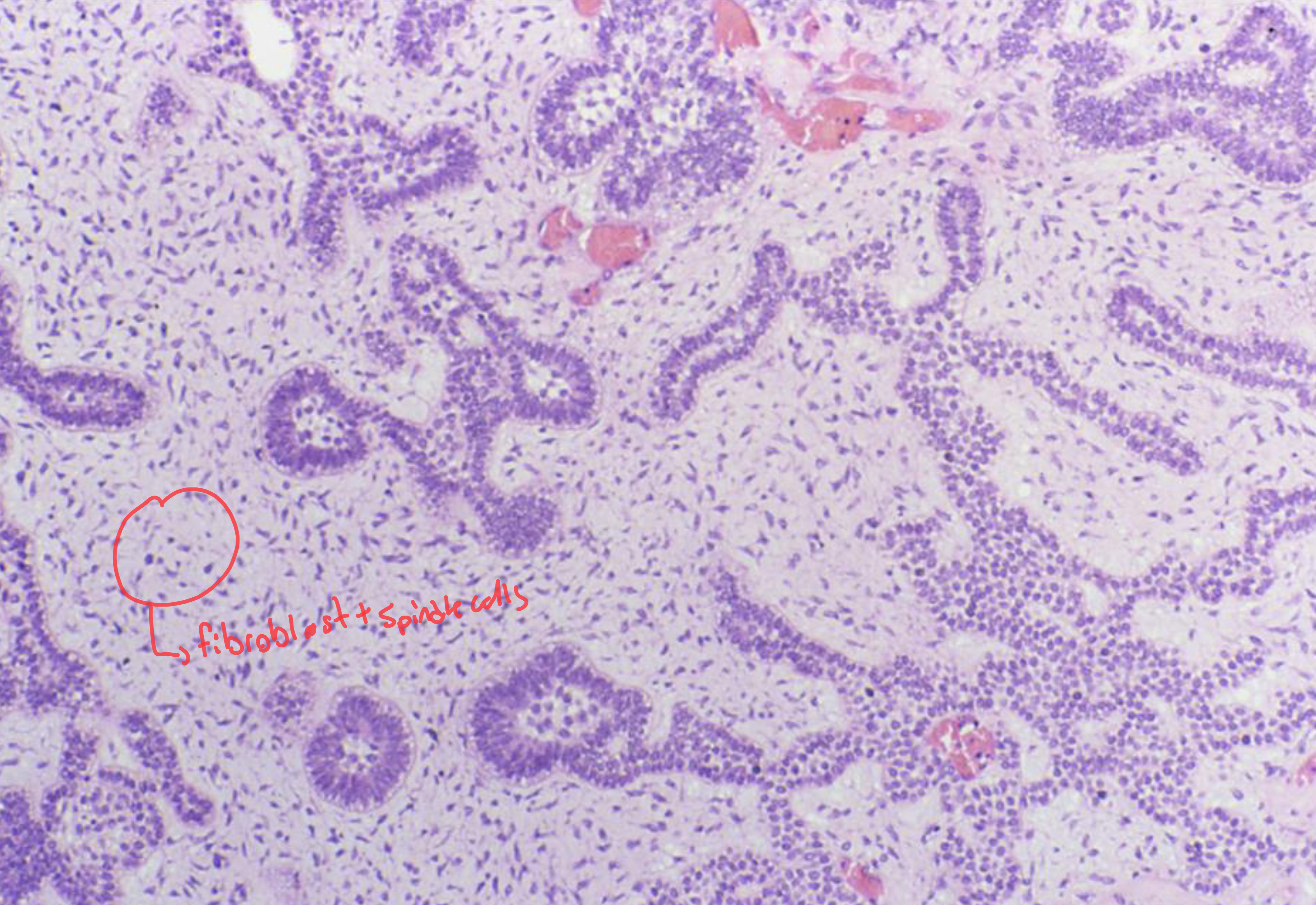
- Mixoid = less collagen fibers and more ecm (i.e. Glycogen)
-
**Odontoma
Compound odontome
- odontoma will remain attached to the tooth
Complex odontome
- The empty spaces will be enamel
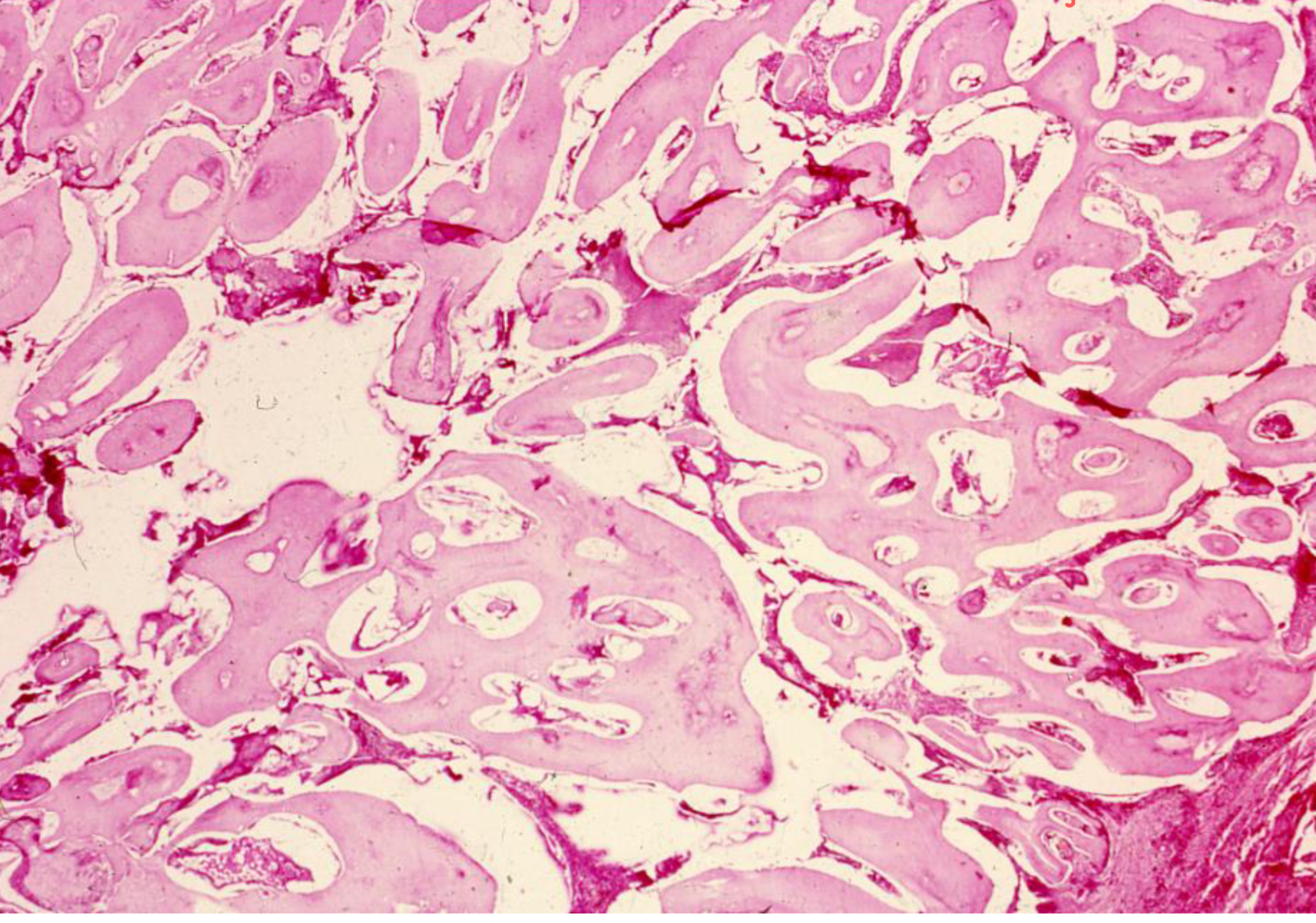
Compound odontome
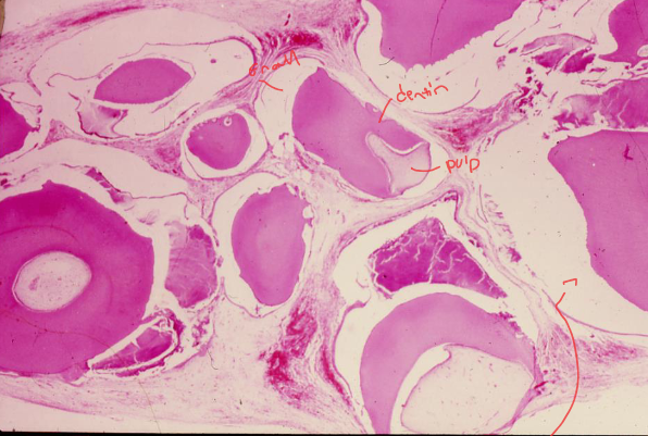
- The prescence of ordered dental tissue is what gives the odtomoe the “compound name” it is a defining characteristic
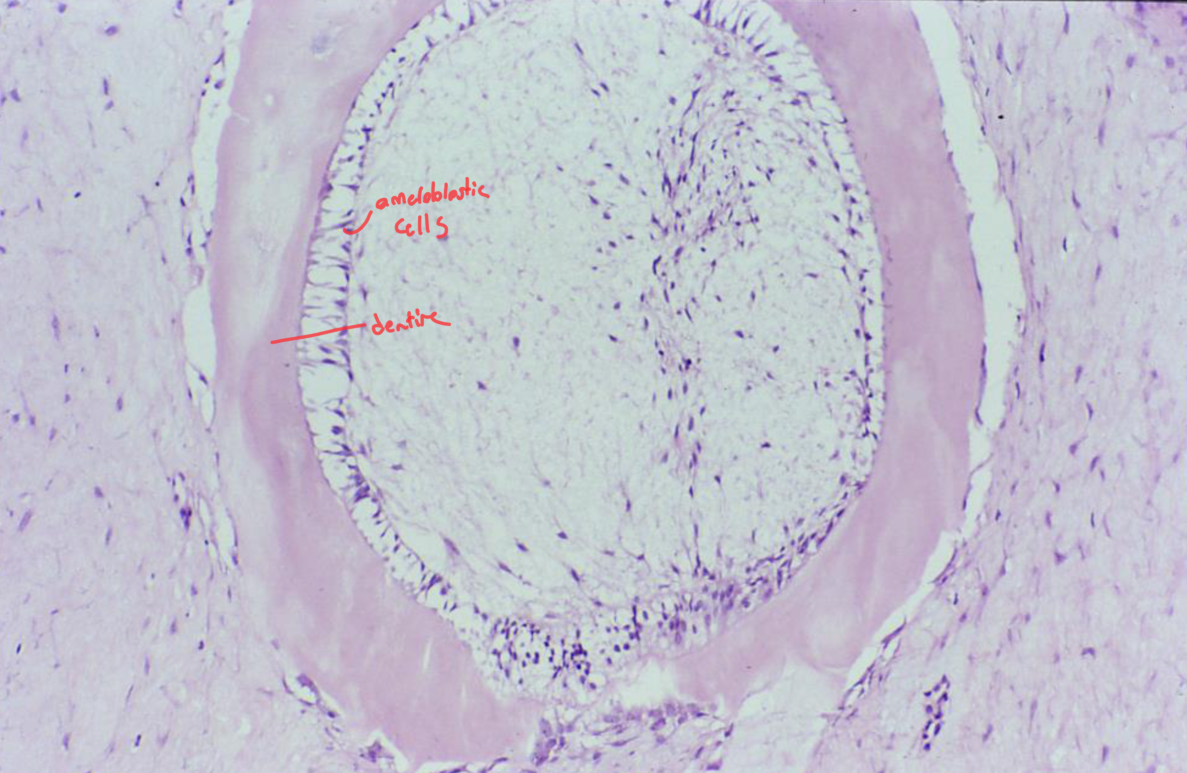
Ameloblastic fibro odontome and ameloblastic fibrodentinoma
- You are going to see, enamel, dentine and cementum structure but with random organization
- This is essentially a mix between ameloblastic tumour and an odontome
- If it shows all layers of tooth (i.e. enamel , dentine cementum) then we call it an ameloblastic fibro odontome
- If it shows only dentine then we call it an ameloblastic fibrodentinoma
- Note that some classifications just combine this together
Odontogenic fibroma
-
Usually odontogenic fibromas form a large radiolucent area which may cause root resorption
-
Radiographically they appear like cysts
-
The defining characteristic of an odontogenic fibroma is the epithelial islands of odontogenic epithelium, without these they would just be fibroma
- We are not expecting to see cellular atypa(i.e. changes to morphology of the cells which causes them to be malignant (i.e. abnormal variations of nucleus and cell size)
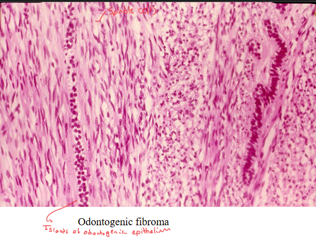
- We are not expecting to see cellular atypa(i.e. changes to morphology of the cells which causes them to be malignant (i.e. abnormal variations of nucleus and cell size)
Odontogenic myxoma
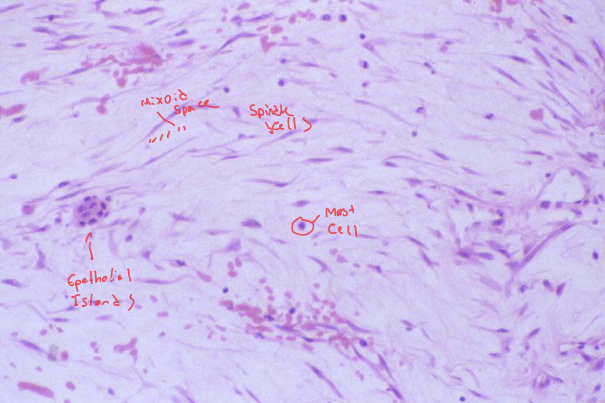
- Usually appear in the young population
- they have large spaces of myxoid tissue with less collagen fibers
- spindle shaped fibroblasts with a slightly elongated and wavy nuclues
Cementoblastoma
-
Consists of cementoblasts proliferating and producing cementum
- Sometimes they can cause resopriton of the root
- In some caes they are well defined and they show a mixed radoiopaque and radiolucency
- The radiograph is dependent on the amount of mineralization and calcified materials
-
microscopically you can see cementoblasts with “cementum-like” material
Case Report: Journal of Oral and Maxillofacial Surgery
- Recurrent cementoblastoma associated with a developing lower second molar.
- Benign odontogenic neoplasm forming cementum-like tissue continuous with the tooth root, usually mandibular premolars or first molars.
- Presents as tender, sometimes painful swelling at buccal and lingual/palatal alveolus.
- Affects mainly individuals under 30, more common in males (1.2:1).
- Radiographically shows as a radiopaque mass with a thin radiolucent rim; shape may be rounded or irregular and mottled.
- Can cause tooth resorption, root outline loss, and obliteration of periodontal ligament space.
- Histology reveals cementum-like calcified tissue with reversal lines, sometimes[ [Pagetoid Appearance]] appearance, containing cementoblasts and cementoclasts.
Malignant odontogenic tumours (carcinomas)
Odontogenic carcinoma - pathol. fracture
- One of the side effects is that they cause bone necrosis, this can cause fracture of the jaw which can even happen without trauma
- Microscopic
- Atypical mitosis among other malignant features
- Because their appearance resembling odontogenic cells and the production of little calcified materials we can call them an odontogenic carcinoma
Ameloblastic carcinoma
-
Frequent mitoses
-
The center becomes necrotic because it doesn’t receive nutritional support!
-
staining identifies the cells which are currently in mitosis
- Those are the brown cells in this image
-
Therefore the tumour can be said to have a high proliferation index
CCOC
- Keratin 19, a marker for odontogenic epithelium
- We can use the different types of cytokeratin biomarkers to identify the different types of epithelium
- Cytokeratin 19 can label odontogenic epithelia