Frontal Facial Analysis Exercises
About these exercises
Full-face (frontal) judgment practice, compiled from the OrthoInstruction module 01 - Facial Form Analysis. For each patient, make your call on the four points below from the photos, then expand the Answer to compare. Profile (lateral) cases and the combined facial self-test are in Profile Facial Analysis Exercises. Underlying theory: Level II Unit B Summary.
For each patient, judge:
- Facial symmetry — within normal limits or asymmetric; if present, location of asymmetry
- Incisor tooth display — OK, too little, or too much (excess gingiva)
- Buccal corridor width — OK, too little, or too much
- Smile arc — OK, flat, or excessive
How to read the full-face view
A quick guide to how to make the judgments below — what to construct on the photo and the proportions to expect. Method follows the DMD3S1 Workbook (primary); norms from Contemporary Orthodontics 6e, Ch.6.
1. Construct your reference lines on a well-oriented frontal photo (patient in natural head position):
- a midsagittal vertical (glabella → nasal tip → chin) — to check symmetry and the facial vs dental midlines
- vertical fifths — five equal columns across the face
- horizontal thirds — hairline → brow, brow → base of nose, base of nose → chin
- glance for an occlusal-plane cant (is the smile line level, or tipped?)
2. Check the proportions (norms):
- Fifths — the central, medial, and lateral fifths should be roughly equal; the interpupillary distance ≈ width of the mouth, and the width of the nose ≈ the central fifth (Fig 6.9).
- Thirds — the three facial thirds should be roughly equal (in modern Caucasians the lower third is often slightly longer); within the lower third, the mouth sits ≈ ⅓ of the way from the base of the nose to the chin (Fig 6.13).
- Midlines — facial and dental midlines should be coincident and centred; a deviation greater than ~3 mm becomes noticeable.
3. Then make the four judgments (facial symmetry, incisor tooth display, buccal corridor width, smile arc) as listed above.
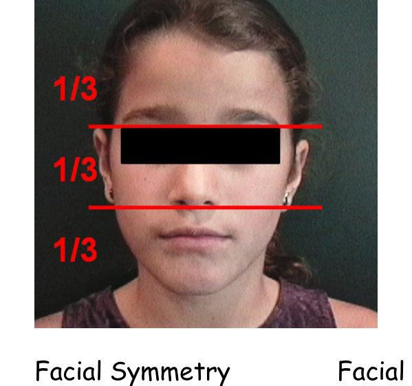 Facial thirds. Facial thirds. | 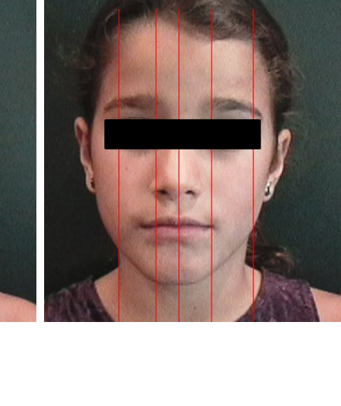 Facial fifths. Facial fifths. |
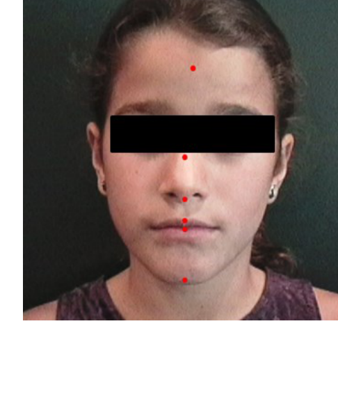 Facial symmetry. Facial symmetry. | 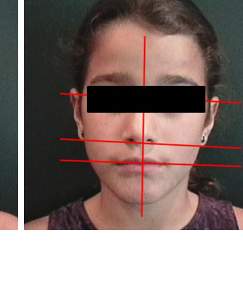 Occlusal-plane cant. Occlusal-plane cant. |
Patient 1
 Image 1 Image 1 |  Image 2 Image 2 |
Answer
- Symmetry: significant asymmetry — her face is larger on the right side
- Tooth display: relatively normal on the left side, affected by the asymmetry on the right
- Buccal corridor: excessive on the right, OK on the left
- Smile arc: abnormal because of the asymmetry
Patient 2
 Image 1 Image 1 |  Image 2 Image 2 |
Answer
- Symmetry: mild mandibular asymmetry, with his chin off to the left — but probably not enough to be a problem
- Tooth display: normal incisor display
- Buccal corridor: excessive width — too much space between the posterior teeth and the cheeks
- Smile arc: normal
Patient 3
 Image 1 Image 1 |  Image 2 Image 2 |
Answer
- Symmetry: significant mandibular asymmetry. Compare him to Patient 1 — here the asymmetry is just in the lower face (due to excessive growth of the mandible on the left side), instead of affecting the entire face.
- Tooth display: inadequate display of upper incisors; displays lower incisors that should not be seen at this age
- Buccal corridor: not seen well enough to evaluate
- Smile arc: flattened (upper teeth are hardly visible and don’t follow the lip line)
Patient 4
 Image 1 Image 1 |  Image 2 Image 2 |
Answer
- Symmetry: no asymmetry
- Tooth display: too little display of maxillary incisors
- Buccal corridor: normal width
- Smile arc: flattened
Patient 5
 Image 1 Image 1 |  Image 2 Image 2 |
Answer
- Symmetry: mild chin asymmetry, not enough to be a problem
- Tooth display: marginally adequate maxillary tooth display, some display of lower incisors
- Buccal corridor: minimal width, perhaps too little
- Smile arc: slightly flattened
Self-test
The module’s multiple-choice self-test covers both frontal and profile facial analysis. It lives in Profile Facial Analysis Exercises > Self-Test.