L10 Dento-Alveolar Trauma 3 - Tissue Responses to Injury
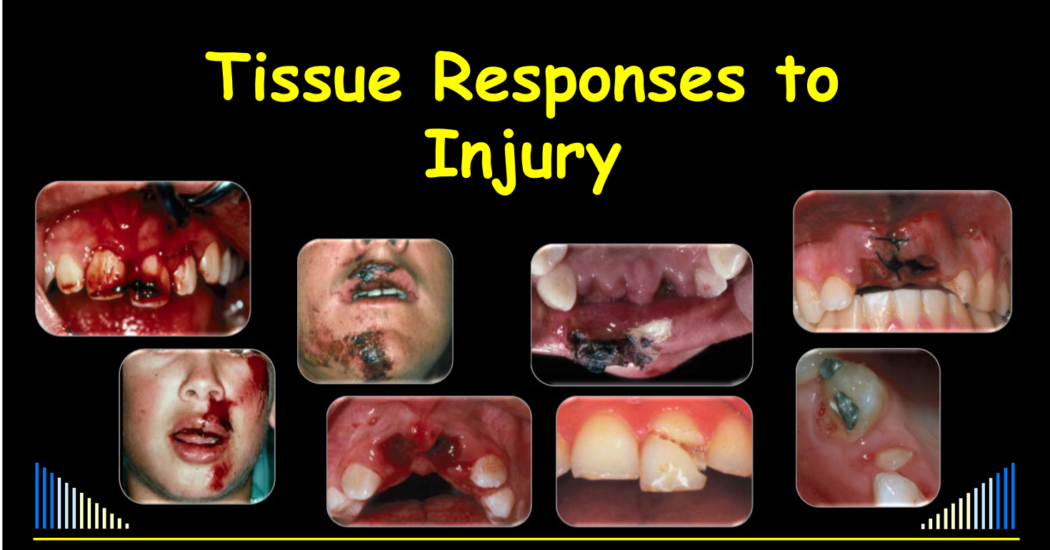
Tissue Responses to Injury12
Prof. Paul V. Abbott AO
BDSc, MDS, FRACDS(Endo), FPFA, FADI, FICD, FACD, FIADT
Specialist Endodontist
Winthrop Professor of Clinical Dentistry
UWA Dental School
The University of Western Australia
DMD - 3rd Year - 2020
Strategies to Minimise the Consequences of Trauma to the Teeth
Abbott PV, Castro Salgado J. J Oral Health & Dental Management 2014; 13: 229-42.
Prevention and Management of External Inflammatory Resorption following Trauma to Teeth
Abbott PV. Aust Dent J 2016; 61 (1 Suppl): 82-94.
 |  |
 |  |
Introduction
- Potential consequences of trauma
- Tissue responses to trauma
- Pulp
- Peri-radicular tissues
- Gingiva and oral mucosa
- Factors that affect healing
Definition of Trauma3
An injury or wound to the tissues. Can be caused by:
- An accident
- e.g. MVA, fall, fight, etc
- Chemical means
- e.g. Erosion
- Mechanical means
- e.g. Abrasion, Attrition
- Bacteria
- e.g. Caries
Lecturer — Accidental Trauma
The lecture focuses on accidental trauma: events generally outside the person’s control and outside the dentist’s control when they occur.
- Bacteria were described as a form of long-term trauma, particularly to the pulp and eventually to the periapical and peri-radicular tissues.
- Understanding tissue responses is essential for selecting appropriate management for different dental injuries.
- Reference sources mentioned include the Dental Trauma Guide, Textbook and Color Atlas of Traumatic Injuries to the Teeth, the Dental Traumatology Journal, and articles concerning the consequences of trauma to teeth.
Key Considerations About Dental Trauma
Trauma is a common occurrence4
- One in every 2 children by the age of 14 years
- 30% of children — primary dentition trauma
- 22% of children — permanent dentition trauma
- Boys twice as often as girls
Peak age ranges
-
2–4 years old
- Primary dentition
- Underlying developing permanent teeth
- May be damaged and affect their development
- Trauma to primary teeth can affect the development and eruption of the underlying permanent teeth.
- Treatment may be less involved because the primary teeth may not need to remain for much longer.
-
8–10 years old
- Permanent dentition
- Incompletely developed teeth
- May be problematic if pulp necrosis occurs
- Immature roots may be advantageous because pulp revascularization is more likely.
- If revascularization does not occur and pulp necrosis develops, root canal treatment may be more difficult.
-
Trauma often occurs “after hours”
-
Trauma is an “emergency”
- Patients often present to hospital clinics (usually no dentist)
-
General dentists do not treat trauma regularly
- Can lead to uncertainty about treatment
-
The long-term prognosis depends largely on the “emergency management”
- Correct treatment is essential to help reduce the consequences of dental trauma
Lecturer — Emergency Management
Patients may initially attend a hospital clinic, where there may be no dentist available.
- Management may therefore be provided by a medical practitioner or nurse with limited dental-trauma experience.
- Severe trauma can involve multiple teeth, bone fractures, soft-tissue lacerations, and significant bleeding.
- Regular continuing professional development, including conferences and workshops, was recommended to maintain readiness to manage dental trauma.
Outline5
- Introduction
- Potential consequences of trauma
- Tissue responses to trauma
- Pulp
- Peri-radicular tissues
- Gingiva and oral mucosa
- Factors that affect healing
Lecturer — Lecture Scope
The lecture also addressed the timing of tissue responses and the importance of follow-up.
- Mechanical and biological factors affecting healing were included in the discussion.
Potential Consequences of Trauma6
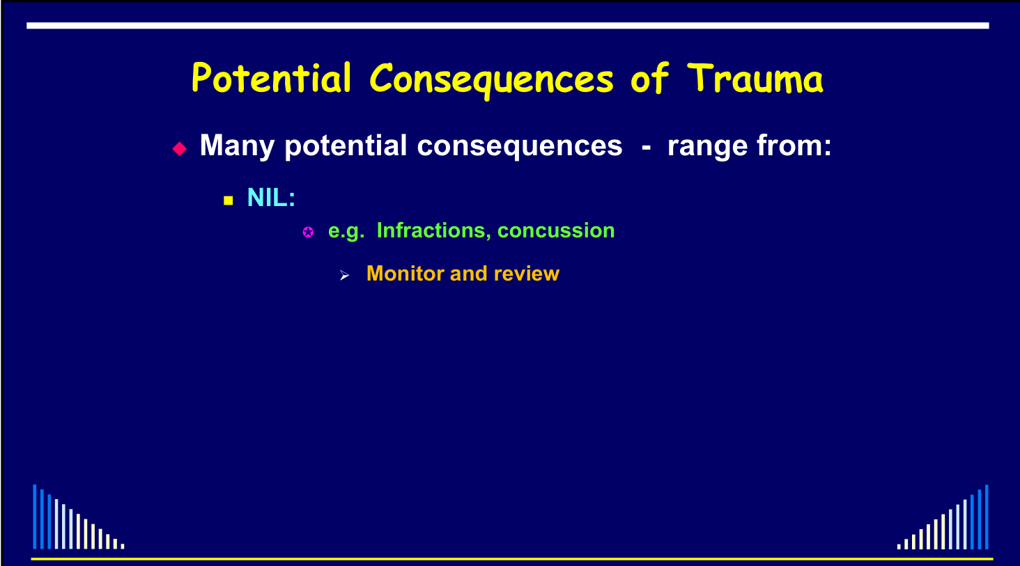
Many potential consequences - range from:
- NIL
- e.g. Infractions, concussion
- Monitor and review
- e.g. Infractions, concussion
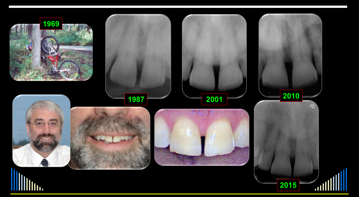
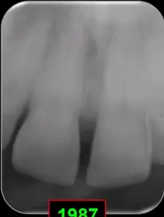
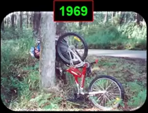
Photographs: 1969, 1987, 2001, 2010, 2015.
Long-Term Monitoring After Crown Fractures
A 12-year-old boy fell from a bicycle in 1969 and sustained minor, uncomplicated crown fractures to both central incisors.
- No specific treatment was provided apart from monitoring.
- Later radiographs and photographs showed the small enamel fractures remained visible as slightly rough incisal edges.
- The fractured edges were smoothed, and no other consequences developed.
- Continued monitoring, including another radiograph and pulp test, remained necessary.
 |  |
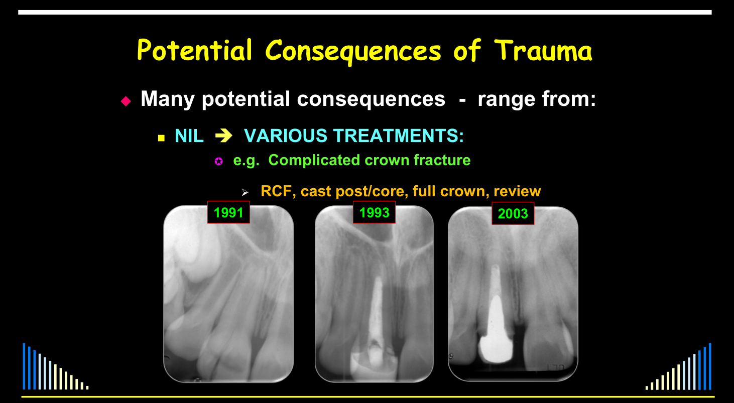
Range of Outcomes From Nil to Extraction78
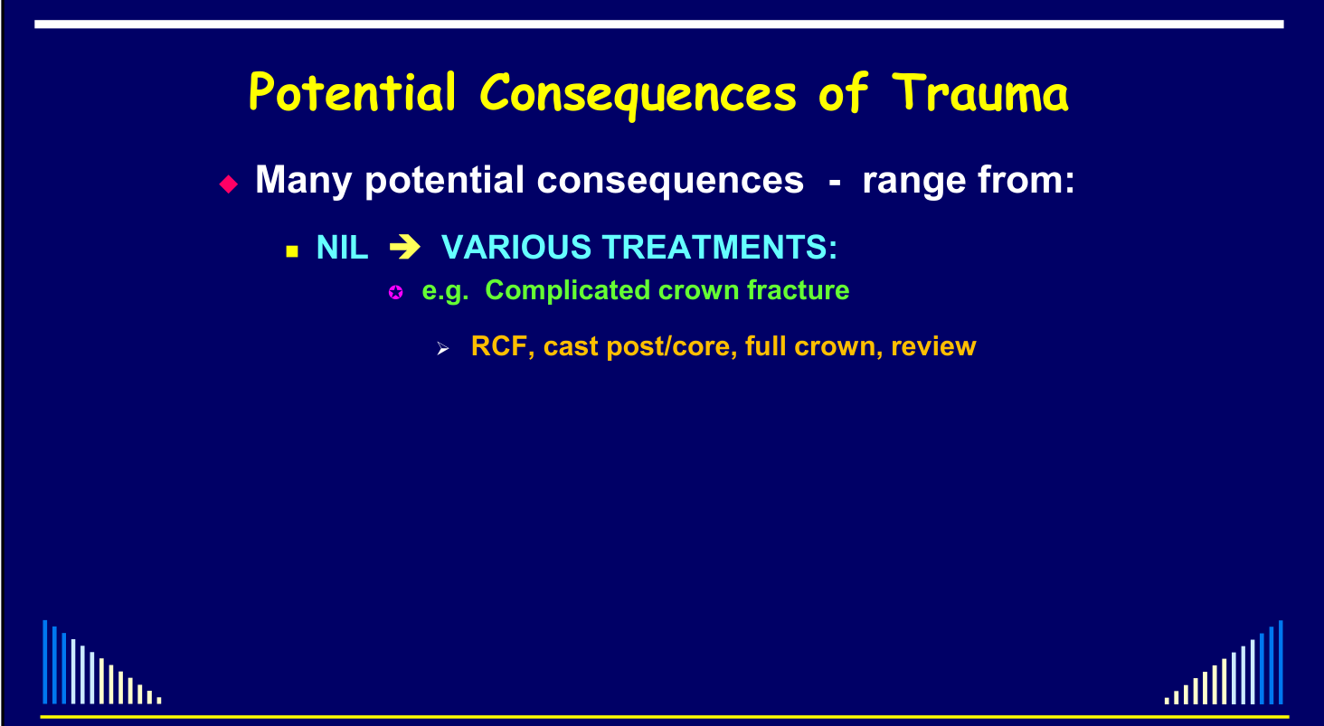
Many potential consequences - range from:
- NIL → VARIOUS TREATMENTS
- e.g. Complicated crown fracture
- RCF, cast post/core, full crown, review
- e.g. Complicated crown fracture
Photographs: 1991, 1993, 2003.
Lecturer — Lifelong Trauma Effects
A tooth affected by trauma should be regarded as having a lifelong experience.
- Consequences may develop one or two years after the injury, or even 10, 20, 30, or 40 years later.
- Treatment does not end when a restoration is placed because restorations have a finite lifespan.
- The root canal system may later become inflamed or infected again.
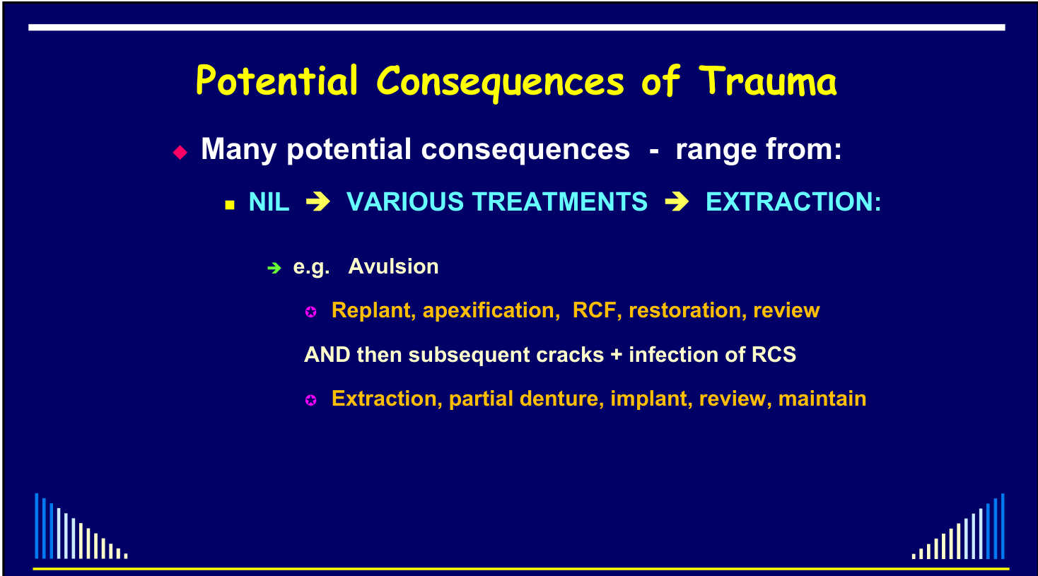
Many potential consequences - range from:
- NIL → VARIOUS TREATMENTS → EXTRACTION
- e.g. Avulsion
- Replant, apexification, RCF, restoration, review
- AND then subsequent cracks + infection of RCS
- Extraction, partial denture, implant, review, maintain
- Replant, apexification, RCF, restoration, review
- e.g. Avulsion
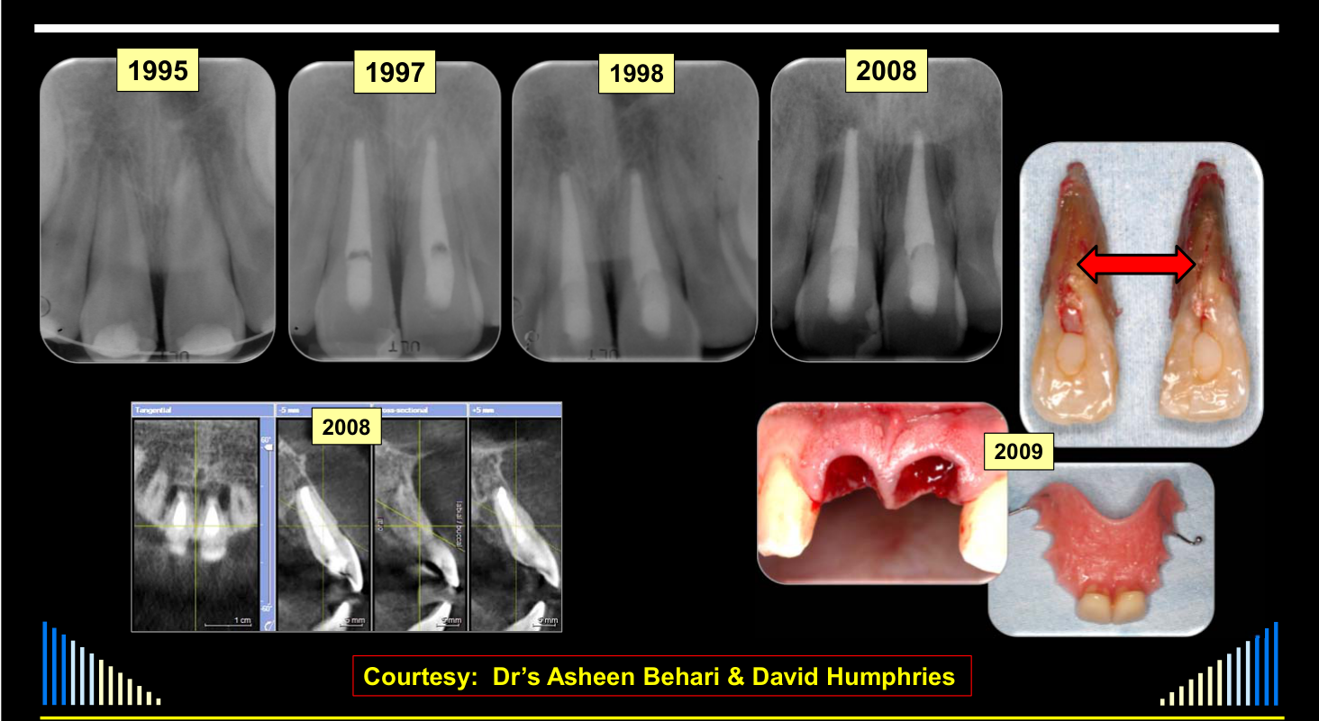
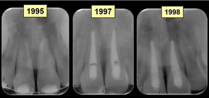
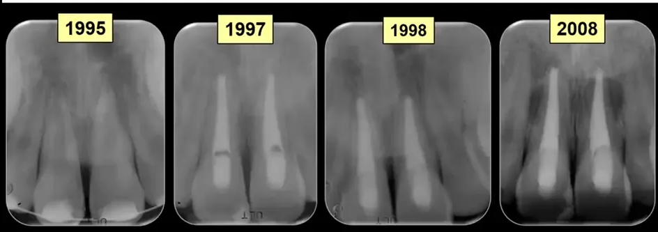
Photographs: 1995, 1997, 1998, 2008, 2008, 2009. Courtesy: Dr’s Asheen Behari & David Humphries.
Avulsed Incisors Requiring Implants
A young boy avulsed both central incisors in 1995, and the teeth later developed pulp necrosis managed with long-term apexification.
- Hard-tissue barriers formed across the ends of the roots, and the teeth were reviewed in 1998 and again approximately 10 years later.
- The patient later reported palatal tenderness; deep, narrow periodontal pockets and transillumination demonstrated vertical cracks.
- Radiographs and CT scans showed radiolucencies associated with the teeth and their apical regions.
- Both teeth were extracted because of the cracks.
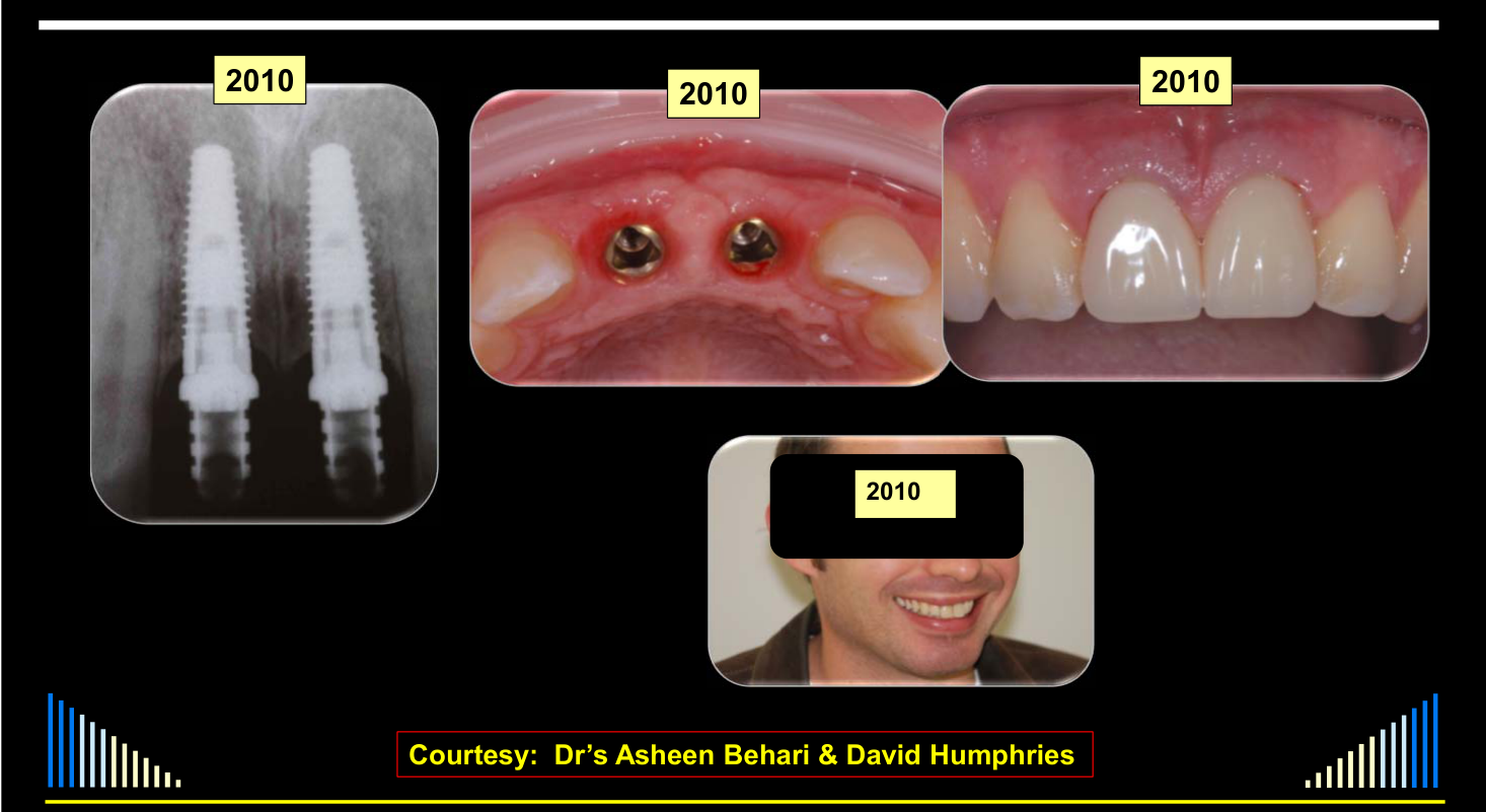
- A temporary partial denture was followed by two implants and crowns, which also required regular monitoring and maintenance.
Photographs: 2010, 2010, 2010, 2010. Courtesy: Dr’s Asheen Behari & David Humphries.
 |  |
 |  |
Outline9
See - Lecture outline
Lecturer — Necrosis Pathways
Pulp necrosis may result directly from trauma, but it may also result from bacterial contamination.
- Bacteria may enter through infractions extending into dentine.
- Fractures with exposed dentine or contamination of an avulsed tooth outside the mouth may also allow bacterial entry.
Lecturer — Pulp Fibrosis
Recovery and return to a normal state is the preferred response, and a damaged pulp may repair with fibrotic tissue comparable to scar tissue. Pulp fibrosis cannot be diagnosed clinically because the tooth may appear normal and respond normally to pulp tests.
Lecturer — Canal Calcification
A radiographically complete pulp canal calcification does not mean that the canal is completely absent. Histologically, a very narrow canal may remain even though it is too narrow to be visible radiographically, and calcification may be a favorable sign because it indicates pulp survival.
Root Fracture with Reversible Pulpitis
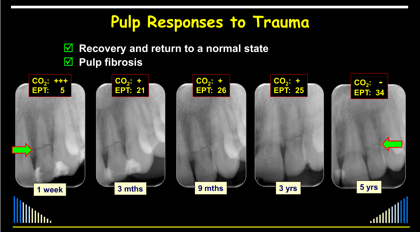
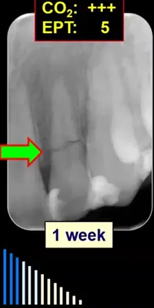
A patient was struck in the mouth by a squash racket, causing a root fracture of a lateral incisor that was splinted to adjacent teeth with composite resin. One week later, sensitivity to hot and cold indicated reversible pulpitis caused by the trauma, but no pulp treatment was required because the tooth was stabilized and the pulpitis was expected to resolve.
- Symptoms resolved within one or two weeks, and the splint was retained for approximately three months with monthly reviews.
- After splint removal, the tooth was reviewed at three, nine, and twelve months, then annually for up to five years.
- Later canal calcification reduced the carbon dioxide response, particularly coronally, while electric pulp testing remained normal.
- Radiographs showed no peri-apical or peri-radicular changes, so the pulp was considered to have recovered, although some fibrosis may have been present.
Lecturer — Apical Breakdown
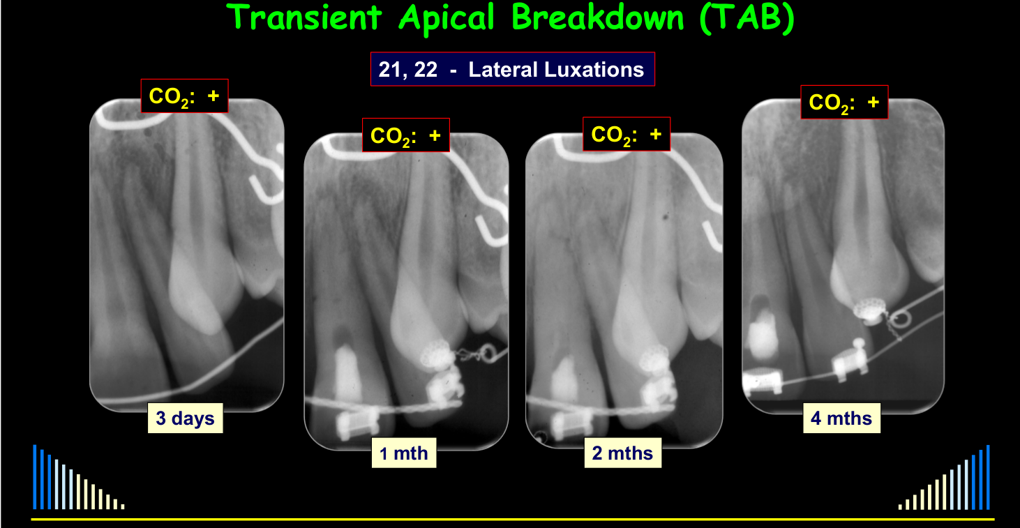
Transient apical breakdown is a temporary remodeling process rather than true permanent breakdown. A radiolucency may resemble pulp necrosis with root canal infection, but continued pulp-test responses support transient apical breakdown instead; the apical region may remodel, the foramen may widen initially, and the canal may later calcify and narrow.
Lateral Luxation During Orthodontics
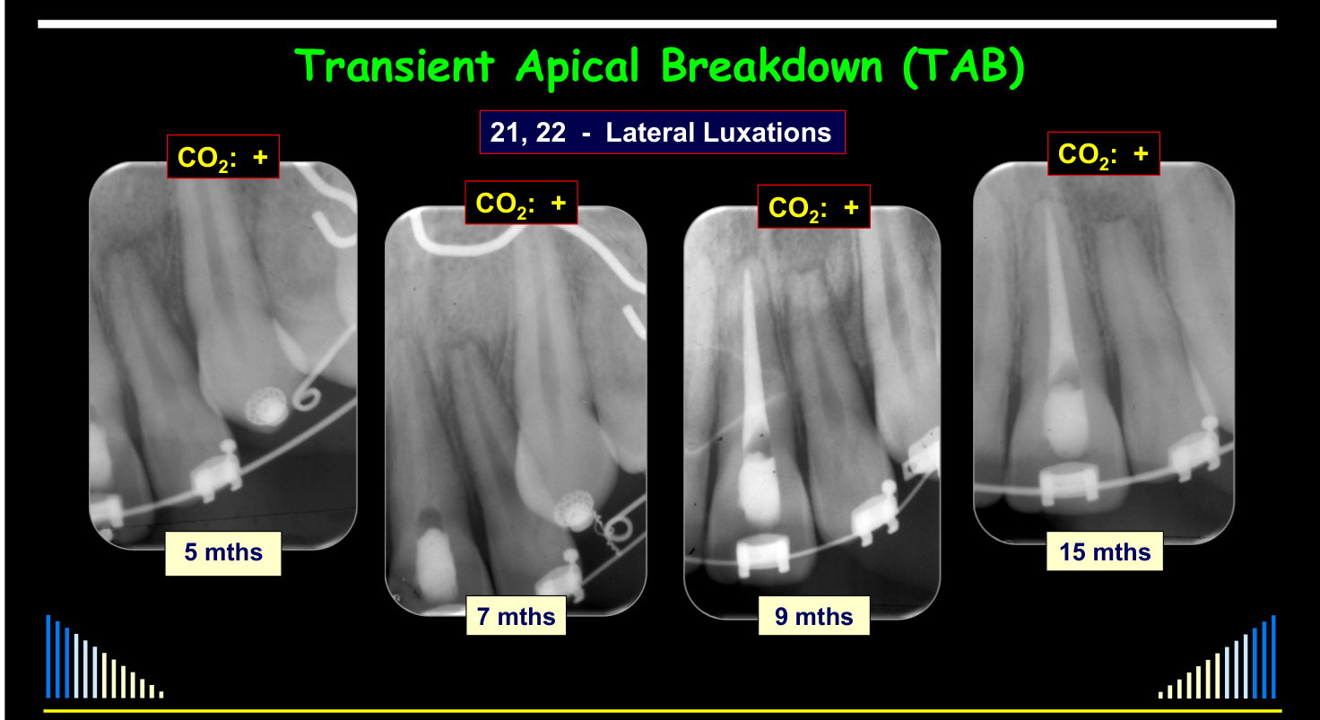
A young boy undergoing orthodontic treatment fell from a new bicycle and sustained lateral luxations of a central and lateral incisor. The teeth were repositioned and splinted; the central incisor became necrotic within one month, whereas the lateral incisor continued responding to carbon dioxide despite progressive root-end changes.
- The lateral incisor developed root-end blunting, widening of the apical foramen, later narrowing and calcification of the canal, and rounding of the root end.
- Brackets and wires prevented electric pulp testing, so carbon dioxide testing was used.
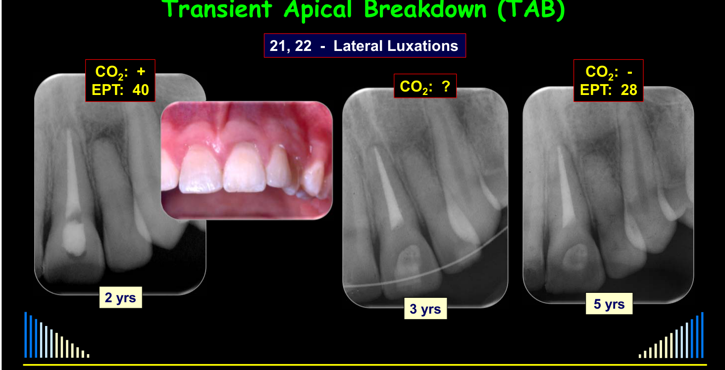
- At approximately two years, the canal appeared almost completely calcified radiographically and the open apex had disappeared.
- After three and five years, the tooth remained stable; it no longer responded to carbon dioxide but responded normally to electric pulp testing.
Lecturer — External Resorption
External surface resorption may not be visible clinically or radiographically. If it extends into dentine and the root canal becomes infected, external inflammatory resorption may develop; external replacement resorption may occur with ankylosis, while external invasive resorption may be associated with trauma without trauma being necessary in every case.
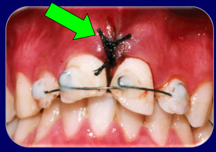
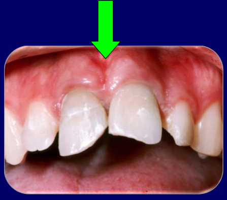
Lecturer — Soft Tissue Assessment
Lacerated tissue may appear attached after a luxated tooth is repositioned even when it is not actually attached. The palatal tissue is particularly important to assess after lateral luxation, intrusion, avulsion, or extrusion; if it is unattached, it should be sutured, typically with sutures between the teeth to hold it in position.
Lecturer — Late Necrosis
Pulp necrosis may occur many years after trauma when restorations break down, infractions permit bacterial entry, or other injuries have compromised the tooth. Teeth with infractions, fractures, or restorations therefore require long-term review and may eventually need restorative, endodontic, prosthetic, or implant-related management.
Tissue Responses to Trauma
Andreasen - Dent Traumatol 2012.
Topic guides
- Trauma Mechanisms and Tissue Response for the L3 mechanics and L10 healing/TAB connection.
- Intrusion and Avulsion for this healing and timing context alongside L12 protocols.
Pulp Responses to Trauma
Trauma and Healing Responses10
- Traumatic dental injuries are complex
- 6 types of luxations and 9 types of fractures
- Each is a unique injury to hard & soft tissues
- Often concurrent luxations + fractures
- Hence 6 x 9 = 54 healing scenarios
- 19 cellular systems in the dental organ
- All with different healing potentials
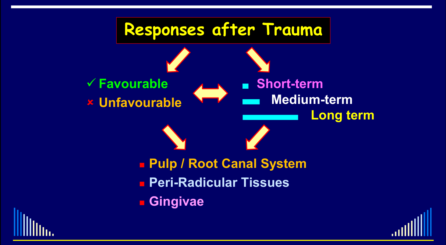
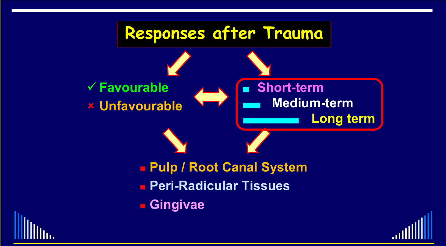
Responses after Trauma
✓ Favourable / × Unfavourable
Assessed over the short-term, medium-term and long term for:
- Pulp / Root Canal System
- Peri-Radicular Tissues
- Gingivae
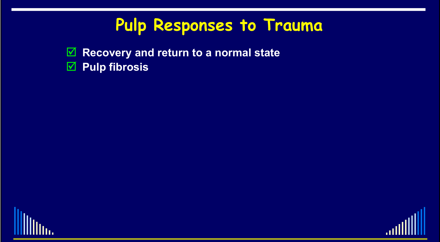
Recovery and return to a normal state — Pulp fibrosis11
Soft-Tissue Management After Avulsion
Two avulsed teeth were replanted, but the lacerated labial tissues were not repositioned and sutured. Gingival recession subsequently developed around both teeth, associated with bone loss and loss of attachment.
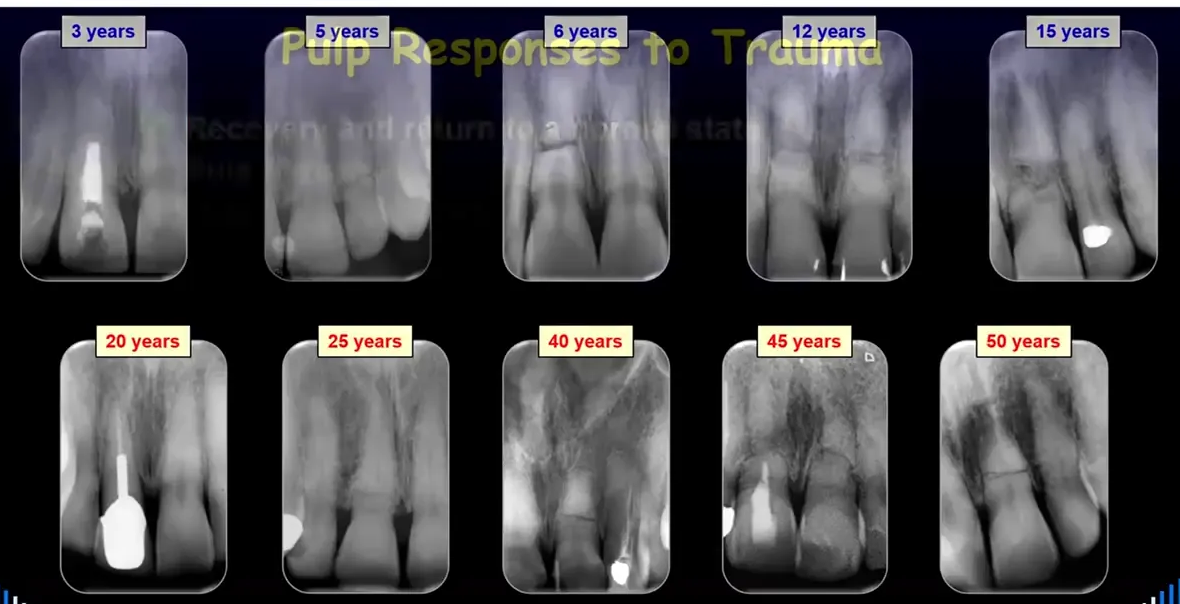
Serial test results over time:
- 1 week: CO₂: +++, EPT: 5
- 3 mths: CO₂: +, EPT: 21
- 9 mths: CO₂: +, EPT: 26
- 3 yrs: CO₂: +, EPT: 25
- 5 yrs: CO₂: -, EPT: 34
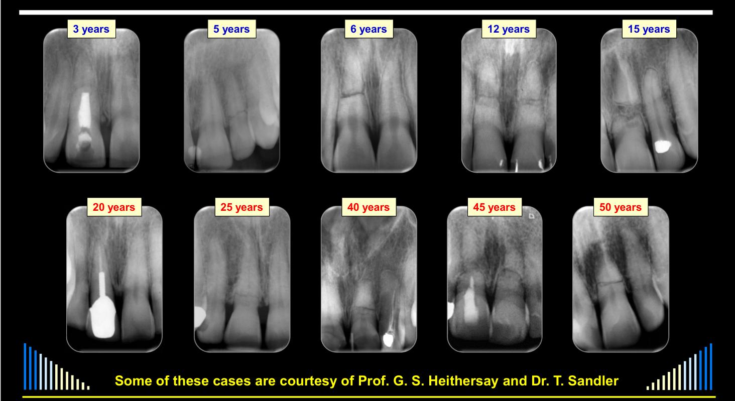
Long-term follow-up images at: 3 years, 5 years, 6 years, 12 years, 15 years, 20 years, 25 years, 40 years, 45 years, 50 years.
 |  |
Pulp Responses to Trauma12
- Recovery and return to a normal state
- Pulp fibrosis
- Pulp Canal Calcification (PCC)
- Partial
- “Complete” - i.e. radiographically → PCC - can usually (be diagnosed) within the first year
Andreasen FM
- Recovery and return to a normal state
- Pulp fibrosis
- Pulp Canal Calcification (PCC)
- Partial
- “Complete” - i.e. radiographically
- Pulp necrosis
- Infection of the pulp space
- PN - can usually diagnose within 3 mths after luxations and root fractures
Andreasen FM et al - EDT 1985.
Pulp Responses to Trauma (continued)13
See - Pulp response categories
Recovery, pulp fibrosis and PCC are marked favourable; pulp necrosis/infection and internal resorption are unfavourable. Responses may occur simultaneously or sequentially over time. Full categories: Summary of Tissue Responses.
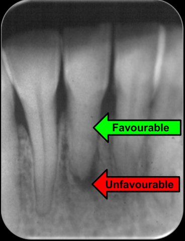
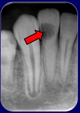
Lateral Luxation + Infractions - 8 years ago14
Favourable / Unfavourable
- Initially - the Pulp survived
- Then - Pulp Canal Calcification
- Partial
- “Complete” - i.e. radiographically
- Later - Pulp Necrosis & Infection of the Root Canal System
- Chronic Apical Periodontitis
- 2° Acute Apical Periodontitis
Favourable / Unfavourable
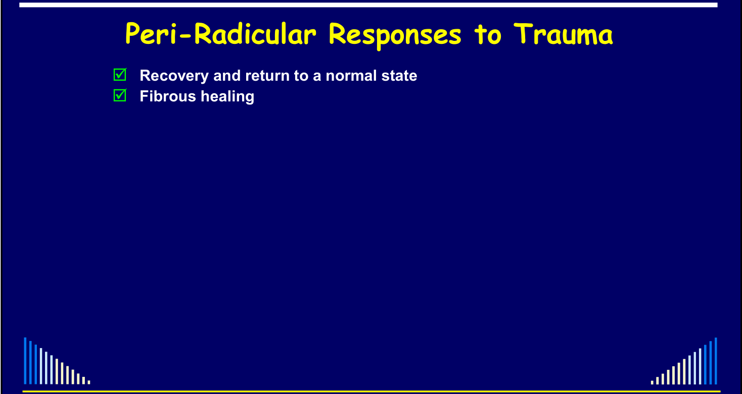
Peri-Radicular Responses to Trauma15
Recovery and return to a normal state:
- Fibrous healing
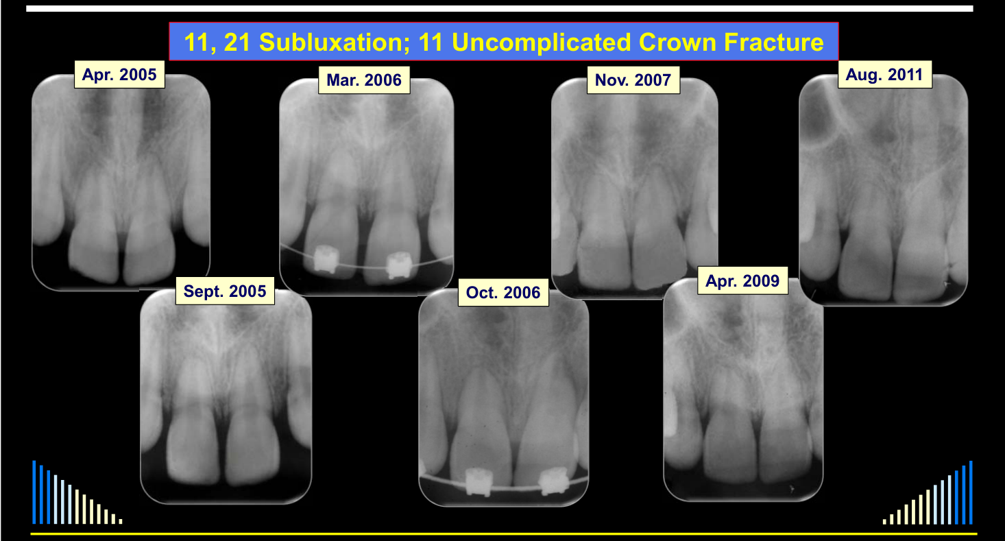
Serial radiographs: Apr. 2005, Sept. 2005, Mar. 2006, Oct. 2006, Nov. 2007, Apr. 2009, Aug. 2011.
Recovery and return to a normal state:
- Fibrous healing
- Transient Apical Breakdown (TAB) — Andreasen FM et al, EDT 1986
Frances M. Andreasen — “Pulpal healing after tooth luxation and root fractures in the permanent dentition”.
 |  |
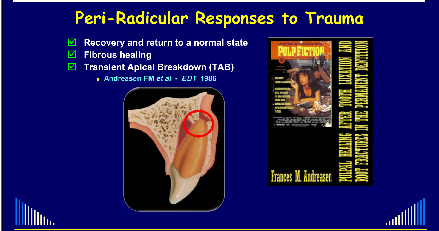
Transient Apical Breakdown1617
Transient Apical Breakdown (TAB)
Slides 21, 22 — Lateral Luxations:
- 3 days — CO₂: +
- 1 mth — CO₂: +
- 2 mths — CO₂: +
- 4 mths — CO₂: +
Slides 33–34: clinical/radiographic images only — no text content.
Recovery and return to a normal state:
- Fibrous healing
- Transient Apical Breakdown (TAB)
- Cessation of root development
- Disturbances to root development
 |  |
 |  |
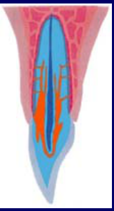
Bone and External Resorption18192021
Bone resorption:
- Marginal (crestal)
- Apical
- Lateral
Recovery and return to a normal state:
- Fibrous healing
- Transient Apical Breakdown (TAB)
- Cessation of root development
- Disturbances to root development
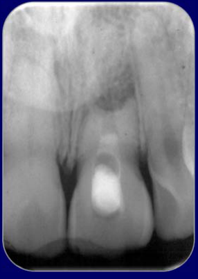
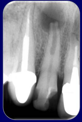
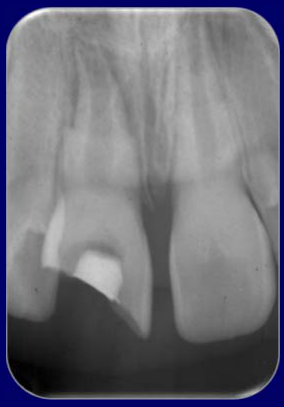
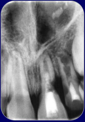
- Bone resorption
- Marginal (crestal)
- Apical
- Lateral
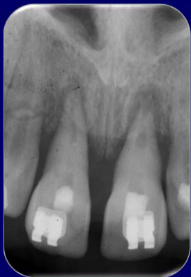
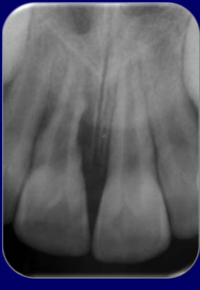
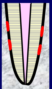
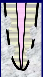
- External resorption
- Surface
- Inflammatory
External Inflammatory Resorption & Bone Resorption
- Combinations of various responses
- Simultaneously
- Sequentially over time

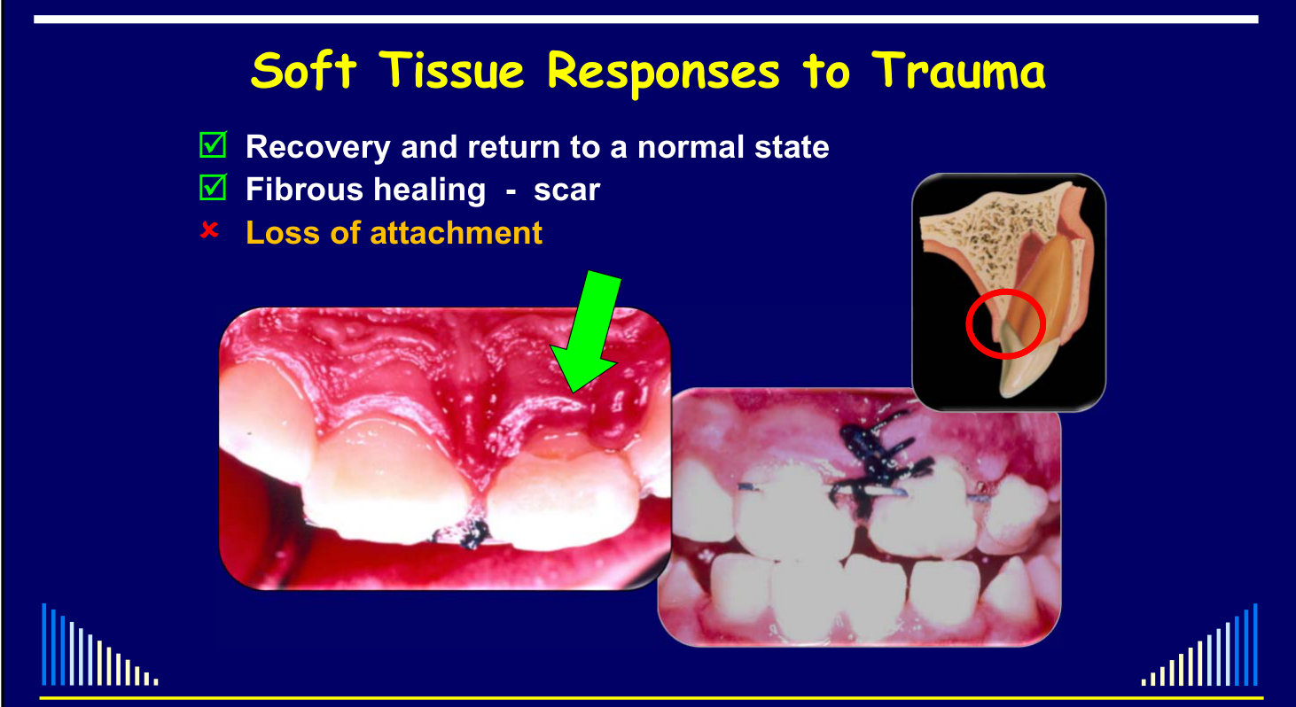
Soft Tissue Responses to Trauma2223
- Recovery and return to a normal state
- Fibrous healing — scar
Possible soft tissue responses to trauma:
- Recovery and return to a normal state
- Fibrous healing — scar
- Loss of attachment
- Gingival recession
W/Prof. Paul V. Abbott AO — Tissue Responses to Injury
 |  |
 |  |
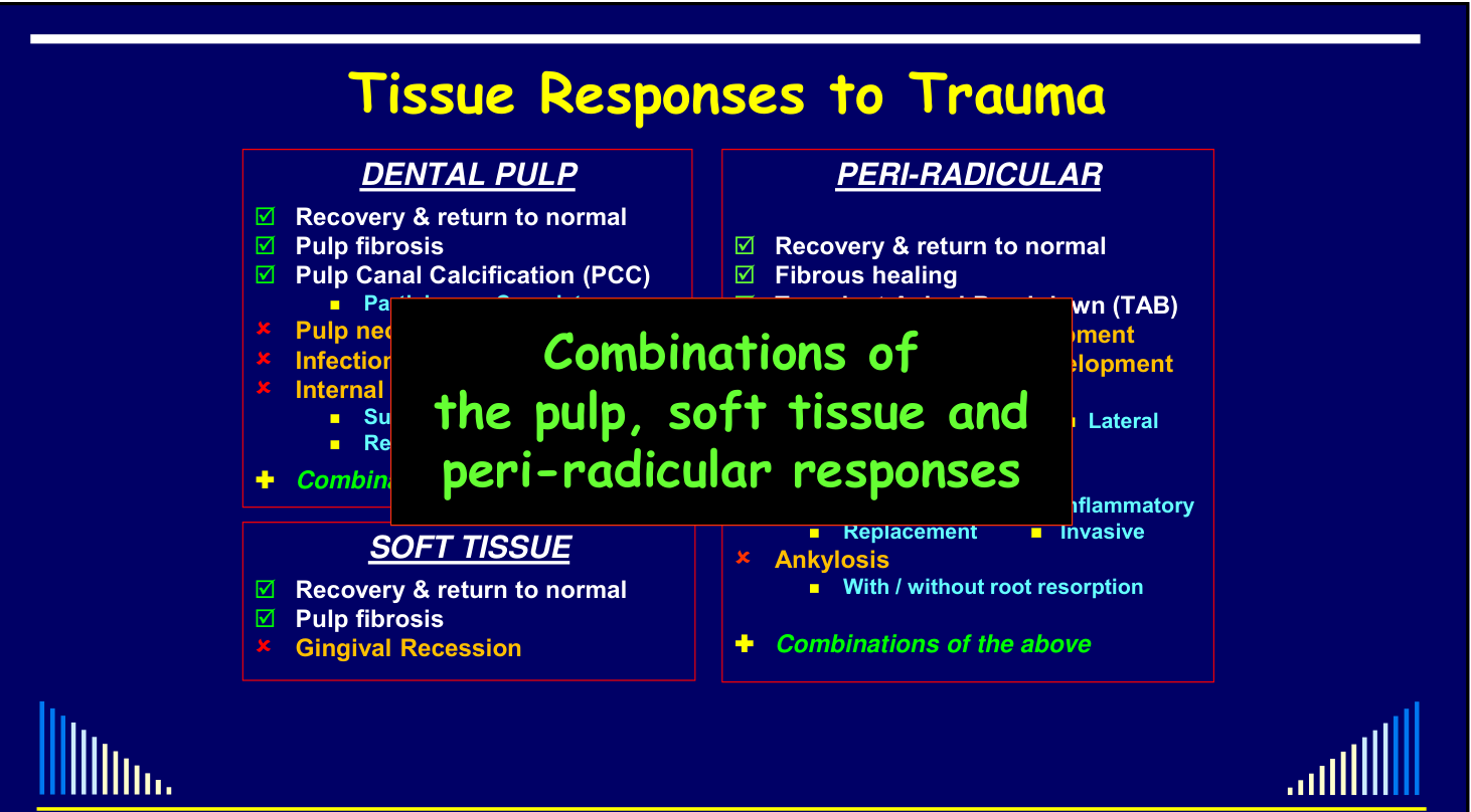
Summary of Tissue Responses
Dental Pulp24
- Recovery & return to normal
- Pulp fibrosis
- Pulp Canal Calcification (PCC)
- Partial
- Complete
- Pulp necrosis
- Infection of the pulp space
- Internal resorption
- Surface
- Inflammatory
- Replacement
- Combinations of the above
Peri-Radicular
- Recovery & return to normal
- Fibrous healing
- Transient Apical Breakdown (TAB)
- Cessation of root development
- Disturbances to root development
- Bone resorption
- Crestal
- Apical
- Lateral
- Gingival recession
- External root resorption
- Surface
- Inflammatory
- Replacement
- Invasive
- Ankylosis
- With / without root resorption
- Combinations of the above
Soft Tissue
- Recovery & return to normal
- Pulp fibrosis
- Gingival Recession
Combinations of the pulp, soft tissue and peri-radicular responses can also occur.
Responses can be favourable (✓) or unfavourable (×), and are considered over three time frames:
- Short-term
- Medium-term
- Long-term
These responses apply to the:
- Pulp / Root Canal System
- Peri-Radicular Tissues
- Gingivae
Timing of Responses
Pulp Survival After Lateral Luxation — Permanent Dentition25
L10 labels the 1985-cited chart “Time for the Various Responses”; it compares lateral-luxation pulp survival at 1, 5 and 10 years by open versus closed apex and gives no denominator. Full chart and original page: Andreasen Trauma Healing Study 1985.
Lecturer — Pulp Survival
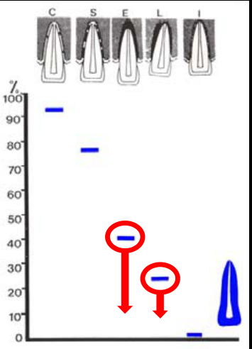
Pulp survival depends on the type and severity of luxation and whether the root is immature or fully developed. Immature teeth have a greater chance of revascularization, but survival decreases with increasingly severe luxation; intrusion and avulsion had the poorest outcomes, with no pulps surviving those injuries in fully developed teeth in the data discussed.
Andreasen & Vestergaard Pedersen — Endod Dent Traumatol 1985.
Time for the Various Responses26
- No clear definition in the literature
- My definitions:
- Short-term → up to 3 months
- Medium-term → 3 months to 1 year
- Long-term → more than 1 year
Short-term (up to 3 months) Responses
See - Tissue response categories
The same pulp, peri-radicular and soft-tissue categories are covered here: Summary of Tissue Responses.
Medium-term (3 months to 1 year) Responses27
See - Tissue response categories
The same pulp, peri-radicular and soft-tissue categories are covered here: Summary of Tissue Responses.
Long-term (more than 1 year) Responses
See - Tissue response categories
The same pulp, peri-radicular and soft-tissue categories are covered here: Summary of Tissue Responses.
Lecturer — Apical Diameter
A wider apical foramen and shorter root indicate an earlier stage of root development, and the less developed the root, the greater the likelihood of revascularization. Measuring 0.7 mm precisely on a radiograph is difficult, but an apical diameter of approximately 0.5 to 1 mm was described as an important practical range.
Factors Affecting Healing After Trauma28
- Introduction
- Potential consequences of trauma
- Tissue responses to trauma
- Pulp
- Peri-radicular tissues
- Gingiva and oral mucosa
- Factors that affect healing
 |  |
Mechanical Factors2930
- Biological Factors
- Direct or Indirect trauma
- Energy of impact - Mass and Velocity (NB: E = mc²)
- Resiliency of the impacting object
- Shape of the impacting object
- Direction of the impacting force
Lecturer — Trauma Mechanics
Direct trauma occurs when the teeth themselves are struck and commonly affects anterior teeth, whereas indirect trauma occurs when the lower teeth strike the upper teeth after an impact to the jaw or beneath the jaw. The energy of impact depends on mass and velocity, with velocity being the more important factor; an intervening object such as a mouthguard also affects resiliency.
These factors will determine the:
- Types of injuries
- Severity of injuries
→ Affect healing responses
Obtain a thorough history about how the accident occurred — helps predict the injuries that are likely to be present.
Lecturer — Trauma History
The dentist is not present when the accident occurs, so a detailed history from the patient, parent, or accompanying person is essential. Even though trauma is an emergency, this is not a reason to omit a thorough examination.
Biological Factors
- Stage of root development
- Extent of pulp involvement
- Degree of displacement of tooth
- Concurrent injuries to the same tooth
→ MUST ALL be assessed after trauma ↳ Via: the history, clinical examination, pulp sensibility tests, radiographs, etc.
Stage of Root Development31
Incompletely Developed Teeth
Two charts show % Pulp Healing (y-axis: 0–100%) against Pulp WIDTH (< 1.5 mm, 1.5 – 2.75 mm, > 2.75 mm) and Pulp LENGTH (< 17 mm, 17 – 19.5 mm, > 19.5 mm).
Andreasen et al 1992
-
AIM: Always wait for pulp revascularisation
- Allows further root development
- Improves long-term prognosis of tooth
-
Prognostic factor: 0.7mm apical diameter
- Andreasen FM et al - EDT 1986
Management
- Reposition and stabilise
- Reassess regularly over time - with:
- Pulp sensibility tests - CO₂ and electric tests
- Radiographs - especially look for:
- Further root development
- Pulp canal calcification
Lecturer — Immature Tooth Management
Initial management of an immature traumatized tooth should be conservative: reposition a luxated tooth, replant an avulsed tooth, stabilize it, and monitor it regularly. Revascularization means that the pulp blood supply is re-established and the pulp survives.
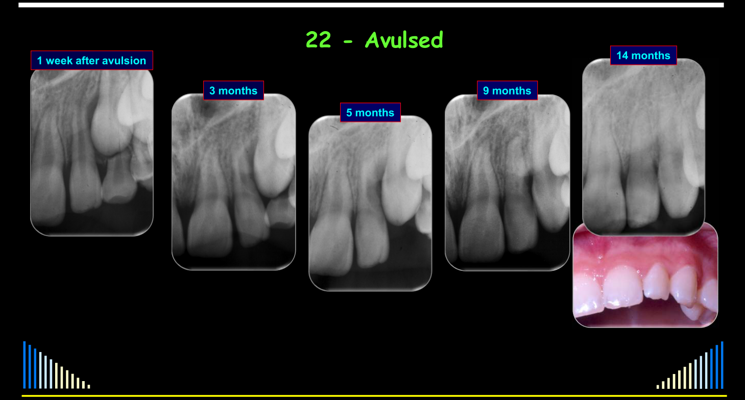
22 - Avulsed — series shown at: 1 week after avulsion, 3 months, 5 months, 9 months, 14 months.
Incompletely Developed Teeth
Endodontics - ONLY IF very definite signs of pulp necrosis AND infection - e.g.:
-
Inflammatory root resorption
-
Periapical radiolucency
-
Draining sinus
-
Swelling
-
Pain, etc
-
Endodontics - ONLY IF very definite signs of pulp necrosis AND infection
- Then: need APEXIFICATION
- Or other procedures - such as:
- An “Apical Plug”
- A “Repair” procedure
- Commonly / ?? incorrectly called “regeneration”
Managing Infected RCS’s in Incompletely Developed Teeth
| Apexification | ”Repair” | Apical “Plug” |
|---|---|---|
Radiographic and clinical outcomes of the treatment of immature permanent teeth by revascularization or apexification: a pilot retrospective cohort study. Alobaid et al - J Endod 2014; 40: 1063-1070
Lecturer — Repair Procedures
The term “regeneration” was considered inappropriate because these procedures do not regenerate a normal pulp. Repair may produce connective or fibrous tissue and hard tissue described as cementum-like or bone-like tissue rather than actual pulp.
Study Results
L10 reports no significant root-length or root-width difference, and that repair was “not superior”, in this small retrospective cohort: a combined Ca(OH)₂-apexification/MTA-plug group of 12 teeth (mean follow-up 22 months) versus 19 repair teeth (15 months). The combined comparator and unequal follow-up establish neither superiority, inferiority nor equivalence. Full survival, clinical-success and adverse-event results: Alobaid et al 2014 - Revascularization and Apexification Pilot Cohort.
Lecturer — Immature Tooth Fracture
A major problem with all three procedures is the thin dentine, particularly in the cervical region, which leaves the tooth weak and susceptible to fracture. The lecture expressed a preference for long-term calcium hydroxide apexification because it is predictable and not particularly difficult to perform.
Follow-up Requirements
BUT - Teeth with Incomplete Root Development need early and regular follow-up:
- Especially in the first few months after luxation / avulsion
- Monitor for signs of External Inflammatory Resorption
- As this resorption can develop and progress rapidly!!
11 - Avulsion; 21 - Intrusion; 12, 22 - Lat. Luxation
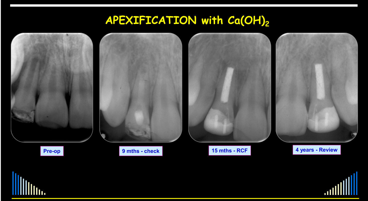
APEXIFICATION with Ca(OH)₂ — Pre-op; 9 mths - check; 15 mths - RCF; 4 years - Review.
Degree of Displacement3233
PROGNOSIS: For BOTH pulp and peri-radicular tissue healing
- Concussion → Best
- Subluxation
- Extrusion
- Lateral Luxation
- Intrusion } Worst
- Avulsion
Lecturer — Displacement Severity
The greater the displacement, the greater the tissue damage and the lower the chance of healing. Periodontal ligament healing also worsens with increasing injury severity, with more severe injuries associated with surface resorption, inflammatory resorption, ankylosis, and replacement resorption.
Relationship between Luxation Injury, Pulp Healing and Stage of Development
(Andreasen 1989)
Chart: % Teeth with Pulp Healing across categories C, S, E, LL, I (Concussion, Subluxation, Extrusion, Lateral Luxation, Intrusion).
AVULSION
- Immature - 34% of pulps survived
- Mature - NO pulps survived
Andreasen et al - 1995
Periodontal Ligament Healing in the Permanent Dentition
In L10’s 1985-cited permanent-dentition charts, outcomes after concussion/subluxation, extrusion/lateral luxation and intrusion are compared by open versus closed apex: normal PDL, surface resorption, ankylosis and inflammatory resorption. The source descriptions give no denominators. Full chart details and original pages: Andreasen Trauma Healing Study 1985.
Periodontal Ligament Healing in the Permanent Dentition
See - Additional PDL charts
After Avulsion — chart of % (0–100%) against minutes dry (5, 20, 40, 60, 120).
Andreasen et al 1989
Extent of Pulp Involvement
KEY FACTORS:
- How likely is pulp necrosis?
- Can bacteria enter the root canal system?
How Likely is Pulp Necrosis?
- Depends on the type of injury
- Luxations, avulsions, root fractures
- Can severe the blood supply apically or at fracture line
- Depends on the ability to revascularise
- Luxations, avulsions, root fractures
Lecturer — Vascular Disruption
With minor extrusion, blood vessels may be stretched rather than severed, reducing their internal diameter and decreasing blood flow. With greater extrusion, the vessels may be severed and the blood supply cut off; survival then also depends on repositioning, stabilization, the original pulp health, and the stage of root development.
How Likely is Pulp Necrosis?
- Depends on the type of injury
- Subluxation, concussion, crown fractures
- Apical blood supply will be intact
- Depends on the ability to resist bacterial invasion AND whether any pathway(s) for bacteria to enter the tooth
- Subluxation, concussion, crown fractures
Can Bacteria Enter the Root Canal System?
Bacterial Pathways:
- Infractions
- Uncomplicated Crown Fractures - dentine exposed
- Complicated Crown Fractures - pulp exposed
Depends on the pulp’s ability to resist the bacterial invasion.
Concurrent Injuries to the Same Tooth
- Often have concurrent injuries to one tooth
- Esp. Luxations and Crown Fractures
- More damage to the tissues
- Will affect the outcome of treatment and the overall long-term prognosis
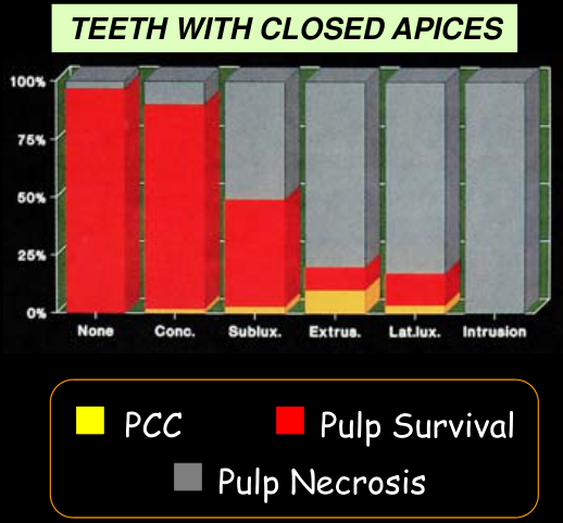
Pulp Survival After Crown Fracture
Chart: Pulp Survival vs Pulp Necrosis (with PCC) across injury categories: None, Conc., Sublux., Extrus., Lat.lux., Intrusion (C, S, E, L, I).
Andreasen et al 1989, 1992
Pulp Survival After Crown Fracture
Chart: Pulp Survival vs Pulp Necrosis (with PCC) across injury categories: None, Conc., Sublux., Extrus., Lat.Lux., Intrusion.
Andreasen et al 1989, 1992
Lecturer — Concurrent Injury Outcomes
In immature teeth, crown fracture alone was associated with survival of almost all pulps, but adding concussion, subluxation, extrusion, lateral luxation, or intrusion progressively reduced pulp survival. When intrusion and crown fracture occurred together, no pulps were expected to survive; in fully developed teeth, adding a crown fracture further reduced survival after subluxation, extrusion, or lateral luxation.
Mechanical Factors
See - Healing factors
Summary34
- Be available to manage trauma - i.e. ASAP !!!
- Understand the injuries - the mechanisms and damage
- Understand ALL the potential tissue responses
- Understand the factors that affect the tissue responses
- Follow up your cases
- To ensure:
- Pulp healing
- Peri-radicular healing
- Gingival and oral mucosa healing
- To manage any problems that develop
- To ensure:
- Perform a thorough history and examination.
- Use pulp tests and radiographs to assess healing.
- Provide appropriate emergency treatment.
- Continued review is necessary to identify later complications and manage them when they develop.
- Pulp outcomes may include recovery, fibrosis, pulp canal calcification, necrosis and infection, or internal resorption.
- Peri-radicular outcomes may include recovery, fibrosis, transient apical breakdown, bone or external resorption, ankylosis, or disturbed root development.
- Soft-tissue outcomes may include recovery, scarring, loss of attachment, gingival recession, or bone loss.
- Tissue responses may occur in combination and may change over time.
- Healing is affected by the mechanical features of the injury, root-development stage, degree of displacement, pulp involvement, and concurrent injuries.
Lecturer — Trauma Consequences
Dental trauma is common and can have consequences ranging from no immediate treatment to extraction and tooth replacement. Emergency management strongly influences the long-term prognosis.
Clinical Cases
Case: Minor crown fractures in a 12-year-old boy
Question
Scenario: A 12-year-old boy fell off his bike and sustained minor uncomplicated crown fractures of his two central incisors.
What’s shown: Radiographs and clinical photographs taken over several years, showing the incisal edges of the central incisors.
Consider: What are the long-term consequences and appropriate management for minor uncomplicated crown fractures?
Answer
Observations:
- The radiographs and photographs show enamel fractures of the incisal edge that remain evident over time, with slightly rough surfaces.
- There are no other pathological consequences or changes in the teeth.
Reasoning: Minor injuries such as uncomplicated crown fractures often have nil or very little long-term consequences. They do not require active treatment at the emergency appointment, but because trauma is a lifelong experience for the patient, the teeth must be monitored regularly in case consequences arise years later.
Outcome: The teeth were monitored on a regular basis over many years. The only treatment required was smoothing over the fractured edges.
Takeaway: Minor dental trauma may not require immediate active treatment but necessitates lifelong monitoring, as complications can arise decades later.
Case: Avulsed central incisors managed with apexification
Question
Scenario: A young boy avulsed his two central incisors, which were subsequently managed with long-term calcium hydroxide apexification after the pulps necrosed.
What’s shown: Initial radiographs showing hard tissue barriers, followed by later radiographs and CT scans showing radiolucencies, and clinical images of the extracted teeth.
Consider: What long-term complications can arise years after successful apexification of avulsed teeth?
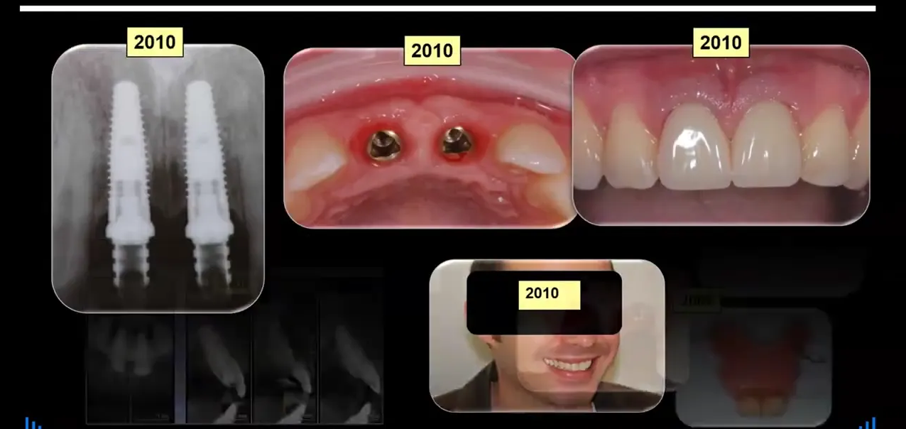
Answer
Observations:
- Initial radiographs showed nice hard tissue barriers forming across the ends of the roots.
- Ten years later, the patient presented with tenderness on the palatal aspect.
- Clinical examination revealed very deep, narrow periodontal pockets suggestive of cracks.
- Transillumination clearly showed vertical cracks on the teeth.
- Radiographs and CT scans revealed radiolucencies associated with both teeth and surrounding the apical parts.
Reasoning: The deep narrow pockets, visible cracks on transillumination, and periapical radiolucencies indicate that the teeth had cracked and the root canal system had become infected, necessitating extraction.
Outcome: The teeth were extracted. The patient was provided with a temporary partial denture and subsequently received two implants with crowns. The implants require ongoing regular monitoring.
Takeaway: Even when avulsed teeth are successfully managed with apexification and hard tissue barriers form, they can develop cracks and infections years later, emphasizing the need for lifelong monitoring.
Case: Root fracture of a lateral incisor from sports trauma
Question
Scenario: A patient was hit in the mouth with a squash racket, resulting in a root fracture of a lateral incisor. The tooth was splinted to the adjacent teeth with composite resin by his dentist.
What’s shown: A sequential series of radiographs from one week up to five years post-injury, along with pulp testing results over time.
Consider: How does the pulp respond to a root fracture, and what is the appropriate follow-up protocol?
Answer
Observations:
- At one week, the tooth was sensitive to hot and cold, and pulp testing showed a painful response to cold, indicating reversible pulpitis.
- The sensitivity resolved within a week or two.
- Over time, the tooth stopped responding to the CO2 cold test but continued to respond normally to the electric pulp test.
- Radiographs at 3, 9, and 12 months, and yearly up to 5 years, showed no periapical or periradicular changes, but pulp canal calcification occurred, particularly in the coronal part.
Reasoning: The initial sensitivity was reversible pulpitis due to trauma, which resolved without further intervention since the tooth was already stabilized. The normal electric pulp test response and lack of periradicular pathology indicate that the pulp recovered and returned to a normal state, likely with some fibrosis and calcification.
Outcome: The splint was kept on for three months with monthly reviews, then removed. The patient was monitored at 3, 9, and 12 months, and yearly up to five years. The pulp recovered successfully.
Takeaway: The pulp can recover from severe injuries like root fractures, demonstrating favorable healing responses such as recovery and pulp canal calcification, which must be confirmed through regular clinical and radiographic monitoring.
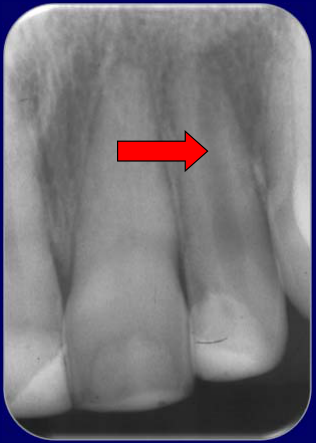
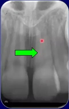
Case: Upper central and lateral incisors following trauma
Question
Scenario: A patient with a history of trauma affecting the upper central and lateral incisors.
What’s shown: A periapical radiograph displaying the roots of the upper central and lateral incisors.
Consider: Identify the different tissue responses occurring in each of the traumatized teeth based on the radiographic appearance.
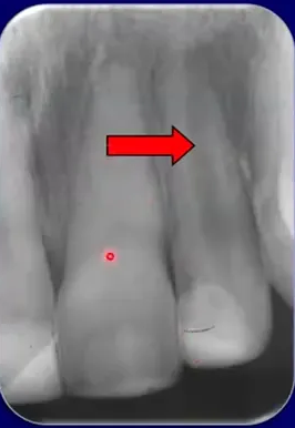
Answer
Observations:
- The central incisor shows narrowing of the root canal down to the apical level and particularly in the pulp chamber.
- The lateral incisor shows no narrowing of the canal and a developing periapical radiolucency.
Reasoning: The central incisor exhibits a favorable response with pulp canal calcification. The lateral incisor, which sustained an uncomplicated crown fracture, exhibits an unfavorable response with pulp necrosis and infection of the root canal system, indicated by the periapical radiolucency.
Outcome: The central incisor demonstrates pulp canal calcification, while the lateral incisor has developed pulp necrosis and a periapical radiolucency.
Takeaway: Different teeth in the same patient can exhibit contrasting tissue responses to trauma, ranging from favorable calcification to unfavorable necrosis and infection.
Case: Lower incisor with sequential tissue responses
Question
Scenario: A patient presents eight years after sustaining a lateral luxation and enamel infractions to a lower incisor.
What’s shown: A periapical radiograph of a lower incisor.
Consider: Evaluate the chronological tissue responses and the current status of the tooth.
Answer
Observations:
- Radiographic evidence of pulp canal calcification.
- A periapical radiolucency.
Reasoning: The initial favorable response was pulp survival leading to pulp canal calcification. Subsequently, bacteria likely entered through the infractions that reached the dentine, leading to pulp necrosis, infection of the root canal system, and chronic apical periodontitis.
Outcome: The patient presented for treatment due to the development of a secondary acute apical periodontitis.
Takeaway: Tissue responses to trauma can occur sequentially over time, with early favorable responses potentially followed by late unfavorable responses if bacteria gain access to the root canal system.
Case: Subluxated and fractured upper incisors at six-year follow-up
Question
Scenario: A patient involved in a car accident resulting in subluxation of the upper central incisors and an uncomplicated crown fracture of one of them.
What’s shown: A chronological series of radiographs from the time of injury up to six years post-trauma.
Consider: Assess the long-term periapical and pulpal outcomes of these injuries.
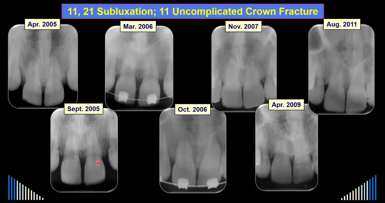
Answer
Observations:
- Initial presentation shows subluxation of teeth 11 and 21, with an uncomplicated crown fracture on tooth 11.
- Follow-up radiographs at six months and six years show healthy periapical tissues.
Reasoning: The teeth were stabilized and the crown fracture was restored with composite resin. Regular follow-up and pulp testing confirmed the pulp remained vital and the periapical tissues returned to a normal state, indicating recovery and possible fibrous healing.
Outcome: The teeth recovered and returned to a normal state over the six-year follow-up period. The patient also successfully completed orthodontic treatment.
Takeaway: Minor injuries such as subluxation and uncomplicated crown fractures can have a favorable prognosis, returning to a normal state with appropriate stabilization, restoration, and monitoring.
Case: Lateral incisor with transient apical breakdown following lateral luxation
Question
Scenario: A young boy undergoing orthodontic treatment fell off his bicycle, sustaining lateral luxation injuries to his maxillary central and lateral incisors. His orthodontist repositioned the teeth and placed a wire splint on the day of the injury. The patient was reviewed by the lecturer a few days later.
What’s shown: A chronological series of clinical photographs and periapical radiographs of the maxillary anterior teeth over a 5-year follow-up period.
Consider: Evaluate the radiographic and clinical changes in the lateral incisor over time, determine the differential diagnosis for the periapical findings, and identify the final outcome.
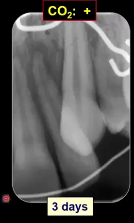
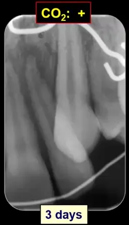
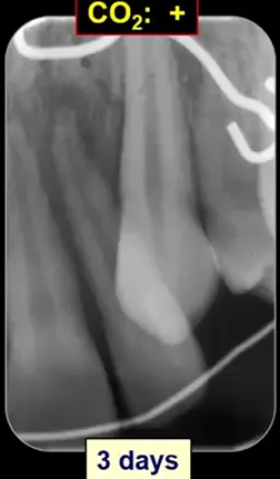
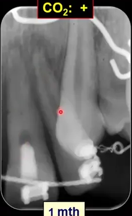
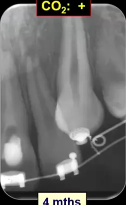
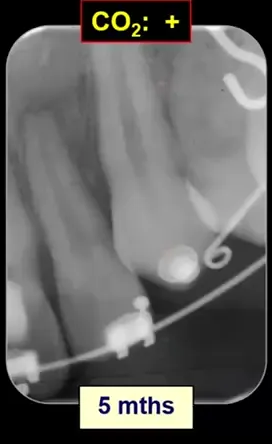
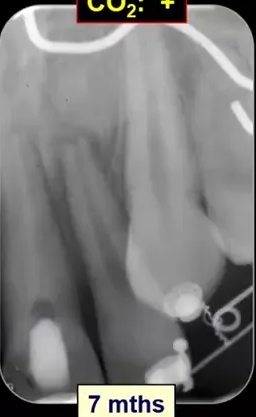
Answer
Observations:
- The central incisor developed pulp necrosis and required root canal treatment at one month.
- The lateral incisor initially responded normally to a CO2 pulp test.
- At 1 to 4 months, the lateral incisor developed a periapical radiolucency, the root end appeared blunted, and the apical foramen widened.
- The lateral incisor continued to respond to CO2 testing (electric pulp testing was not possible due to orthodontic brackets).
- At 15 months, the root canal began to narrow, indicating calcification.
- At 2 years, the canal appeared almost completely calcified radiographically, the root end rounded off, and the open apex disappeared.
- At 3 and 5 years, the tooth remained clinically normal and stable; it was unresponsive to CO2 but responded normally to electric pulp testing.
Reasoning: The widening of the apical foramen and periapical radiolucency raised a differential diagnosis between transient apical breakdown and chronic apical periodontitis secondary to an infected root canal. However, the positive response to the CO2 pulp test indicated the pulp was still vital, ruling out an infected necrotic pulp and pointing to transient apical breakdown. Over time, the radiographic evidence of root end blunting followed by rounding off, along with canal calcification and the eventual return of normal electric pulp test responses, confirmed the remodeling process characteristic of this condition.
Outcome: The lateral incisor underwent transient apical breakdown with subsequent pulp canal calcification and remodeling of the root apex, remaining stable and vital at the 5-year review. The central incisor required root canal treatment due to pulp necrosis.
Takeaway: Transient apical breakdown is a favorable, temporary periapical response to trauma (often lateral luxation) characterized by apical radiolucency and root end blunting; it can be distinguished from infectious apical periodontitis by a positive pulp vitality test and typically resolves with root canal calcification and remodeling over time.
Case: Soft tissue loss of attachment after trauma
Question
Scenario: A patient presents following dental trauma where the soft tissues were not managed well.
What’s shown: An intraoral view focusing on the palatal aspect of the maxillary anterior teeth.
Consider: How to properly assess soft tissue attachment after trauma and the appropriate management if it is compromised.
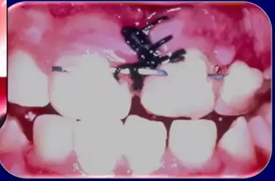
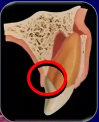
Answer
Observations:
- The palatal tissues appear to be attached because they are sitting against the tooth.
- Upon checking with an instrument, the tissues are found to be detached.
Reasoning: After injuries such as lateral luxation, intrusion, avulsion, or extrusion, torn tissues may simply sit against the repositioned tooth and look acceptable. However, they may not be truly attached. It is necessary to use an instrument, such as a probe or a flat plastic instrument, to check for actual attachment. If the tissue is not attached, it must be sutured, typically between the teeth, to hold it back in place.
Outcome: Failure to properly manage and suture the tissues leads to loss of attachment.
Takeaway: Always verify soft tissue attachment with an instrument after dental trauma, as tissues can appear attached when they are not, and suture them if they are detached to prevent loss of attachment.
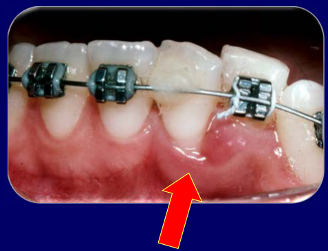
Case: Gingival recession after avulsion and replantation, first case
Question
Scenario: A patient who experienced tooth avulsion and replantation, where the labial soft tissues were not managed properly.
What’s shown: A clinical photograph of a replanted tooth exhibiting gingival recession on the labial aspect.
Consider: The clinical consequences of failing to reposition and suture lacerated labial tissues following tooth replantation.
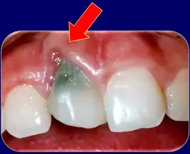
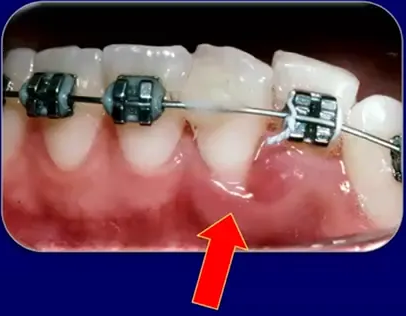
Answer
Observations:
- Gingival recession is visible on the labial aspect of the replanted tooth.
Reasoning: The lacerated tissues on the labial aspect were not repositioned and sutured after the tooth was replanted. This lack of soft tissue management directly resulted in gingival recession.
Outcome: The patient developed gingival recession, accompanied by bone loss and loss of attachment.
Takeaway: Lacerated labial tissues must be repositioned and sutured after tooth replantation to prevent gingival recession and subsequent attachment and bone loss.
Case: Gingival recession after avulsion and replantation, second case
Question
Scenario: A second patient who experienced tooth avulsion and replantation, with improper management of the labial soft tissues.
What’s shown: A clinical photograph of another replanted tooth exhibiting gingival recession on the labial aspect.
Consider: The clinical consequences of failing to reposition and suture lacerated labial tissues following tooth replantation.
Answer
Observations:
- Gingival recession is visible on the labial aspect of the replanted tooth.
Reasoning: Similar to the first case, the lacerated tissues on the labial aspect were not repositioned and sutured after replantation, which consequently led to gingival recession.
Outcome: The patient developed gingival recession, along with bone loss and loss of attachment.
Takeaway: Lacerated labial tissues must be repositioned and sutured after tooth replantation to prevent gingival recession and subsequent attachment and bone loss.
Case: Avulsed lateral incisor in a young girl, monitored over 14 months
Question
Scenario: A young girl avulsed her lateral incisor at school. The tooth was replanted immediately and stabilized with a composite resin splint, which fell off within a week. The patient was evaluated one week post-avulsion.
What’s shown: A composite series of five periapical radiographs tracking the tooth over time.
Consider: What is the appropriate management and expected radiographic outcome for this avulsed, incompletely developed tooth when no active endodontic treatment is performed?
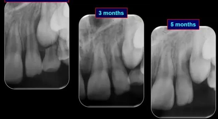
Answer
Observations:
- The composite splint fractured off within a week, which was not critical as flexible splinting is sufficient for initial periodontal ligament healing.
- At three and five months, the root canal began to show signs of narrowing and calcification.
- By 14 months, root development had proceeded and was complete, with the root canal system narrowing into a calcified state, though a small canal remained radiographically visible.
Reasoning: For an incompletely developed avulsed tooth that is replanted and monitored without active treatment, the pulp can recover, leading to continued root development and pulp canal calcification. Active treatment like root canal therapy is only indicated if definite signs of pulp necrosis and infection (e.g., external inflammatory resorption, periapical radiolucency, draining sinus, swelling, or pain) develop.
Outcome: The tooth was simply monitored over 14 months, resulting in complete root development and pulp canal calcification without any active endodontic treatment.
Takeaway: Incompletely developed traumatized teeth should be managed conservatively with repositioning, flexible splinting, and close monitoring, as revascularization and continued root development can occur without active intervention.
Case: Severe anterior trauma with external inflammatory resorption at 5 weeks
Question
Scenario: A patient presented with a severe maxillary anterior injury involving an avulsion, an intrusion, and two laterally luxated teeth.
What’s shown: Clinical intraoral photographs of the anterior maxillary region at 5 weeks post-trauma.
Consider: What radiographic complication is observed in this severe injury case, and why is it particularly concerning in incompletely developed teeth?
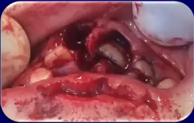
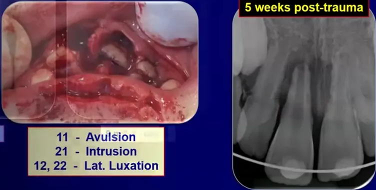
Answer
Observations:
- At five weeks post-trauma, signs of external inflammatory resorption are visible on at least three of the injured teeth.
Reasoning: Severe displacement injuries (like avulsion and intrusion) cause significant tissue damage. In incompletely developed teeth, the dentine is very thin, meaning bacteria or toxins can easily travel from the root canal to the peripheral tissues through wide dentine tubules. This allows external inflammatory resorption to develop and progress very rapidly.
Outcome: The case demonstrates the rapid onset of external inflammatory resorption at just 5 weeks post-trauma in severely injured immature teeth.
Takeaway: Immature teeth with severe displacement injuries must be monitored very carefully in the early stages, as external inflammatory resorption can develop and progress rapidly due to thin dentine and wide dentine tubules.
Case: Tooth with open apex and crown fracture, radiographic series
Question
Scenario: A tooth with an open apex became infected as a result of a crown fracture.
What’s shown: A series of four periapical radiographs arranged horizontally.
Consider: Identify the treatment approach demonstrated and evaluate the radiographic outcome over time.
Answer
Observations:
- The radiographs document the application of long-term calcium hydroxide.
- A hard tissue barrier forms across the apex.
- The final radiograph at four years shows a stable periapical region.
Reasoning: For infected immature teeth with open apices, the preferred management is long-term calcium hydroxide treatment (apexification). The sequential radiographs demonstrate the successful formation of a calcified barrier across the open apex, confirming the biological response to the treatment.
Outcome: The treatment resulted in a well-formed hard tissue barrier across the apex, which remained stable at the four-year follow-up. The lecturer notes this procedure is quite predictable and not particularly difficult to perform.
Takeaway: Long-term calcium hydroxide apexification is a predictable and effective treatment for infected immature teeth, leading to the formation of a stable hard tissue barrier.
Footnotes
-
Original PDF page 1: L10 Dento-Alveolar Trauma 3 - Tissue Responses to Injury, p.1 ↩
-
Original PDF page 2: L10 Dento-Alveolar Trauma 3 - Tissue Responses to Injury, p.2 ↩
-
Original PDF page 3: L10 Dento-Alveolar Trauma 3 - Tissue Responses to Injury, p.3 ↩
-
Original PDF page 4: L10 Dento-Alveolar Trauma 3 - Tissue Responses to Injury, p.4 ↩
-
Original PDF page 5: L10 Dento-Alveolar Trauma 3 - Tissue Responses to Injury, p.5 ↩
-
Original PDF page 6: L10 Dento-Alveolar Trauma 3 - Tissue Responses to Injury, p.6 ↩
-
Original PDF page 7: L10 Dento-Alveolar Trauma 3 - Tissue Responses to Injury, p.7 ↩
-
Original PDF page 8: L10 Dento-Alveolar Trauma 3 - Tissue Responses to Injury, p.8 ↩
-
Original PDF page 9: L10 Dento-Alveolar Trauma 3 - Tissue Responses to Injury, p.9 ↩
-
Original PDF page 10: L10 Dento-Alveolar Trauma 3 - Tissue Responses to Injury, p.10 ↩
-
Original PDF page 11: L10 Dento-Alveolar Trauma 3 - Tissue Responses to Injury, p.11 ↩
-
Original PDF page 12: L10 Dento-Alveolar Trauma 3 - Tissue Responses to Injury, p.12 ↩
-
Original PDF page 13: L10 Dento-Alveolar Trauma 3 - Tissue Responses to Injury, p.13 ↩
-
Original PDF page 14: L10 Dento-Alveolar Trauma 3 - Tissue Responses to Injury, p.14 ↩
-
Original PDF page 15: L10 Dento-Alveolar Trauma 3 - Tissue Responses to Injury, p.15 ↩
-
Original PDF page 16: L10 Dento-Alveolar Trauma 3 - Tissue Responses to Injury, p.16 ↩
-
Original PDF page 17: L10 Dento-Alveolar Trauma 3 - Tissue Responses to Injury, p.17 ↩
-
Original PDF page 18: L10 Dento-Alveolar Trauma 3 - Tissue Responses to Injury, p.18 ↩
-
Original PDF page 19: L10 Dento-Alveolar Trauma 3 - Tissue Responses to Injury, p.19 ↩
-
Original PDF page 20: L10 Dento-Alveolar Trauma 3 - Tissue Responses to Injury, p.20 ↩
-
Original PDF page 21: L10 Dento-Alveolar Trauma 3 - Tissue Responses to Injury, p.21 ↩
-
Original PDF page 22: L10 Dento-Alveolar Trauma 3 - Tissue Responses to Injury, p.22 ↩
-
Original PDF page 23: L10 Dento-Alveolar Trauma 3 - Tissue Responses to Injury, p.23 ↩
-
Original PDF page 24: L10 Dento-Alveolar Trauma 3 - Tissue Responses to Injury, p.24 ↩
-
Original PDF page 25: L10 Dento-Alveolar Trauma 3 - Tissue Responses to Injury, p.25 ↩
-
Original PDF page 26: L10 Dento-Alveolar Trauma 3 - Tissue Responses to Injury, p.26 ↩
-
Original PDF page 27: L10 Dento-Alveolar Trauma 3 - Tissue Responses to Injury, p.27 ↩
-
Original PDF page 42: L10 Dento-Alveolar Trauma 3 - Tissue Responses to Injury, p.42 ↩
-
Original PDF page 28: L10 Dento-Alveolar Trauma 3 - Tissue Responses to Injury, p.28 ↩
-
Original PDF page 29: L10 Dento-Alveolar Trauma 3 - Tissue Responses to Injury, p.29 ↩
-
Original PDF page 32: L10 Dento-Alveolar Trauma 3 - Tissue Responses to Injury, p.32 ↩
-
Original PDF page 36: L10 Dento-Alveolar Trauma 3 - Tissue Responses to Injury, p.36 ↩
-
Original PDF page 38: L10 Dento-Alveolar Trauma 3 - Tissue Responses to Injury, p.38 ↩
-
Original PDF page 43: L10 Dento-Alveolar Trauma 3 - Tissue Responses to Injury, p.43 ↩