L12 Dento-Alveolar Trauma 5 - Emergency Management of Traumatic Dental Injuries
Emergency Management of Traumatic Dental Injuries1
W/Prof. Paul V. Abbott AO
Emergency Management of Traumatic Dental Injuries
Prof. Paul V. Abbott AO BDSc, MDS, FRACDS(Endo), FPFA, FADI, FICD, FACD, FIADT
- Specialist Endodontist
- Winthrop Professor of Clinical Dentistry
- UWA Dental School
- The University of Western Australia
ORAL HEALTH CENTRE OF WESTERN AUSTRALIA
DMD - 3rd Year - 2020
 |  |
General Principles for Managing Trauma
- History, examination and assessment
- Establish priorities
- Protection
- Pulp (if exposed)
- Root surface (e.g. during repositioning)
- Reposition — immediately: teeth, bone, soft tissues
- Stabilisation
- Bone and teeth: splint (rigid or flexible)
- Soft tissues: sutures
- Temporary restorations
- Medications
- Systemic: tetanus, antibiotics, analgesics, anti-inflammatory
- Local: intra-canal dressings, chlorhexidine mouthwash, gel
- Follow-up
Lecturer — Initial Assessment
Emergency assessment should include more than establishing priorities. It also involves documenting how, when, why, and where the accident occurred, performing pulp sensibility testing, and taking the complete trauma radiograph series.
- Management should account for the mechanical factors that produced the injury.
- Biological factors include root development, pulp exposure, concurrent injuries, and the condition of the periodontal ligament and surrounding tissues.
Lecturer — Tissue Repositioning
The objective is to return displaced teeth to their original position and to reposition and stabilize bone. Teeth and bone can often be stabilized together with a splint, while displaced or lacerated soft tissues should be sutured.
Lecturer — Healing and Treatment
Healing must be allowed to occur before irreversible treatment is undertaken whenever possible. Periodontal ligament healing may take approximately three to six months, while pulp healing may take several months and failure may only become evident after a couple of years.
- Immediate root canal treatment is limited to injuries where external inflammatory resorption is likely, or where pulp removal is required for a complicated crown or crown-root fracture.
- In immature teeth, preserving the pulp is preferred so root development can continue.
Lecturer — Temporary Treatment
Temporary or initial restorations should generally be maintained while the pulp response becomes clear. Irreversible treatment and definitive restorations should be deferred unless they are necessary; regular reviews should include pulp tests, percussion, mobility assessment, and radiographs.
Emergency Management of Traumatic Dental Injuries — Injury Types
-
Infractions
-
Crown fractures
-
Crown : root fractures
-
Root fractures
-
Concussion + subluxation
-
Extrusive luxation
-
Lateral luxation
-
Intrusive luxation
-
Avulsion
-
Root fractures are excluded from this lecture and will be discussed separately.
Broader injury classification and supporting-bone context: Trauma Classification and Supporting Bone. DMD — 3rd Year — 2020
Infractions
- Crown Fractures
- Crown : Root Fractures
- Root Fractures
- Concussion + Subluxation
- Extrusive Luxation
- Lateral Luxation
- Intrusive Luxation
- Avulsion
Infractions2
- Coat tooth surface with light-cured unfilled resin bonding agent
- To minimise chances of bacterial ingress while pulp possibly damaged or inflamed from trauma
- Love 1996
- To minimise chances of bacterial ingress while pulp possibly damaged or inflamed from trauma
 |  |
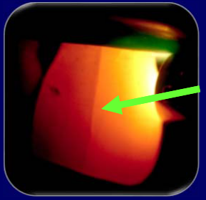 |
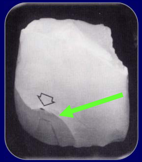
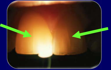
Bacterial Penetration Study After Simulated Trauma
Important Aspect Following Trauma to Teeth3
- 1.7 – 3.5% of teeth with enamel infractions demonstrated pulp necrosis (Ravn 1985a, b)
- Bacteria in the pulp system will prevent revascularisation (Cvek et al 1990)
Love EDT 1996 — Bacterial Penetration of the Root Canal of Intact Incisor Teeth after a Simulated Traumatic Injury
Outline of Study4
Full experimental sequence, methods, and source pages: Love 1996 - Bacterial Penetration After Simulated Trauma.
Summary and Conclusions5
In Love’s in-vitro model, simulated trauma enabled bacterial penetration and the tested unfilled-resin seal blocked it; clinical pulp healing was not measured.
- Unfilled resin: may help prevent infection of the pulp system:
- Apply as soon as possible after trauma to minimise bacterial penetration
- May help to allow healing of the compromised pulp
- Resin will wear away — so this effect is not permanent
Lecturer — Resin Protection
The resin wears away relatively quickly through tooth cleaning, eating, and normal use, so its protective effect may last only a few days.
- Despite being temporary, the procedure is simple, non-invasive, and quick.
- Infractions
- Crown Fractures
- Crown : Root Fractures
- Root Fractures
- Concussion + Subluxation
- Extrusive Luxation
- Lateral Luxation
- Intrusive Luxation
- Avulsion
Lecturer — Infractions
Infractions are small cracks, essentially in enamel, that are not thought to extend into dentine.
- Their depth cannot always be determined clinically, so dentine involvement is possible.
- If limited to enamel, no major treatment is usually required.
- Infractions should be followed using the usual trauma review schedule.
- <mark>Cervical pulpotomy may be used in some cases.</mark>
- <mark>Pulp capping may be appropriate for a small exposure in suitable circumstances.</mark>
- <mark>In an immature tooth, retaining the pulp is generally preferred.</mark>
- <mark>In a fully developed tooth with a reasonably large exposure, pulp removal and root canal treatment are usually most appropriate.</mark>
- <mark>The treatment decision depends on the overall clinical situation rather than pulp exposure alone.</mark>
- <mark>The tooth can then be restored with composite.</mark>
Lecturer — Fracture Follow-Up
The first restoration does not necessarily need to be definitive.
- A definitive restoration may be delayed for several days or weeks while the pulp response becomes clear.
- If the fractured fragment is available, bonding it back can rapidly restore aesthetics and may remain effective for a long time.
- Crown fractures require routine follow-up.
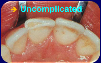 | 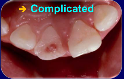 |
Crown Fractures
Protect the pulp6
- If pulp IS exposed
- Partial pulpotomy or pulpectomy
- Depends on stage of root development
- Partial pulpotomy or pulpectomy
- If pulp is NOT exposed
- Indirect pulp cap / lining
- Use glass ionomer
- “Base” or “dentine replacement”
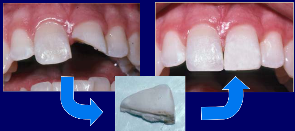
Restore the fractured portion of the tooth
- Composite resin “bandage” / GIC / bond fragment
- Definitive restoration later
- When pulp status clear
Crown-Root Fractures78
- Infractions
- Crown Fractures
- Crown : Root Fractures
- Root Fractures
- Concussion + Subluxation
- Extrusive Luxation
- Lateral Luxation
- Intrusive Luxation
- Avulsion
 |  |  |
 | 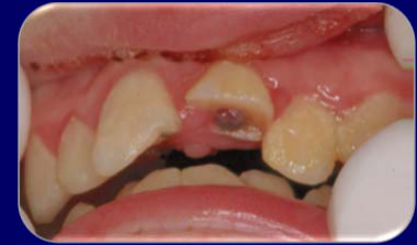 |
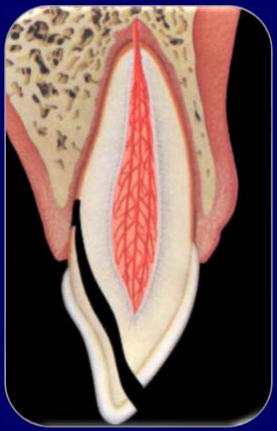
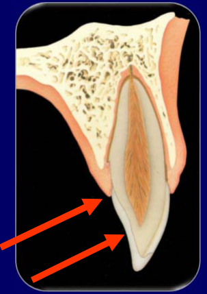
Crown : Root Fractures9
-
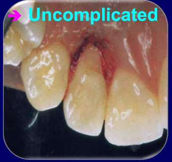
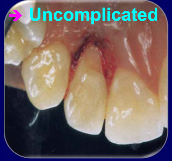
Uncomplicated
- Minor involvement of root
- Restore as for crown fracture
-
Uncomplicated
- Minor involvement of root
- Restore as for crown fracture
-
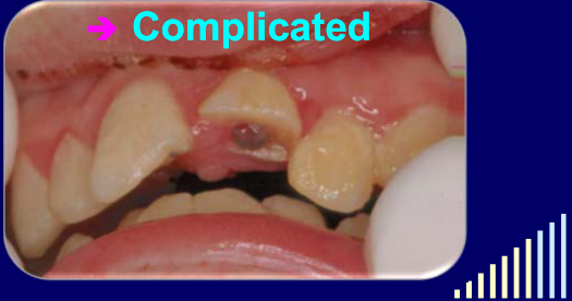
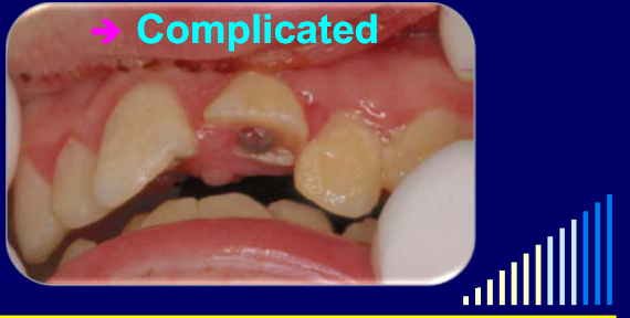
Complicated
- Major involvement of root
- Treat as for a supra-crestal root fracture in the coronal third of the root
-
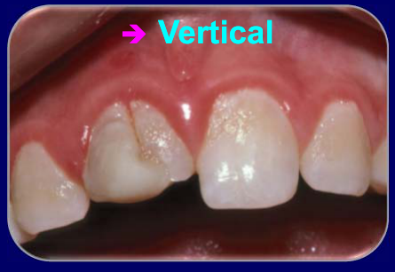
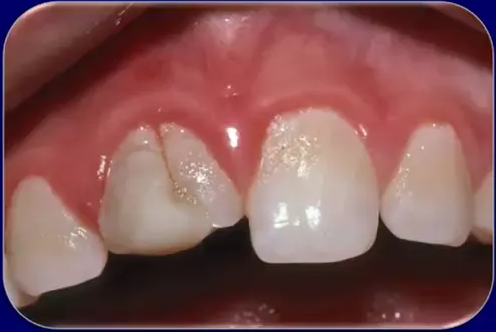
Vertical crown:root fractures
- Extraction
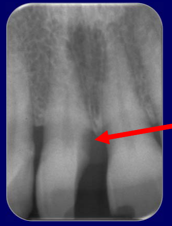
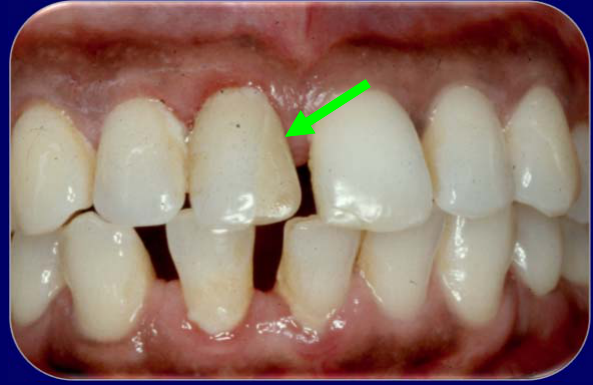
Uncomplicated Crown-Root Fracture
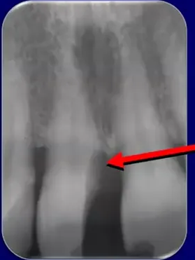
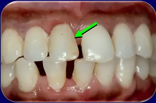
An uncomplicated crown-root fracture was restored with glass ionomer while the tooth was monitored. The pulp became necrotic and infected within a few months, requiring root canal treatment, but the glass ionomer remained intact during treatment and stable approximately six months after placement.
See separate lecture re: Root Fractures
Lecturer — Uncomplicated Fractures
An uncomplicated crown-root fracture generally involves only a small part of the root and has a relatively shallow subgingival margin. The tooth should be restored to approximately where the cementoenamel junction was located before the fracture.
- This allows cementum formation across exposed dentine and tissue reattachment.
- Extending restorative material to the fractured margin would prevent tissue attachment and could result in a permanent periodontal defect.
- Vertical crown:root fractures
- Extraction
 |  |  |
 |  |
Emergency Management of Traumatic Dental Injuries10
- Infractions
- Crown Fractures
- Crown : Root Fractures
- Root Fractures — See separate lecture
- Concussion + Subluxation
- Extrusive Luxation
- Lateral Luxation
- Intrusive Luxation
- Avulsion
Vertical Split Molar
A soldier fainted and struck his chin on bitumen, creating indirect trauma. A mandibular molar already weakened by extensive caries split vertically when the mandible was forced against the maxilla.
Lecturer — Injury Definitions
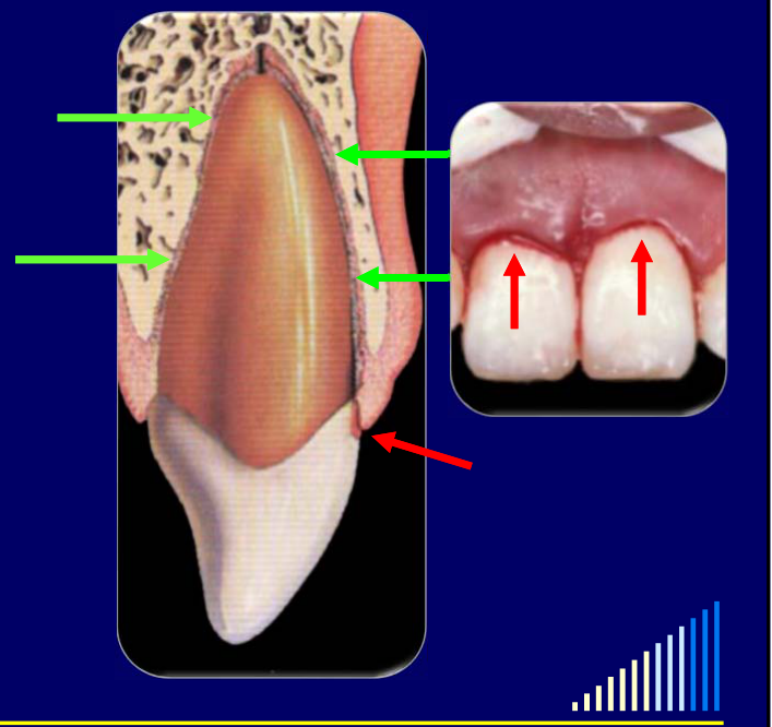
Concussion is tenderness to percussion without tooth looseness, whereas subluxation is mobility without displacement.
- Subluxation reflects greater periodontal-ligament stretching and may cause bleeding in the gingival sulcus.
- A very loose subluxated tooth may receive a short-term simple splint, particularly when mobility interferes with occlusion.
- Occlusal relief should remove the injured tooth from centric, lateral, and protrusive contacts, using articulating paper.
Concussion and Subluxation
- Infractions
- Crown Fractures
- Crown : Root Fractures
- Root Fractures
- Concussion + Subluxation
- Extrusive Luxation
- Lateral Luxation
- Intrusive Luxation
- Avulsion
Concussion & Subluxation11
- Splinting is NOT required
- Unless the tooth is very loose (i.e. only a severe subluxation)
- Relieve the occlusion
- Check with articulating paper
- Review
- As per recommended review schedule if no other injuries
Prognosis and Outcome Figures12
Prognosis — is generally very good
Depends on the many mechanical and biological factors — such as the stage of root development, pulp involvement, concurrent injuries, etc. — as previously discussed.
- These figures are a general guide ONLY
- They are based on retrospective analyses of many teeth
- They should NOT be used to predict the outcome for an individual tooth
- They are NOT the probability of any of the listed outcomes / sequelae
 | 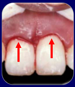 |
Concussion13
Prognosis — is generally very good
Depends on the many mechanical and biological factors — such as the stage of root development, pulp involvement, concurrent injuries, etc. — as previously discussed.
| Concussion | |
|---|---|
| Pulp Necrosis | 2 % |
| PCC | 2.2 % |
| TAB | 0.1 % |
| Resorption | - |
- These figures are a general guide ONLY
- They are based on retrospective analyses of many teeth
- They should NOT be used to predict the outcome for an individual tooth
- They are NOT the probability of any of the listed outcomes / sequelae
Subluxation
Prognosis — is generally very good
Depends on the many mechanical and biological factors — such as the stage of root development, pulp involvement, concurrent injuries, etc. — as previously discussed.
| Subluxation | |
|---|---|
| Pulp Necrosis | 6 - 47 % |
| PCC | 26 % |
| TAB | 2.2 % |
| Resorption | 4 % |
- These figures are a general guide ONLY
- They are based on retrospective analyses of many teeth
- They should NOT be used to predict the outcome for an individual tooth
- They are NOT the probability of any of the listed outcomes / sequelae
Lecturer — Prognosis Language
General terms such as good, fair, and poor are preferred to overly precise numerical predictions for individual teeth.
Lecturer — Concussion Outcomes
Pulp canal calcification may occur after concussion, while transient apical breakdown is rare.
- Resorption is very rare but cannot be considered impossible.
- If pulp necrosis and infection develop, apical inflammatory resorption may occur.
Lecturer — Subluxation Outcomes
Subluxation has a generally good prognosis, but it is less favorable than concussion.
- Pulp necrosis occurs more often when subluxation is combined with a crown fracture, and the higher reported figure of approximately 47% was particularly associated with concurrent crown fractures.
- Pulp canal calcification is more common than in concussion, and transient apical breakdown may occur.
- Resorption is typically external inflammatory resorption secondary to pulp necrosis and infection.
Extrusive Luxation
- Infractions
- Crown Fractures
- Crown : Root Fractures
- Root Fractures
- Concussion + Subluxation
- Extrusive Luxation
- Lateral Luxation
- Intrusive Luxation
- Avulsion
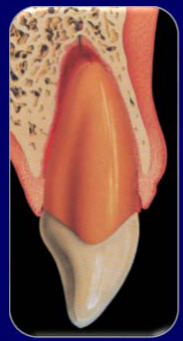
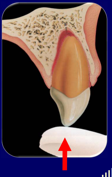
Management14
- Reposition immediately
- Mould bone back into position
- Splint
- Flexible
- → 7 - 10 days
- Flexible
- Suture soft tissues
- Review regularly
Lecturer — Repositioning Details
Early repositioning is usually simple, but delay can make the procedure more difficult.
- Gentle finger pressure is used to push the tooth back into position; after several days, a blood clot at the socket base may require orthodontic repositioning.
- The palatal gingival attachment must be probed for a hidden degloving-type injury, and detached tissue should be sutured back into position.
- Sutures may be removed after approximately five to six days, or when the splint is removed at around seven days.
- Follow-up should assess periodontal ligament and pulp healing through clinical tests and radiographs.
Prognosis - is generally good
Depends on the many mechanical and biological factors - such as the stage of root development, pulp involvement, concurrent injuries, etc. - as previously discussed.
Lecturer — Pulp Outcomes
Pulp necrosis is the principal concern after extrusive luxation, particularly when extrusion exceeds a couple of millimeters.
- Apical blood supply may be severed, and revascularization is less likely in fully developed teeth.
- With a concurrent crown fracture, pulp recovery is even less likely, with pulp necrosis figures approaching 90–98% in the more severe situations described.
- A recovering pulp may develop pulp canal calcification, while transient apical breakdown can resemble periapical disease from an infected root canal system or apical inflammatory resorption.
- Close follow-up is required during the first few months.
 |  |
 |
Expected Outcomes15
| Outcome | Frequency |
|---|---|
| Pulp Necrosis | 64 - 98 % |
| PCC | 24 % |
| TAB | 11 % |
| Resorption | 7 % |
| Bone loss | - |
- These figures are a general guide ONLY
- They are based on retrospective analyses of many teeth
- They should NOT be used to predict the outcome for an individual tooth
- They are NOT the probability of any of the listed outcomes / sequelae
Lecturer — Resorption and Bone Loss
External inflammatory resorption may occur when pulp necrosis and infection develop.
- External replacement resorption is uncommon because the root surface and periodontal ligament are not usually severely damaged.
- Bone loss is also uncommon because there is generally no fracture or cervical crushing of the alveolar bone.
Lecturer — Lateral Luxation Treatment
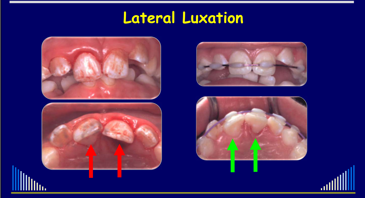
Repositioning should occur immediately or as soon as possible because delayed treatment allows a blood clot to form and makes repositioning more difficult.
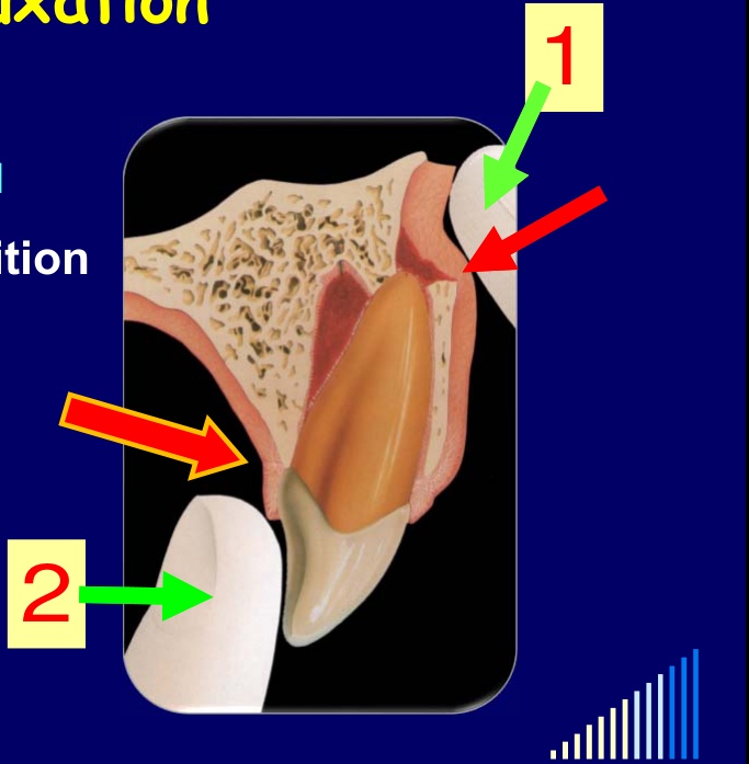
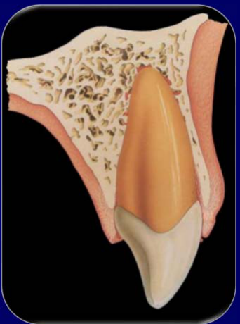
- The first movement is to push down from the apical end to disengage the root from the bony lock created by the socket-wall fracture; the crown is then pushed back toward its original position.
- Simply pushing from the palatal or labial aspect without first disengaging the root can cause further apical damage and increase the risk of external replacement resorption.
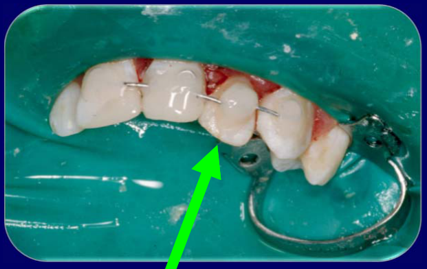
- A more rigid splint, such as wire and composite, is used for approximately six weeks in an adult and four weeks in a child because a bone fracture is present.
- Soft tissues should be sutured, with particular attention to possible palatal degloving.
- Follow-up includes early suture review and continued assessment of splint stability, oral hygiene, pulp healing, periodontal ligament healing, and radiographic changes.
- Root canal treatment may need to begin immediately when external inflammatory resorption is likely, such as in a fully developed tooth with a concurrent crown fracture.
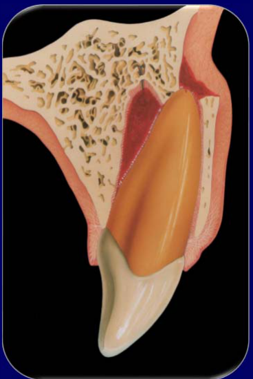
Lateral Luxation16
- Reposition immediately
- TWO movements required
- Mould bone back into position
- NB: fractured socket wall
- Splint — Rigid
- Since also bone fracture → 6 weeks
- Suture soft tissues
- Review regularly
 |  |
 |
Prognosis17
Prognosis is generally good. It depends on many mechanical and biological factors — such as the stage of root development, pulp involvement, concurrent injuries, etc. — as previously discussed.
- Pulp Necrosis: 58%
- PCC: 24%
- TAB: 12%
- Resorption: 7%
- Bone loss: 6–31%
These figures are a general guide ONLY. They are based on retrospective analyses of many teeth. They should NOT be used to predict the outcome for an individual tooth, and they are NOT the probability of any of the listed outcomes / sequelae.
Lecturer — Lateral Luxation Prognosis
Several sequelae have characteristic patterns in lateral luxation.
- Pulp necrosis occurs in approximately 50–60% of cases and may be higher when a crown fracture is also present.
- If the pulp survives, pulp canal calcification commonly develops.
- Transient apical breakdown is more frequent than with the other injuries discussed because the apical root area can be damaged as it scrapes over the socket-wall ledge.
- External inflammatory resorption is associated with pulp necrosis and infection, whereas external replacement resorption may occur at the apex because of mechanical damage.
- External replacement resorption may also occur in the cervical or coronal third of the palatal root where the root, periodontal ligament, cementum, and bone are crushed.
 |  |
Intrusive Luxation
Related topic index: Intrusion and Avulsion.
Emergency Treatment of Intrusive Luxation1819
-
Infractions
-
Crown Fractures
-
Crown : Root Fractures
-
Root Fractures
-
Concussion + Subluxation
-
Extrusive Luxation
-
Lateral Luxation
-
Intrusive Luxation
-
Avulsion
-
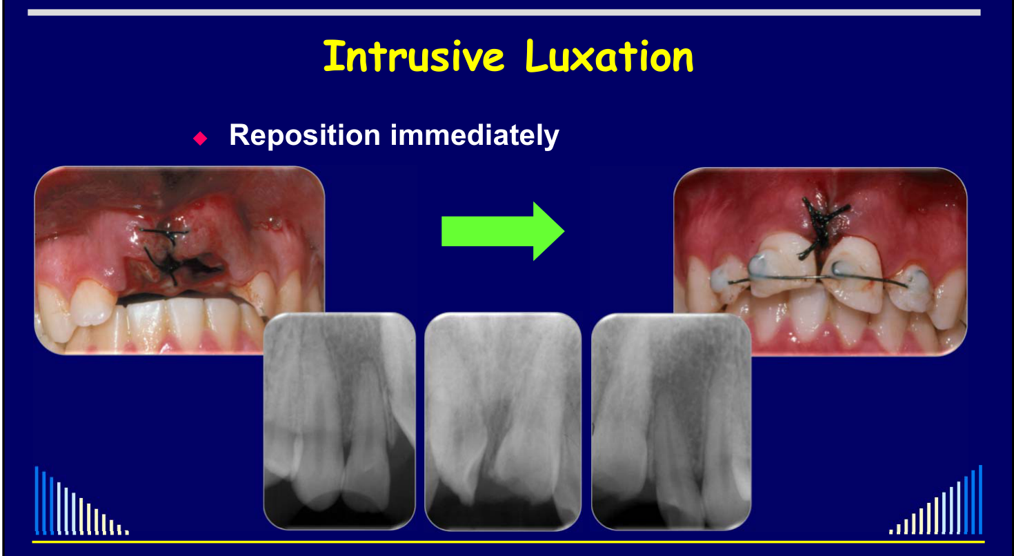
Reposition immediately
- Other options:
- Allow to re-erupt spontaneously
- Orthodontic extrusion
- Other options:
-
Reposition immediately
- Other options:
- Allow to re-erupt spontaneously
- Only likely if incompletely developed tooth
- Orthodontic extrusion
- Later - if no re-eruption spontaneously
- BUT this complicates other early management
- Endodontic treatment
- Allow to re-erupt spontaneously
- Other options:
 |  |
 |
Reasons for Immediate Repositioning20
- Reposition immediately
- Reasons:
- Avoids pressure necrosis of the PDL
- Reduce chances of external replacement resorption
- Fully developed teeth are not very likely to erupt
- Allows access to the root canal
- NB: Some teeth need IMMEDIATE endodontic treatment to prevent inflammatory resorption
- All MATURE teeth
- IMMATURE teeth with concurrent crown fracture
- NB: Some teeth need IMMEDIATE endodontic treatment to prevent inflammatory resorption
- Avoids surgery for orthodontic extrusion
- To attach brackets, wires
- Avoids pressure necrosis of the PDL
- Reasons:
Technique Considerations
- Reposition immediately
- Be careful not to damage the CEJ region
- Avoid using extraction forceps in this region !!
- HINT: use diamond-tipped tweezers
- Be careful not to damage the CEJ region
Intensiv — “Tweezers with diamond-tipped points” — Code: 1001A
- Reposition immediately
- Be careful not to damage the CEJ region
- Avoid using extraction forceps in this region !!
- HINT: use diamond-tipped tweezers
- Acid-etch/bond a small amount of composite to the labial and palatal surfaces to allow forceps or tweezers to hold the tooth without slipping
- Often intruded teeth are also rotated
- May be able to hold the Mesial and Distal surfaces of the crown without slipping as they are almost parallel
- Be careful not to damage the CEJ region
Associated Management Steps21
- Reposition immediately
- Mould bone back into position
- NB: fractured socket wall
- Splint - Rigid
- Since also have a bone fracture
- 6 weeks
- Since also have a bone fracture
- Suture soft tissues
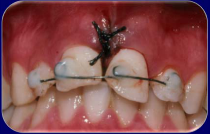
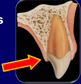
Lecturer — Palatal Degloving
Palatal tissues should be checked for degloving after repositioning.
- If the tissue moves or its attachment is uncertain, place sutures.
- Failure of healing may result in a permanent periodontal defect.
 |  |
Endodontic Treatment22
- Endodontic treatment
- Immediately - to prevent external inflammatory resorption - for:
- MATURE teeth
- IMMATURE teeth WITH a crown fracture
- ALSO: Antibiotics for these cases (for 1 week)
- Immediately - to prevent external inflammatory resorption - for:
- If IMMATURE tooth and NO crown fracture
- Monitor the pulp to assess if it recovers / heals
- Review regularly
Lecturer — Immature Recovery
In an immature tooth without a crown fracture, root canal treatment is not started immediately.
- The tooth is monitored for revascularization and healing.
- Approximately 35–40% may recover.
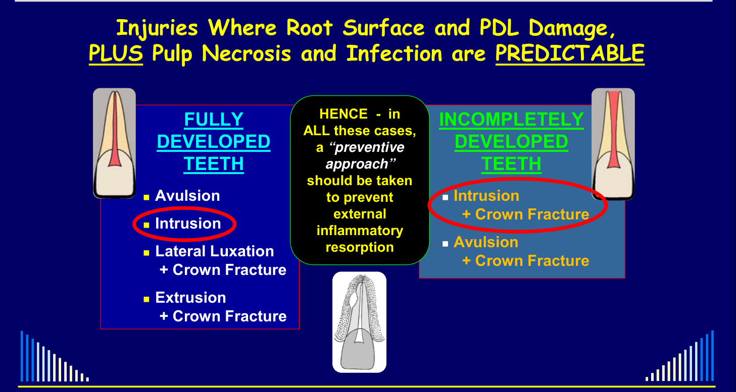
Injuries Where Root Surface and PDL Damage, PLUS Pulp Necrosis and Infection are PREDICTABLE
FULLY DEVELOPED TEETH
- Avulsion
- Intrusion
- Lateral Luxation + Crown Fracture
- Extrusion + Crown Fracture
HENCE - in ALL these cases, a “preventive approach” should be taken to prevent external inflammatory resorption
INCOMPLETELY DEVELOPED TEETH
- Intrusion + Crown Fracture
- Avulsion + Crown Fracture
Preventing External Inflammatory Resorption
Preventive Management — Mature and Immature Teeth23
- IMMEDIATELY after the emergency treatment
- Start Systemic antibiotics - Tetracycline preferred
- e.g. Doxycycline 100mg, 2 tabs. on 1st day, then 1 tab. daily for 1 week
- If tetracycline cannot be used, phenoxymethyl penicillin or amoxicillin is the second choice
- Start Systemic antibiotics - Tetracycline preferred
- IMMEDIATELY after replanting / repositioning & splinting
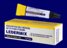
- Remove the pulp
- Prepare the canal if time available - otherwise do it at the next visit
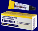
- Place a CS-AB paste dressing
 |  |
Preventive Management — Mature Teeth
- MATURE teeth - After 6 weeks
- Re-dress canal with the CS-AB paste
- MATURE teeth - After another 6 weeks
- Take PA radiograph
- If resorption present: Re-dress canal with the CS-AB paste
- If no resorption: Place new dressing using 50:50 CS-AB + Ca(OH)₂
- Take PA radiograph
- MATURE teeth - After a further 2 months
- Take PA radiograph
- If no resorption: Complete the Root Canal Filling (GP + cement)
- Take PA radiograph
Preventive Management — Immature Teeth24
- IMMATURE teeth - After 4 weeks
- Re-dress canal with the CS-AB paste
- IMMATURE teeth - After a further 4 weeks
- Re-dress canal with the CS-AB paste
- IMMATURE teeth - After another 4 weeks
- Take PA radiograph
- If resorption present: Re-dress canal with the CS-AB paste
- If no resorption: Place new dressing using 50:50 CS-AB + Ca(OH)₂
- Take PA radiograph
- IMMATURE teeth - After a further 2 months
- Take PA radiograph
- If no resorption:
- An apical hard tissue barrier is desirable
- Re-dress canal with Ca(OH)₂ paste
- Change Ca(OH)₂ dressing every 3 months
- Until hard tissue repair / apexification evident
- Complete the Root Canal Filling (GP + cement)
- An apical hard tissue barrier is desirable
- If no resorption:
- Take PA radiograph
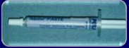
Preventive Management — All Cases25
- ALL Cases
- Clinical and radiographic reviews are essential
- At 6 months after the RCF is completed
- Then every 3-4 years
- For as long as possible !!!
Lecturer — Follow-up Schedule
Intruded teeth should generally be reviewed at six months and twelve months, then yearly for the first five years.
- Longer-term follow-up may be needed depending on the injury.

Prognosis and Clinical Case Examples
Prognosis of Intrusive Luxation
Prognosis - is generally fair
Depends on the many mechanical and biological factors - such as the stage of root development, pulp involvement, concurrent injuries, etc. - as previously discussed
| Outcome | Frequency |
|---|---|
| Pulp Necrosis | 96 % |
| PCC | 4 % |
| TAB | 12% |
| Resorption | 52 % |
| Bone loss | 6 - 31 % |
| Arrested root develᵐ | 16 % |
- These figures are a general guide ONLY
- They are based on retrospective analyses of many teeth
- They should NOT be used to predict the outcome for an individual tooth
- They are NOT the probability of any of the listed outcomes / sequelae
Lecturer — Additional Outcomes
Other possible outcomes include pulp canal calcification and gingival recession. External replacement resorption is a major problem and is related to the damage caused during the injury.
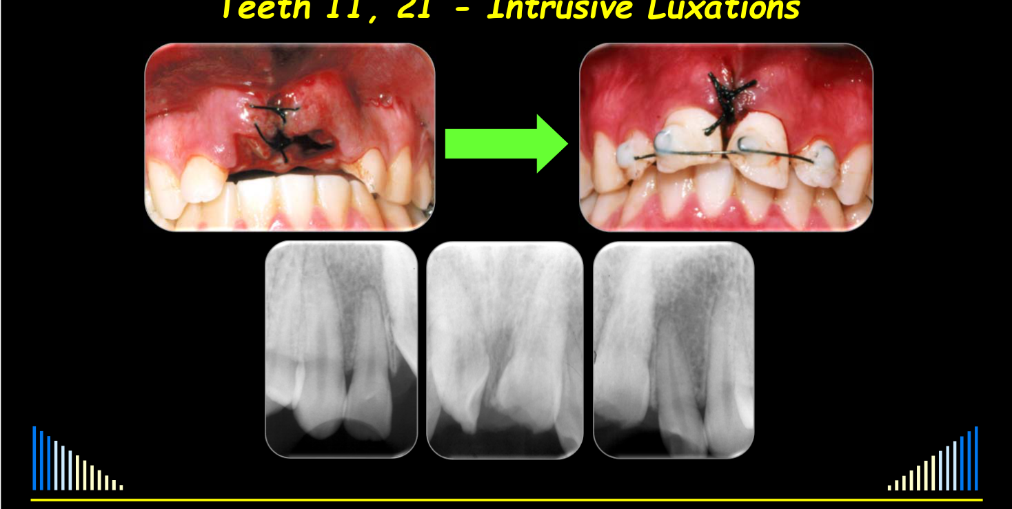
Teeth 11, 21 - Intrusive Luxations26
Teeth 11, 21 - Intrusive Luxations
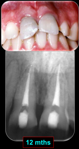
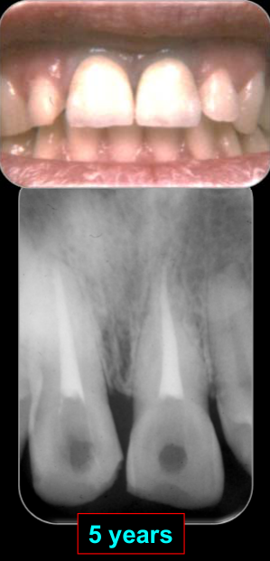
Teeth 11 and 21
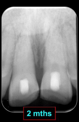
Two fully developed teeth were intruded and rotated and also had uncomplicated crown fractures. The teeth were repositioned, stabilized, and treated with root canal treatment.
- At twelve months and again at five years, there were no signs of external inflammatory resorption or external replacement resorption.
 |  |
 |  |
Avulsion
Related topic index: Intrusion and Avulsion.
Emergency Treatment of Avulsion
- Infractions
- Crown Fractures
- Crown : Root Fractures
- Root Fractures
- Concussion + Subluxation
- Extrusive Luxation
- Lateral Luxation
- Intrusive Luxation
- Avulsion
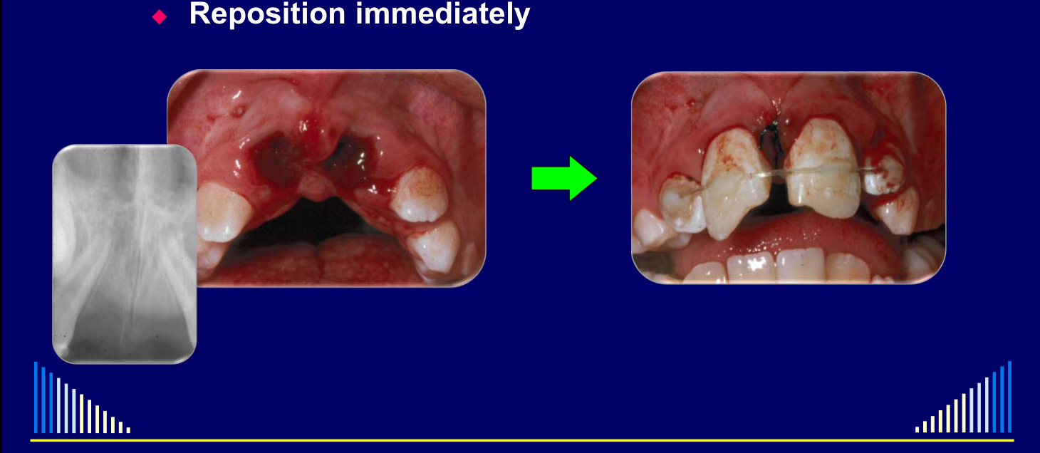
Avulsion27
-
Reposition immediately
-
Reposition immediately
-
Mould bone back into position
- May be socket wall fracture
-
Splint
- Flexible – if no bone fracture
- 7 – 10 days
- Flexible – if no bone fracture
-
Suture soft tissues
Lecturer — Splinting and Tissue Assessment
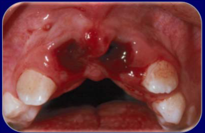
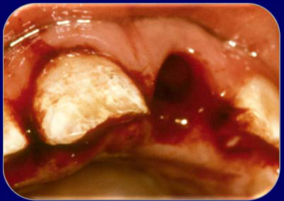
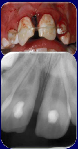
Flexible splinting for approximately seven to ten days permits movement, which helps reduce ankylosis and external replacement resorption.
- Examine the palatal tissues and the tissue between multiple avulsed teeth for tears or detachment.
 |  |
 |
Avulsion — Follow-up Management28
- Endodontic treatment
- Immediately – to prevent external inflammatory resorption – for:
- MATURE tooth
- IMMATURE tooth WITH a crown fracture
- ALSO: Antibiotics for these cases (for 1 week)
- Immediately – to prevent external inflammatory resorption – for:
- If IMMATURE tooth and NO crown fracture
- Monitor pulp to assess if it recovers / heals
- Review regularly
Lecturer — Follow-up Assessment
Patients require regular review according to the trauma schedule.
- Follow-up assesses pulp healing or necrosis, periodontal ligament healing, resorption, bone loss, and splint and soft-tissue healing.
- The one-week review may include removal of the flexible splint and sutures.
- In a fully developed tooth, root canal treatment should already have been started by this appointment.
Preventive Protocol for Predictable Pulp Necrosis
Injuries Where Root Surface and PDL Damage, PLUS Pulp Necrosis and Infection are PREDICTABLE29
See - Shared predictable-risk combinations
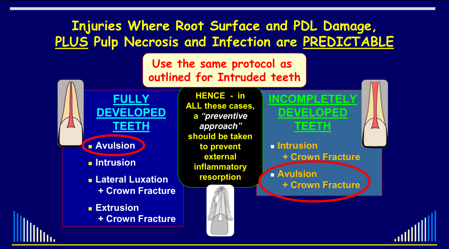 |  |
Fully Developed Teeth
See - Mature-tooth injury combinations
Incompletely Developed Teeth
See - Immature-tooth injury combinations
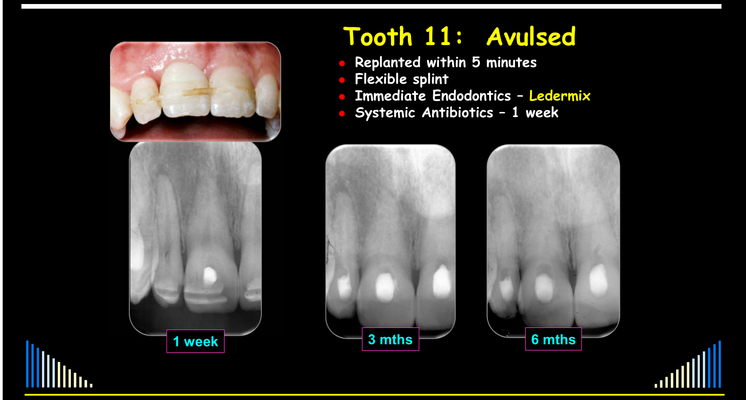
Protocol for Avulsion (Fully Developed Teeth)
- Replanted within 5 minutes
- Flexible splint
- Immediate Endodontics – Ledermix
- Systemic Antibiotics – 1 week
Review schedule: 1 week, 3 mths, 6 mths
Review schedule (continued): 9 mths, 3 years, 5 years
Avulsion — Endodontic Treatment30
Endodontic treatment immediately – to prevent external inflammatory resorption:
- → MATURE tooth
- → IMMATURE tooth WITH a crown fracture
ALSO: Antibiotics for these cases (for 1 week)
Fishing-Line Splint Case
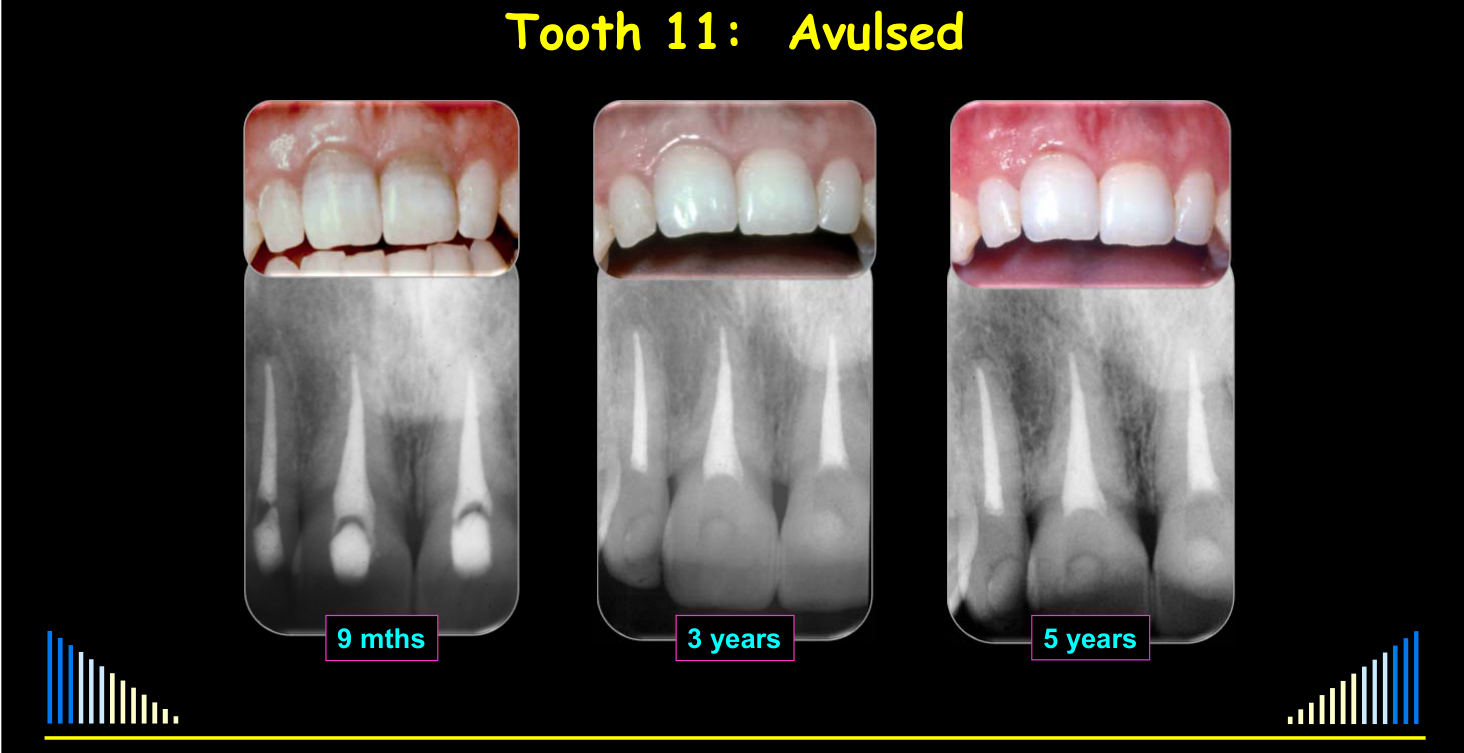
In one case, a general dentist replanted an avulsed central incisor, used a fishing-line and composite flexible splint, started root canal treatment, and administered systemic antibiotics.
- At nine months, discoloration was treated with internal bleaching.
- The tooth was documented at three years and five years.
- It eventually developed external replacement resorption, with early periodontal ligament loss, ankylosis, and progressive replacement resorption.
- Approximately twelve years later, the remaining tooth structure had to be removed and replaced with an implant.
If IMMATURE tooth and NO crown fracture:
- Monitor pulp to assess if it recovers / heals
Lecturer — Immature Tooth Recovery
Approximately 35–40% of immature teeth without a crown fracture may recover through pulp revascularization, particularly when extra-alveolar time is short.
- Teeth that do not recover will eventually require root canal treatment.
Review regularly.
Avulsion Case Examples and Prognosis
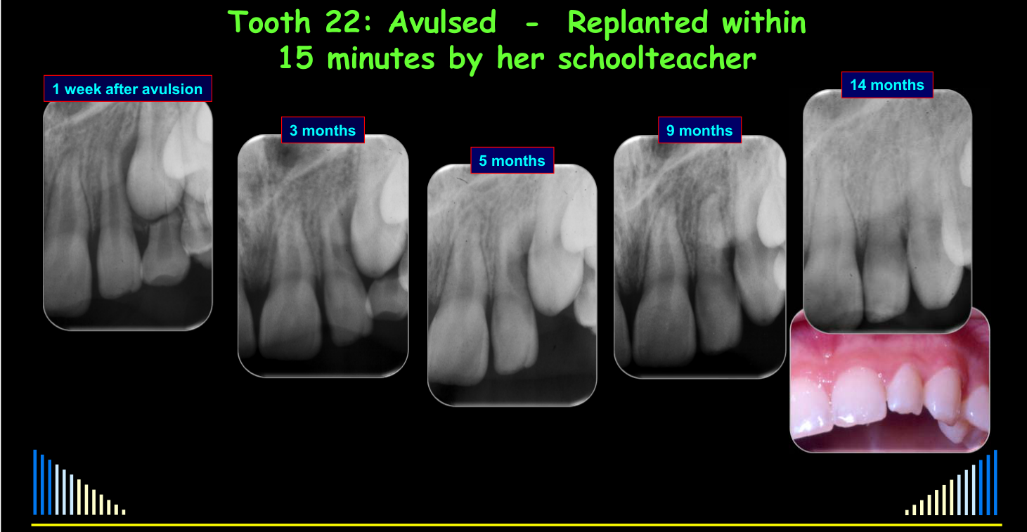
Tooth 22: Avulsed – Replanted within 15 minutes by her schoolteacher31
Follow-up images: 1 week after avulsion, 3 months, 5 months, 9 months, 14 months
Tooth 22 Revascularization
This immature lateral incisor was replanted by a schoolteacher approximately fifteen minutes after avulsion, and a dentist placed a simple splint for one week.
- Because the tooth had an open apex and a wide canal, it was monitored for revascularization.
- Root development continued: canal calcification was beginning by approximately five months, and by fourteen months the canal was substantially narrower with an almost fully developed, closed apex.
- No endodontic treatment was required during the described follow-up.
 | 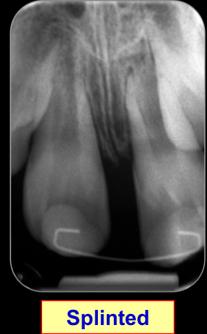 |
Tooth 21: Avulsed – out of the socket for 2 hours – stored in the buccal sulcus
Follow-up images: Replanted, Splinted, 7 Months
Dr. Tony Sandler
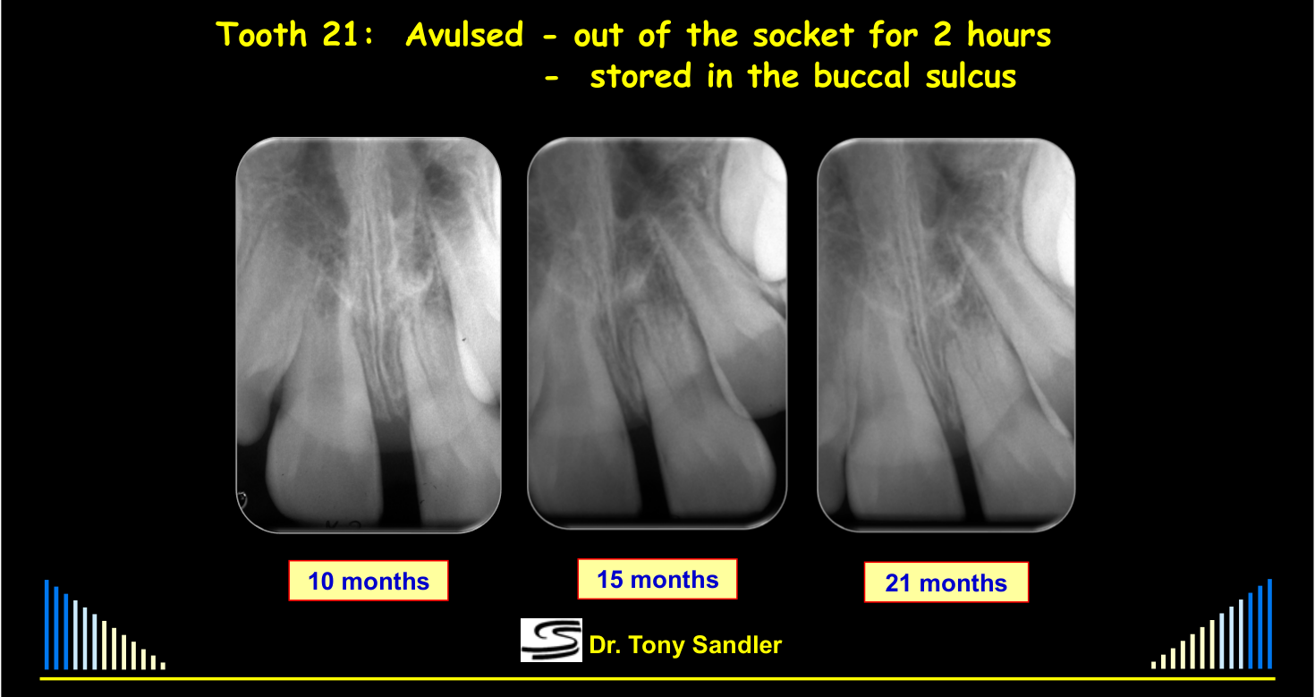
Follow-up images: 10 months, 15 months, 21 months
Dr. Tony Sandler
Tooth 21 Canal Bone Formation
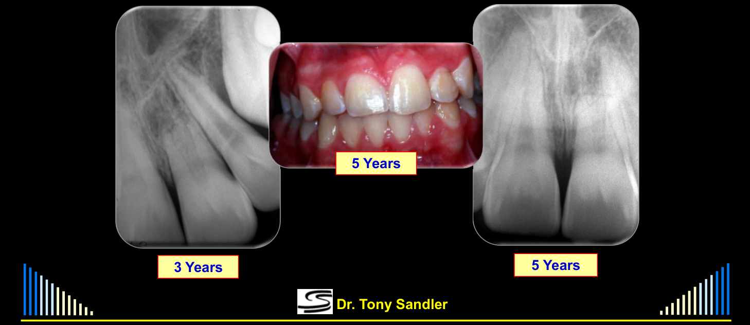
The tooth was stored in the buccal sulcus for two hours, replanted by a general dentist, and stabilized with a flexible wire splint.
- At approximately seven months, bone had grown down into the root canal space, indicating that the pulp had not revascularized but bone had entered the canal.
- The radiographic appearance suggested internal periodontal ligament-like tissue.
- The tooth appeared clinically normal at five years, although some bone loss and a higher gingival margin were present; the root was shorter but had substantial internal and external bone support.
Tooth 21: Avulsed – out of the socket for 2 hours – stored in the buccal sulcus (long-term)32
Follow-up images: 3 Years, 5 Years, 5 Years
Dr. Tony Sandler
Tooth 21 Long-Term Response
Although the pulp did not appear to revascularize, bone formation within the canal allowed the tooth to remain clinically functional and supported.
- The case demonstrated that a replanted tooth may remain in place for a considerable period even when the eventual biological response is not normal.
Prognosis
Prognosis – is fair.
Depends on the many mechanical and biological factors – such as the stage of root development, pulp involvement, concurrent injuries, etc. – as previously discussed.
| Sequela | Frequency |
|---|---|
| Pulp Necrosis | up to 100 % |
| PCC | 4 % |
| Resorption | 74 – 96 % |
| Bone loss | 6 – 31 % |
- These figures are a general guide ONLY
- They are based on retrospective analyses of many teeth
- They should NOT be used to predict the outcome for an individual tooth
- They are NOT the probability of any of the listed outcomes / sequelae
Lecturer — Replantation Rationale
Many avulsed teeth are eventually lost, but replantation remains worthwhile because it preserves the alveolar bone and maintains future options for prosthetic replacement.
- In immature teeth, approximately 35–40% may retain a surviving pulp, and pulp canal calcification commonly occurs when the pulp survives.
- External inflammatory resorption can be managed more effectively when treated early or immediately.
 |  |
Overview of Traumatic Dental Injuries
- Infractions
- Crown Fractures
- Crown : Root Fractures
- Root Fractures — See separate lecture
- Concussion + Subluxation
- Extrusive Luxation
- Lateral Luxation
- Intrusive Luxation
- Avulsion
Lecturer — Immediate Storage
Avulsion, together with intrusion, is among the most severe dental injuries.
- The injured person or someone present, such as a parent, friend, coach, or teacher, should replant the tooth as soon as possible.
- If immediate replantation is not possible, place the tooth in milk, saliva in a container, or plastic wrap.
- On presentation, assess the patient while keeping the tooth in milk or saline.
Summary33
-
Full assessment of the injury is essential
- How, when, why, and where it happened
- History, examination, tests, radiographs
- Assess priorities for treatment
- Establish the history and mechanism, identify likely injuries, and perform a thorough clinical examination
- Complete pulp testing and trauma radiographs
- Determine treatment priorities when multiple injuries are present
-
Reposition and stabilise ALL tissues
- Assess priorities for each tooth / injury
- Stabilize teeth and bone with splints
- Repair soft tissues with sutures
- Assess priorities for each tooth / injury
-
Allow time for healing
- Preserve the pulp whenever possible, especially in immature teeth
- PDL: 3 - 6 months
- Pulp: 3 - 24 months
-
Correct IMMEDIATE and EARLY management of traumatised teeth will significantly improve their long-term prognosis
- Initiate immediate root canal treatment only when necessary, including injuries with predictable external inflammatory resorption or major pulp exposure
- Provide systemic antibiotics when root-surface and periodontal-ligament damage occur with predictable pulp necrosis and infection
-
Correct FOLLOW-UP management is also important to improve prognosis
- Complications may appear months or years after the injury, so follow-up remains essential
-
DEFER all irreversible treatment and final restorations for as long as possible
- To improve the prognosis, and
- To allow the prognosis to be reassessed and confirmed
- Use temporary restorations when appropriate and defer definitive irreversible treatment until the pulp response is known
DMD - 3rd Year - 2020
Clinical Cases
Case: Uncomplicated crown fracture restored with glass ionomer
Question
Scenario: A tooth with an uncomplicated crown/root fracture is evaluated and managed conservatively.
What’s shown: A radiograph and a clinical photo taken about six months after the initial restoration.
Consider: What was the initial management approach, and what was the subsequent clinical outcome for the pulp?
Answer
Observations:
- The tooth was initially restored with a glass ionomer restoration.
- The restoration remained intact over a period of months.
- The pulp necrosed and became infected within a couple of months of the trauma.
Reasoning: The glass ionomer restoration served as a temporary measure while monitoring the tooth, remaining stable even during subsequent root canal treatment.
Outcome: The tooth required root canal treatment due to pulp necrosis and infection a couple of months post-trauma. The glass ionomer restoration remained intact throughout the root canal treatment period.
Takeaway: A simple glass ionomer restoration can remain stable for a long period while monitoring a traumatized tooth, even if root canal treatment later becomes necessary.
Case: Complicated crown root fracture with a mobile palatal fragment
Question
Scenario: A tooth with a complicated crown root fracture involving deep subgingival margins is evaluated.
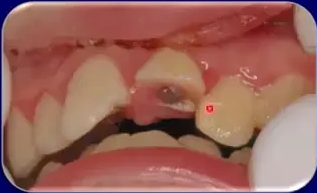
What’s shown: A clinical view showing considerable loss of the crown and a small, mobile piece on the palatal aspect.
Consider: What immediate step is required regarding the mobile palatal fragment to properly assess the injury?
Answer
Observations:
- A considerable amount of the crown is lost.
- A small piece on the palatal aspect is mobile and held in place only by the periodontal ligament.
Reasoning: The mobile fragment needs to be removed to properly determine how deep the fracture margin extends subgingivally.
Outcome: The mobile palatal piece requires removal to assess the fracture depth. The management of this complicated crown root fracture is similar to a supracrestal root fracture in the coronal third of the root.
Takeaway: In complicated crown root fractures, mobile fragments held only by the periodontal ligament must be removed to accurately determine the depth of the fracture margin.
Case: Vertical crown root fracture with a failed composite splint
Question
Scenario: A tooth with a vertical crown root fracture is evaluated.
What’s shown: An example of a vertical split where a previous dentist attempted to splint the fragments together using composite resin.
Consider: Why did the previous restorative intervention fail, and what is the definitive management for this tooth?
Answer
Observations:
- There is a vertical split in the tooth.
- Composite resin was used to splint the pieces together, but it has fractured.
Reasoning: The splint failed because the fracture extends beyond the level where composite can be effectively placed. Movement of the tooth fragments causes the composite to fracture.
Outcome: The previous treatment is ineffective; the only management for this type of vertical crown root fracture is extraction.
Takeaway: Splinting a vertical crown root fracture with composite is ineffective if the fracture extends beyond the restorable level, and extraction is the only treatment.
Case: Lower molar with a vertical split from indirect trauma
Question
Scenario: A soldier fainted on parade, hitting his chin on the bitumen and causing indirect trauma to his dentition.
What’s shown: A lower molar with a complete vertical split.
Consider: What predisposed this tooth to fracture under indirect trauma, and what is the required treatment?
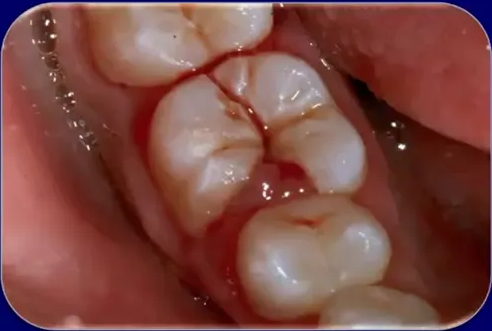
Answer
Observations:
- The mandible was forced against the maxilla during the impact.
- The tooth was weakened by a large carious lesion.
- There is a complete vertical split of the tooth.
Reasoning: The indirect trauma forced the teeth together, and the pre-existing large carious lesion weakened the tooth, leading to a complete vertical split (a complicated crown root fracture).
Outcome: The only way to manage this tooth is extraction.
Takeaway: Indirect trauma can cause a complete vertical split in a tooth weakened by a large carious lesion, necessitating extraction.
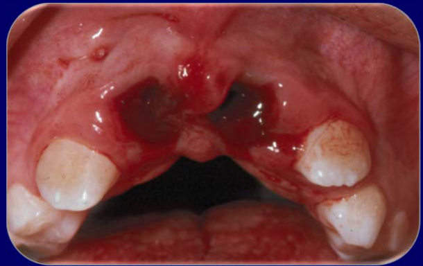
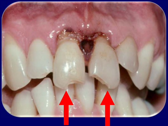
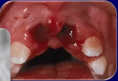
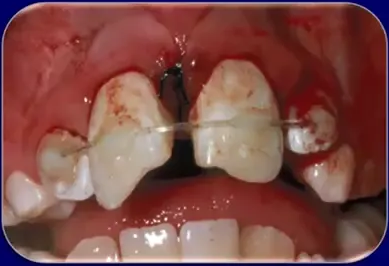
Case: Extrusive luxation with crown fractures presenting 10 days post-injury
Question
Scenario: A patient presents with upper anterior teeth that have been displaced axially out of their sockets, along with uncomplicated crown fractures.
What’s shown: A clinical photograph showing the teeth out of normal position, with the patient having delayed treatment for about 10 days.
Consider: How has the delay in presentation affected the clinical appearance of the surrounding tissues and the treatment options?
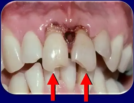
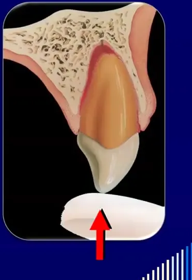
Answer
Observations:
- The teeth are moved in an axial direction out of the socket, appearing longer than adjacent teeth.
- The patient also has uncomplicated crown fractures.
- Soft tissue is in the healing process because the patient did not attend for treatment for about 10 days.
Reasoning: The delayed presentation of 10 days allows the soft tissue to begin healing in the displaced position, and a blood clot forms in the base of the socket, making simple manual repositioning impossible.
Outcome: The teeth are out of normal position with healing soft tissue. Because of the delay, the teeth can only be repositioned orthodontically.
Takeaway: Delayed presentation of extrusive luxation allows soft tissue to heal and a blood clot to form in the displaced position, meaning simple repositioning is no longer possible and orthodontic treatment is required.
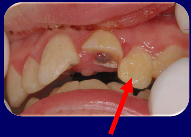
Case: Complicated crown root fracture and lateral luxation of anterior teeth
Question
Scenario: A patient presents with a complicated crown root fracture of a central incisor and a laterally luxated lateral incisor.
What’s shown: Clinical photographs showing the isolated upper anterior teeth, revealing the fractured central incisor and the displaced lateral incisor.
Consider: What is the appropriate emergency management and treatment plan for these injuries?
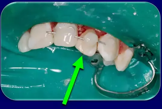
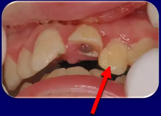
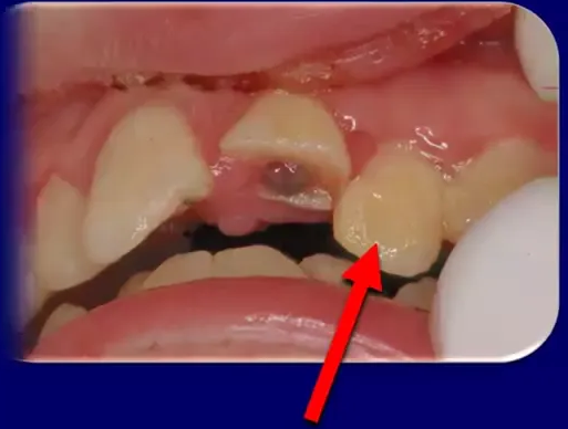
Answer
Observations:
- Complicated crown root fracture of the central incisor.
- Lateral luxation of the lateral incisor.
- The lateral incisor has been repositioned and splinted with a rigid wire and composite resin splint.
Reasoning:
- Lateral luxation requires immediate repositioning to prevent a blood clot from forming in the socket, which would make repositioning difficult.
- Repositioning requires two movements: pushing down from the apical end to disengage the root from the bony lock on the labial socket wall, and then pushing the crown labially.
- Because there is a bone fracture associated with lateral luxation, a rigid splint (wire and composite resin) is required, typically for 4 to 6 weeks.
- In a fully developed tooth with lateral luxation and a concurrent crown fracture, root canal treatment should be commenced immediately to prevent external inflammatory resorption.
Outcome:
- The lateral incisor was repositioned and splinted with a rigid wire and composite resin splint.
- Root canal treatment was commenced on the day of the injury.
Takeaway:
- Lateral luxation requires immediate two-step repositioning to overcome the bony lock, followed by rigid splinting; immediate root canal treatment is indicated in mature teeth with concurrent crown fractures to prevent inflammatory resorption.
Case: Lateral luxation of immature teeth in a young patient
Question
Scenario: A young patient with immature teeth and mixed dentition presents with laterally luxated anterior teeth.
What’s shown: Clinical photographs from an incisal view showing the crowns of the teeth pushed back palatally with bleeding around the gingival margin.
Consider: How should the splinting be managed when a partially erupted tooth is present in the splinting span?
Answer
Observations:
- Immature teeth with lateral luxation (crowns displaced palatally).
- Bleeding around the gingival margin.
- Presence of a partially erupted lateral incisor and deciduous teeth.
- A wire and composite splint placed with the wire anchored on the deciduous canine, avoiding the partially erupted lateral incisor.
Reasoning:
- The teeth need to be repositioned and splinted.
- Bending the splint wire to fit around a partially erupted tooth is complicated and could impede its continued eruption.
- Anchoring the wire on the adjacent deciduous canine creates a slightly longer splint span.
- To compensate for the longer span and maintain rigidity, the composite resin is extended further mesiodistally.
Outcome:
- The teeth were repositioned and splinted using a wire and composite resin, with the wire placed on the deciduous canine to avoid the partially erupted lateral incisor.
Takeaway:
- When splinting in a mixed dentition with partially erupted teeth, the splint can be anchored to adjacent deciduous teeth to allow for continued eruption, and the composite resin can be extended to maintain the rigidity of a longer splint span.
Case: Intrusive luxation of upper anterior teeth with crown fractures
Question
Scenario: A patient presents with intruded upper anterior teeth that were initially left in place by a dentist who did not recognize the injury or take radiographs. The teeth are also rotated and have concurrent crown fractures.
What’s shown: Clinical figures showing the intruded teeth left in their original position, followed by images illustrating the immediate repositioning technique and subsequent stabilization.
Consider: How should this intrusive luxation injury be managed, including repositioning, stabilization, and endodontic treatment, to prevent complications?
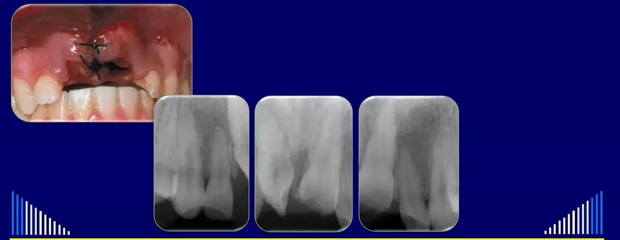
Answer
Observations:
- The teeth are severely intruded into the alveolar bone, rotated, and have concurrent crown fractures.
- The initial dentist failed to recognize the injury and did not take radiographs, leaving the teeth in the intruded position.
- Repositioning requires careful technique to avoid damaging the cementoenamel junction (CEJ).
Reasoning:
- Intrusion involves the entire root being forced against the bone, crushing the periodontal ligament (PDL), cementum, and bone (often fracturing the labial plate).
- Immediate repositioning is preferred over waiting for spontaneous re-eruption (unlikely in mature teeth) or orthodontic extrusion, as it avoids pressure necrosis of the PDL, reduces the risk of external replacement resorption, and allows immediate access for endodontic treatment without needing surgical exposure.
- Extraction forceps should not be used as they can damage the CEJ and predispose to external replacement or invasive resorption; instead, diamond-tipped tweezers or composite bonded to the tooth should be used for grip.
- Because the teeth are mature (or immature with concurrent crown fractures), pulp necrosis is predictable, necessitating immediate root canal treatment to prevent external inflammatory resorption.
Outcome:
- The teeth were immediately repositioned, and the fractured labial bone was moulded back into place.
- A rigid splint was applied (typically 4 weeks for a younger child, 6 weeks for an adult).
- Soft tissues were sutured, with careful attention to the palatal aspect to address any degloving injuries.
- Immediate root canal treatment was initiated (access cavity, pulp removal with a bar broach, irrigation, and calcium hydroxide paste placement) along with a one-week course of systemic antibiotics.
Takeaway:
- Intrusive luxations should be repositioned immediately to prevent PDL pressure necrosis and allow prompt endodontic access, using instruments that protect the CEJ, followed by rigid splinting and preventive root canal treatment for mature teeth or immature teeth with crown fractures.
Case: Intrusive luxation of teeth 11 and 21
Question
Scenario: A patient presents with intrusive luxation of the maxillary central incisors (teeth 11 and 21), which are rotated and have uncomplicated crown fractures, with fully developed roots.
What’s shown: Intraoral clinical photographs showing the intruded and rotated teeth, and radiographs at initial presentation, 12 months, and 5 years.
Consider: The emergency management, splinting, soft tissue care, and endodontic protocol to prevent resorption.
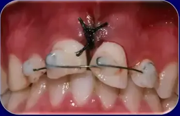
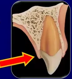
Answer
Observations:
- Teeth 11 and 21 are intruded and rotated.
- Uncomplicated crown fractures are present.
- Roots are fully developed.
- Radiographs at 12 months and 5 years show no signs of external inflammatory or replacement resorption.
Reasoning: Immediate repositioning and moulding of the alveolar bone (especially the labial aspect) is recommended. A rigid splint is used for 4 to 6 weeks due to the likely alveolar bone fracture. Soft tissues, particularly on the palatal aspect, must be checked for degloving and sutured to prevent permanent periodontal defects. Because the teeth are fully developed, root canal treatment is started immediately to prevent external inflammatory resorption, and systemic antibiotics are prescribed.
Outcome: The teeth were repositioned, splinted, sutured, and root canal treatment was initiated. At 12 months and 5 years follow-up, there were no signs of external inflammatory or replacement resorption.
Takeaway: Intrusive luxation requires immediate repositioning, rigid splinting, careful soft tissue management, and immediate root canal treatment for mature teeth to prevent external inflammatory resorption.
Case: Avulsion of two teeth
Question
Scenario: A patient presents with two avulsed teeth and torn soft tissues between them.
What’s shown: Clinical intraoral photographs showing the avulsed teeth and tissue tearing, and a periapical radiograph taken at the one-week review.
Consider: The emergency management of avulsed teeth, including replantation, splinting, soft tissue repair, and endodontic management.
Answer
Observations:
- Two avulsed teeth with tissue tearing between them.
- Radiograph at one week shows root canal treatment has already been started.
Reasoning: The tooth should be replanted immediately or stored in an appropriate medium. Upon presentation, the tooth is replanted and the bone is moulded back into position. A flexible splint is used for 7 to 10 days (if no bone fracture) to allow movement and reduce ankylosis and replacement resorption. Soft tissues are sutured. For fully developed teeth, root canal treatment is started immediately to prevent external inflammatory resorption, and systemic antibiotics are prescribed.
Outcome: The teeth were replanted, splinted, and sutured. Root canal treatment was started on the day of the accident. At the one-week review, splints and sutures were removed, and root canal treatment was ongoing.
Takeaway: Avulsion requires immediate replantation, flexible splinting (unless a bone fracture is present), soft tissue suturing, and immediate root canal treatment for mature teeth to prevent external inflammatory resorption.
Case: Avulsed central incisor followed for 12 years
Question
Scenario: A patient with an avulsed central incisor that was replanted, splinted with a flexible fishing line and composite splint, and had root canal treatment commenced by a general dentist on the day of the accident.
What’s shown: Clinical and radiographic images showing the access cavity, a radiograph at one week, discoloration at nine months, and follow-ups at three and five years, eventually showing loss of the periodontal ligament on the distal aspect.
Consider: What are the long-term outcomes and potential complications of replanting an avulsed tooth?
Answer
Observations:
- Access cavity cut and root canal treatment commenced on the day of the accident, with systemic antibiotics given.
- Radiograph at one week shows continued root canal treatment.
- Discoloration at nine months required internal bleaching.
- Early loss of the periodontal ligament on the distal aspect.
- Progression to ankylosis and external replacement resorption.
Reasoning: The loss of the periodontal ligament led to ankylosis, which progressed to external replacement resorption over time, demonstrating a consequence of avulsion injuries.
Outcome: The tooth underwent external replacement resorption and was eventually removed after 12 years, followed by the placement of an implant.
Takeaway: Replanted avulsed teeth are not necessarily permanent and can undergo external replacement resorption, highlighting the long-term consequences of such injuries.
Case: Avulsed immature lateral incisor with pulp revascularization
Question
Scenario: A patient with an avulsed immature lateral incisor that was replanted within 15 minutes by a school teacher and splinted for a week.
What’s shown: Radiographs showing an open apex and wide canal initially, with follow-up images at five and 14 months showing calcification of the canal and further root development.
Consider: What is the expected outcome when monitoring an avulsed immature tooth with an open apex and no crown fracture?
Answer
Observations:
- Tooth replanted quickly (within 15 minutes) and splinted for a week.
- Open apex and wide canal present initially.
- At five months, calcification of the canal and further root development are evident.
- By 14 months, the canal is narrow and the apex is fully developed and closed.
Reasoning: The quick replantation and open apex allowed for pulp revascularization, leading to continued root development and pulp canal calcification.
Outcome: The tooth was simply monitored and reviewed, resulting in successful revascularization and root development without the need for further intervention.
Takeaway: Immature avulsed teeth with no crown fracture and a short extra-alveolar time can undergo pulp revascularization and continued root development if monitored appropriately.
Case: Avulsed central incisor stored in the buccal sulcus with bone ingrowth
Question
Scenario: A young patient who avulsed a central incisor, which was out of the socket for two hours and stored in the buccal sulcus before being replanted and splinted with a flexible wire splint by a general dentist.
What’s shown: Radiographs showing the replanted tooth with a flexible wire splint, follow-up at seven months, and subsequent radiographs over five years showing bone growing into the root canal space.
Consider: What can happen radiographically when an avulsed tooth is replanted after a prolonged extra-alveolar time and the pulp does not revascularize?
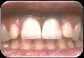
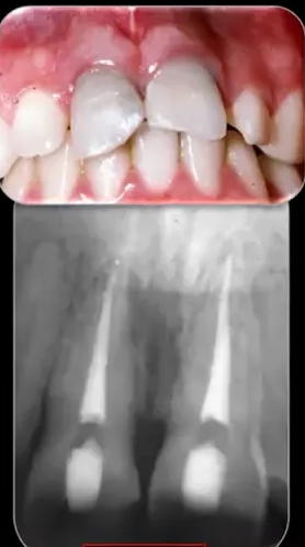
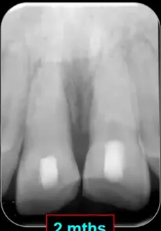
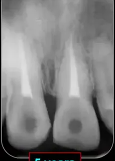
Answer
Observations:
- Tooth stored in the buccal sulcus for two hours before replantation.
- Flexible wire splint placed due to spacing between teeth.
- At seven months, the tooth is in position.
- Radiographs over five years show bone growing down into the root canal space.
- Radiographic appearance of an internal periodontal ligament.
- Clinically normal appearance at five years, though with some gingival margin discrepancy (gingival margin higher than adjacent tooth) and a shorter root.
Reasoning: The pulp did not revascularize, but instead, bone grew into the root canal space, providing internal and external bone support despite the shorter root.
Outcome: The case was followed up for five years with a clinically normal appearance and plenty of bone support internally and externally.
Takeaway: In some cases of avulsed teeth where the pulp does not revascularize, bone can grow into the root canal space, providing a stable long-term result with good bone support.
Footnotes
-
Original PDF page 1: L12 Dento-Alveolar Trauma 5 - Emergency Management of Traumatic Dental Injuries, p.1 ↩
-
Original PDF page 3: L12 Dento-Alveolar Trauma 5 - Emergency Management of Traumatic Dental Injuries, p.3 ↩
-
Original PDF page 4: L12 Dento-Alveolar Trauma 5 - Emergency Management of Traumatic Dental Injuries, p.4 ↩
-
Original PDF page 5: L12 Dento-Alveolar Trauma 5 - Emergency Management of Traumatic Dental Injuries, p.5 ↩
-
Original PDF page 6: L12 Dento-Alveolar Trauma 5 - Emergency Management of Traumatic Dental Injuries, p.6 ↩
-
Original PDF page 7: L12 Dento-Alveolar Trauma 5 - Emergency Management of Traumatic Dental Injuries, p.7 ↩
-
Original PDF page 9: L12 Dento-Alveolar Trauma 5 - Emergency Management of Traumatic Dental Injuries, p.9 ↩
-
Original PDF page 10: L12 Dento-Alveolar Trauma 5 - Emergency Management of Traumatic Dental Injuries, p.10 ↩
-
Original PDF page 8: L12 Dento-Alveolar Trauma 5 - Emergency Management of Traumatic Dental Injuries, p.8 ↩
-
Original PDF page 2: L12 Dento-Alveolar Trauma 5 - Emergency Management of Traumatic Dental Injuries, p.2 ↩
-
Original PDF page 12: L12 Dento-Alveolar Trauma 5 - Emergency Management of Traumatic Dental Injuries, p.12 ↩
-
Original PDF page 13: L12 Dento-Alveolar Trauma 5 - Emergency Management of Traumatic Dental Injuries, p.13 ↩
-
Original PDF page 14: L12 Dento-Alveolar Trauma 5 - Emergency Management of Traumatic Dental Injuries, p.14 ↩
-
Original PDF page 15: L12 Dento-Alveolar Trauma 5 - Emergency Management of Traumatic Dental Injuries, p.15 ↩
-
Original PDF page 16: L12 Dento-Alveolar Trauma 5 - Emergency Management of Traumatic Dental Injuries, p.16 ↩
-
Original PDF page 17: L12 Dento-Alveolar Trauma 5 - Emergency Management of Traumatic Dental Injuries, p.17 ↩
-
Original PDF page 18: L12 Dento-Alveolar Trauma 5 - Emergency Management of Traumatic Dental Injuries, p.18 ↩
-
Original PDF page 19: L12 Dento-Alveolar Trauma 5 - Emergency Management of Traumatic Dental Injuries, p.19 ↩
-
Original PDF page 20: L12 Dento-Alveolar Trauma 5 - Emergency Management of Traumatic Dental Injuries, p.20 ↩
-
Original PDF page 21: L12 Dento-Alveolar Trauma 5 - Emergency Management of Traumatic Dental Injuries, p.21 ↩
-
Original PDF page 22: L12 Dento-Alveolar Trauma 5 - Emergency Management of Traumatic Dental Injuries, p.22 ↩
-
Original PDF page 23: L12 Dento-Alveolar Trauma 5 - Emergency Management of Traumatic Dental Injuries, p.23 ↩
-
Original PDF page 24: L12 Dento-Alveolar Trauma 5 - Emergency Management of Traumatic Dental Injuries, p.24 ↩
-
Original PDF page 25: L12 Dento-Alveolar Trauma 5 - Emergency Management of Traumatic Dental Injuries, p.25 ↩
-
Original PDF page 26: L12 Dento-Alveolar Trauma 5 - Emergency Management of Traumatic Dental Injuries, p.26 ↩
-
Original PDF page 27: L12 Dento-Alveolar Trauma 5 - Emergency Management of Traumatic Dental Injuries, p.27 ↩
-
Original PDF page 28: L12 Dento-Alveolar Trauma 5 - Emergency Management of Traumatic Dental Injuries, p.28 ↩
-
Original PDF page 29: L12 Dento-Alveolar Trauma 5 - Emergency Management of Traumatic Dental Injuries, p.29 ↩
-
Original PDF page 30: L12 Dento-Alveolar Trauma 5 - Emergency Management of Traumatic Dental Injuries, p.30 ↩
-
Original PDF page 31: L12 Dento-Alveolar Trauma 5 - Emergency Management of Traumatic Dental Injuries, p.31 ↩
-
Original PDF page 32: L12 Dento-Alveolar Trauma 5 - Emergency Management of Traumatic Dental Injuries, p.32 ↩
-
Original PDF page 33: L12 Dento-Alveolar Trauma 5 - Emergency Management of Traumatic Dental Injuries, p.33 ↩
-
Original PDF page 35: L12 Dento-Alveolar Trauma 5 - Emergency Management of Traumatic Dental Injuries, p.35 ↩