L8 Vital Pulp Treatment 2
Vital Pulp Treatment12
Dr Mostafa Elkholy
Direct Pulp Capping3
Vital Pulp Treatment Modalities
Dr Mostafa Elkholy, BDS, MSc, PhD
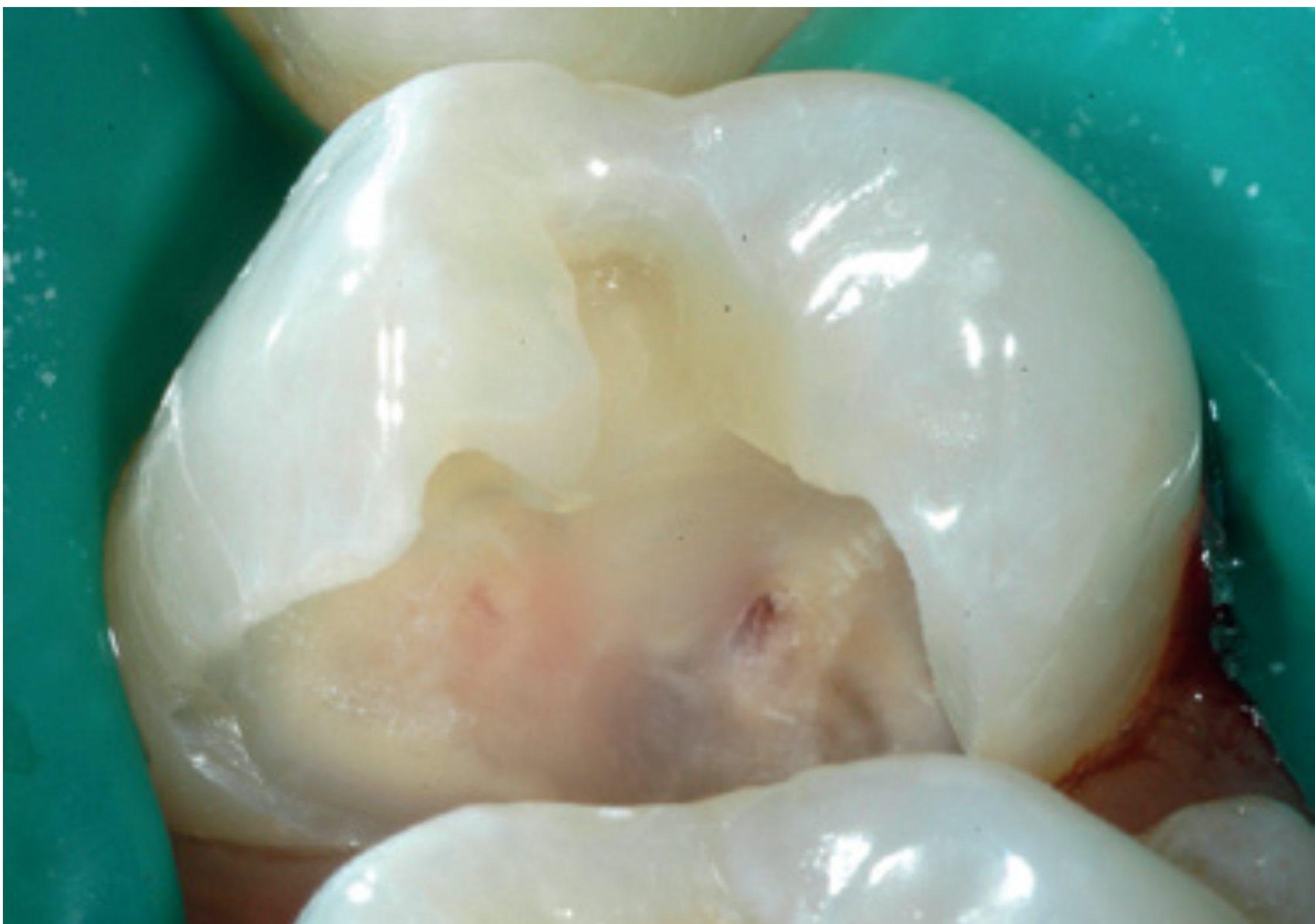 | 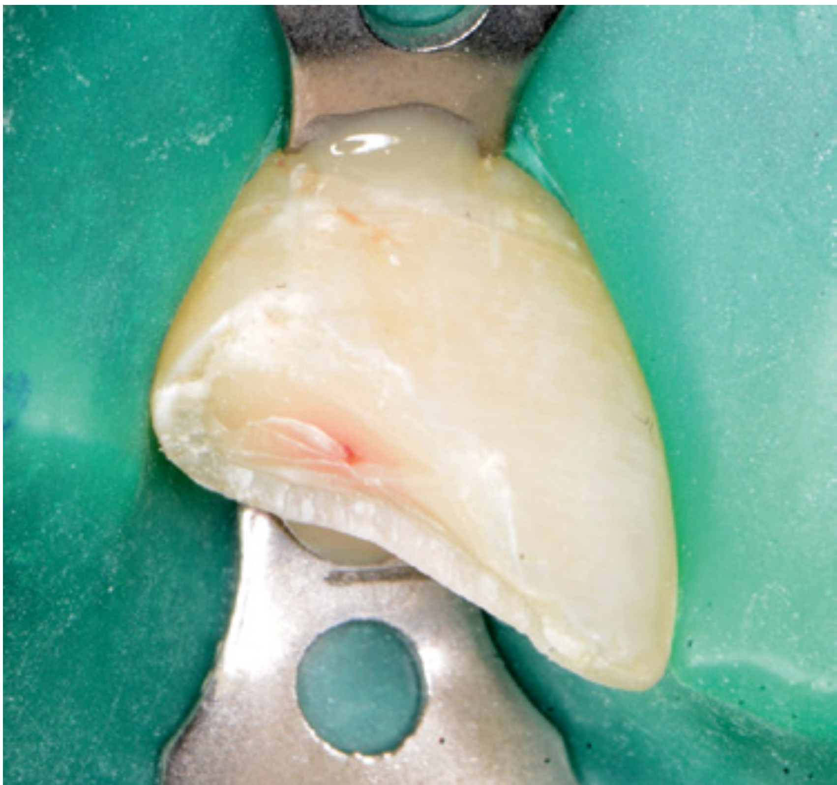 |
Indications4
- No spontaneous pain
- No lingering pain
- Bleeding can be stopped
Factors Affecting Outcomes
Patient Age56
- Patient age is not a contraindication for direct pulp capping, even though pulp tissue regeneration, volume, vascularity, immune defense, and repair mechanisms decrease with age.
- Direct pulp capping can still be successful in older patients as chronological age doesn’t necessarily correlate with pulp healing capacity.
- Biological age, or the tooth’s history (including trauma, extensive restorations, or intrapulpal mineralization), is more relevant for prognosis, with teeth having primary carious defects showing better outcomes.
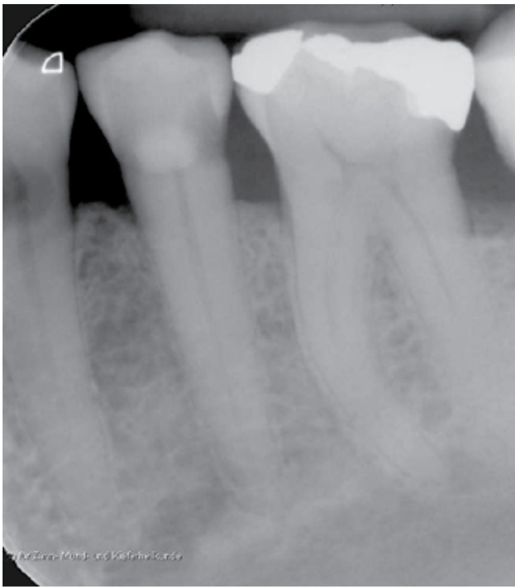 | 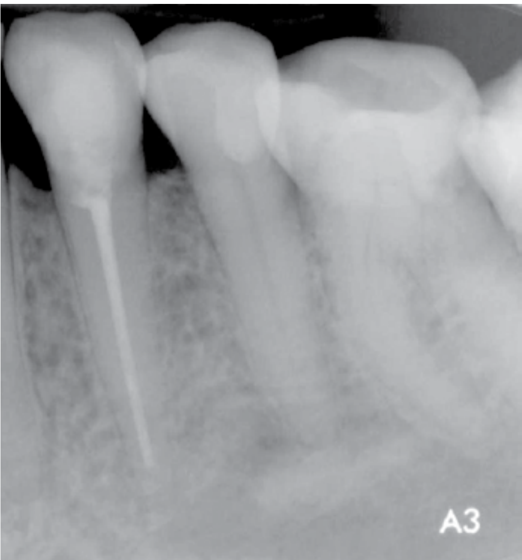 |
 |
Exposure Size7
- Exposure size does not influence the success of direct pulp capping, and the outdated requirement for a maximum exposure area of 1 mm² is no longer valid. Direct pulp capping can be successful after trauma and with large exposure areas, as long as infection is prevented.
- Large-area pulp exposure during caries excavation suggests extensive bacterial penetration into the dentine, which may reduce the chances of success for direct pulp capping. In cases of extensive bacterial contamination, partial pulpotomy can be an alternative treatment.
Location of the Pulp Exposure8
The location of pulp exposure and the specific tooth or jaw group do not significantly influence the prognosis of direct pulp capping.
Microorganisms9
- Only a non-inflamed, bacteria-free pulp can maintain long-term vitality as microorganisms and their metabolites cause pathogenic pulpal changes and periapical diseases.
- Germ-free studies show that pulp can regenerate hard tissue without capping material if microorganisms are absent.
- During carious processes, bacteria in dentinal tubules evade phagocytosis until directly contacting pulp, compromising its regenerative ability as bacterial endotoxins can cause pulp inflammation.
- Direct pulp capping should use sterile instruments, dental dam application, complete caries excavation, cavity disinfection, and bacteria-proof restoration to prevent pulp infection. Pulp shows regenerative capacity when directly capped with calcium hydroxide or hydraulic calcium silicate cement.
Caries Excavation10
Complete caries removal and elimination of all infected tissues to prevent bacterial contamination of the pulp. This involves removing all infected dentine, even if it risks pulp exposure (AAE).
Haemostasis and Cavity Disinfection11
Effective haemostasis is crucial for exposed pulp tissue for several reasons:
- Increased bleeding may indicate higher inflammation, reducing repair ability.
- Capping material must be applied directly onto exposed pulp without blood residue. Blood clots can hinder hard tissue formation and promote pathogenic microorganism growth.
- Blood can release inflammatory mediators, causing chronic inflammation and impaired healing.
- Proper haemostasis ensures good adhesion of capping material to dentine. Moisture and contamination from bleeding can cause leaks and allow microorganism migration into the pulp.
VPT is only successful if you get haemostasis in 10 mins
- Cotton pellet with 3% NaOCl
- If no haemostasis (or try partial pulpotomy, remove upper inflamed pulp & hope lower is vital)
Selection of the Pulp Capping Material12
Calcium Hydroxide
-
Calcium Hydroxide Suspensions: Historically considered the standard for maintaining pulp vitality, they have a high alkaline pH that stimulates pulp cells and enzymes, neutralizes acids and toxins, and promotes pulp repair.
-
Disadvantages of Calcium Hydroxide: Includes poor dentine sealing, mechanical instability, and resorption over time, which can lead to microleakage and bacterial infiltration through defects in the hard tissue.
-
Hard-Setting Calcium Hydroxide Cements: Developed in the 1960s to address the shortcomings of aqueous suspensions. They offer better dentine sealing but have lower antimicrobial effects, slower and less uniform hard tissue formation, and potential long-term dissolution under restorations.
-
Limitations of Hard-Setting Cements: Include reduced hydroxyl ion release, weaker hard tissue regeneration, increased inflammation, and possible toxicity from additives, making them less suitable as a first-choice material for direct pulp capping.
HCSCs
-
Components and Properties: Hydraulic calcium silicate cements (HCSCs) are made from cement powder (containing tri- and/or dicalcium silicate) mixed with water. They release calcium hydroxide and hydroxyl ions upon setting, which promote pulp cell regeneration, hard tissue formation, and maintain a favorable alkaline pH.
-
Advantages: HCSCs stimulate growth factors and cytokines, reducing inflammation and pain. They form a tight bond to dentine, penetrate dentinal tubules, and help trap residual bacteria. They are biocompatible, promote hard tissue repair, and avoid issues like dissolution and mechanical instability seen with calcium hydroxide.
-
Disadvantages: ProRoot MTA, a type of HCSC, can cause tooth discoloration due to heavy metals like bismuth oxide. Other HCSCs with less heavy metal content or different radiographic contrast agents, like zirconia, show minimal discoloration.
-
Comparison with Other Materials: HCSCs offer higher mechanical strength, better sealing, and lower solubility compared to calcium hydroxide products. They are superior for maintaining pulp vitality but are not suitable for pulp capping when mixed with monomers found in light-, dual-, or auto-curing materials.
-
Current Status: Since the introduction of ProRoot MTA, over 40 HCSCs, including Biodentine, are commercially available, with comparable properties to MTA and promising potential for vital pulp maintenance.
Definitive Restoration1314
To prevent contamination of the dentine-pulp complex, a cavity should be restored with a bacteria-proof, definitive filling during the same treatment appointment after direct pulp capping. Ideally, use a dentine-adhesive, resin-based composite. Using only a temporary material, like glass ionomer cement, significantly decreases the success rate of vital pulp therapy (VPT). If a permanent, bacteria-proof filling cannot be placed in the same session, it may be better to reconsider attempting to preserve pulp vitality.
- Patient Age
- Exposure Size
- Location of the Pulp Exposure
- Microorganisms
- Caries Excavation
- Haemostasis and Cavity Disinfection
- Selection of the Pulp Capping Material
- Definitive Restoration
Prerequisite for Successful Direct Pulp Capping15
Vital pulp treatment (VPT) can only be successful if bacterial infection of the pulp can be reliably excluded before, during and after therapy. Therefore, the following must be observed for successful direct pulp capping:
- The tooth should be free of symptoms or should show only mild symptoms of reversible pulpitis beforehand.
- There should be no signs and symptoms indicative of ‘irreversible pulpitis’ present.
- Dental dam and controlled draining from treatment in the pulp-near dentine third.
- Reduce microbes by using sterile instruments.
- Complete caries excavation.
- Bleeding from the exposed pulp can be controlled without difficulty.
- Disinfecting the cavity and sufficient haemostasis with approx. 3% NaOCl.
- Antibacterial wound dressing (ideally HCSC).
- Bacteria-proof final restoration (adhesive composite resin filling) in the same treatment session.
- Patient age, size and location of pulp exposure, and tooth type have no significant effect on success.
Contraindications for Direct Pulp Capping16
- Spontaneous pain
- Severe prolonged bleeding or discharge of serous or purulent exudate from the pulp
- Necrotic pulp tissue
- Existing periradicular changes
- Periodontally damaged teeth
- Radiologically recognizable intrapulpal mineralization
- Lack of retention for a wound dressing (e.g. during crown preparation)
- Lack of retention for an immediate definitive bacteria-proof restoration
- Unable to apply dental dam
- Repeated extensive restorations in close proximity to the pulp (⇒ high biological age of the pulp)
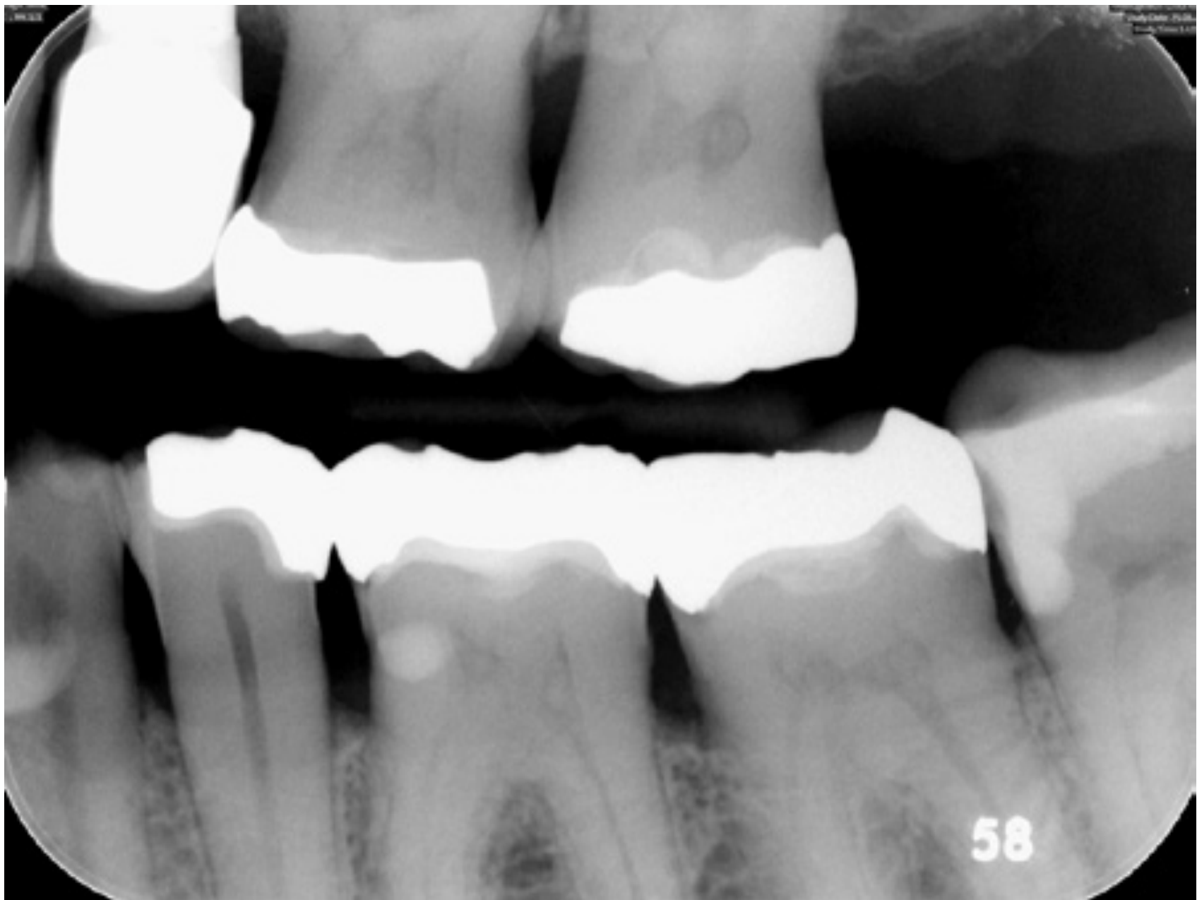
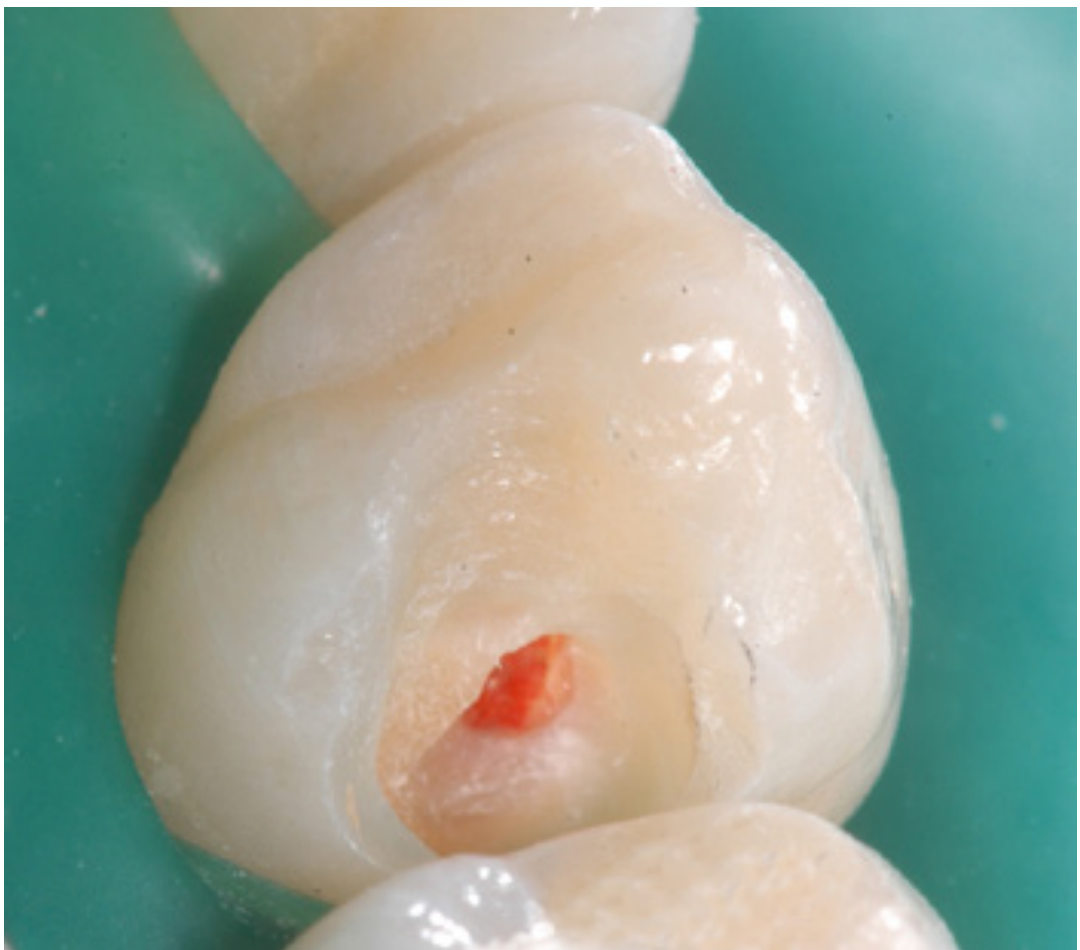
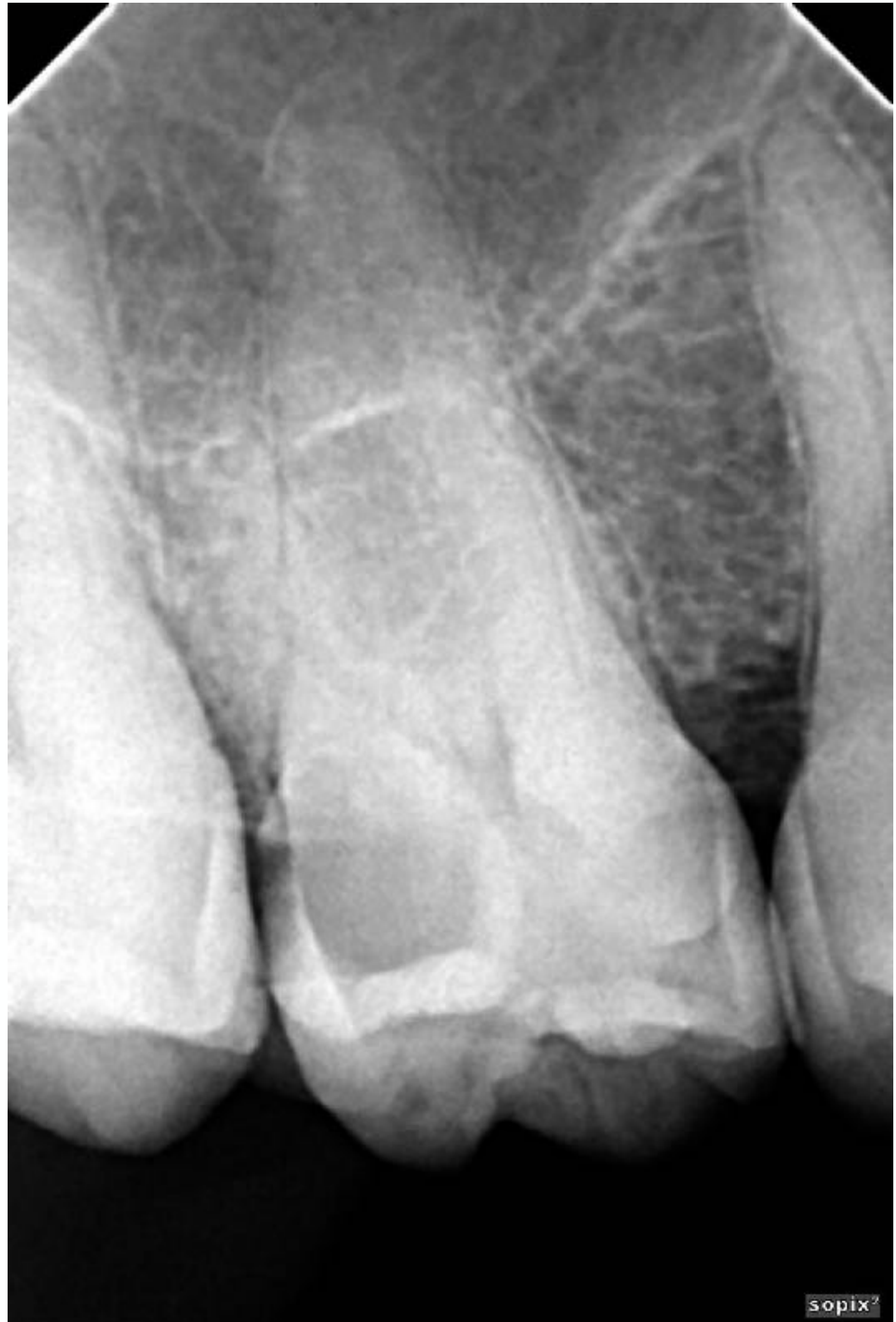
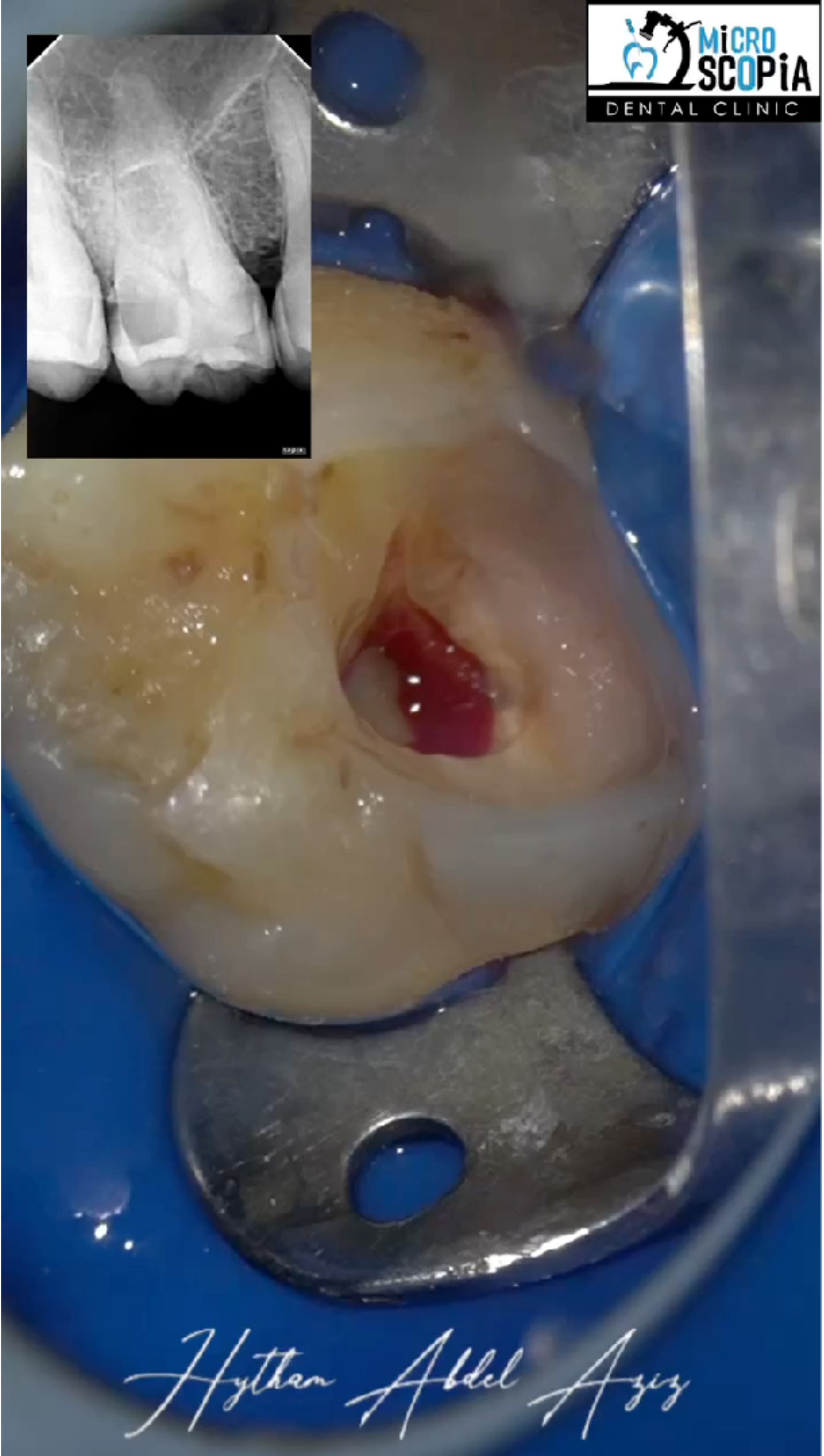
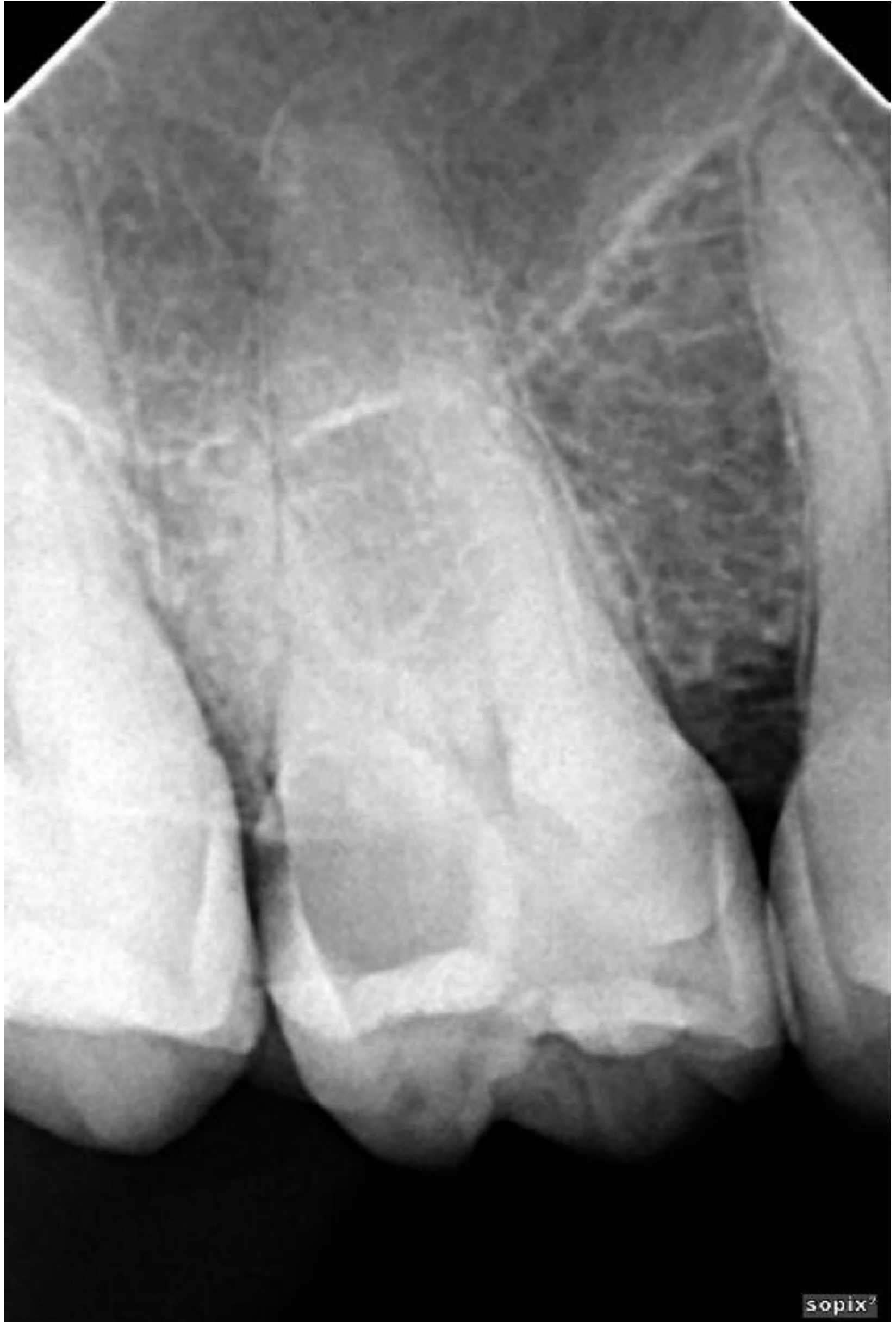
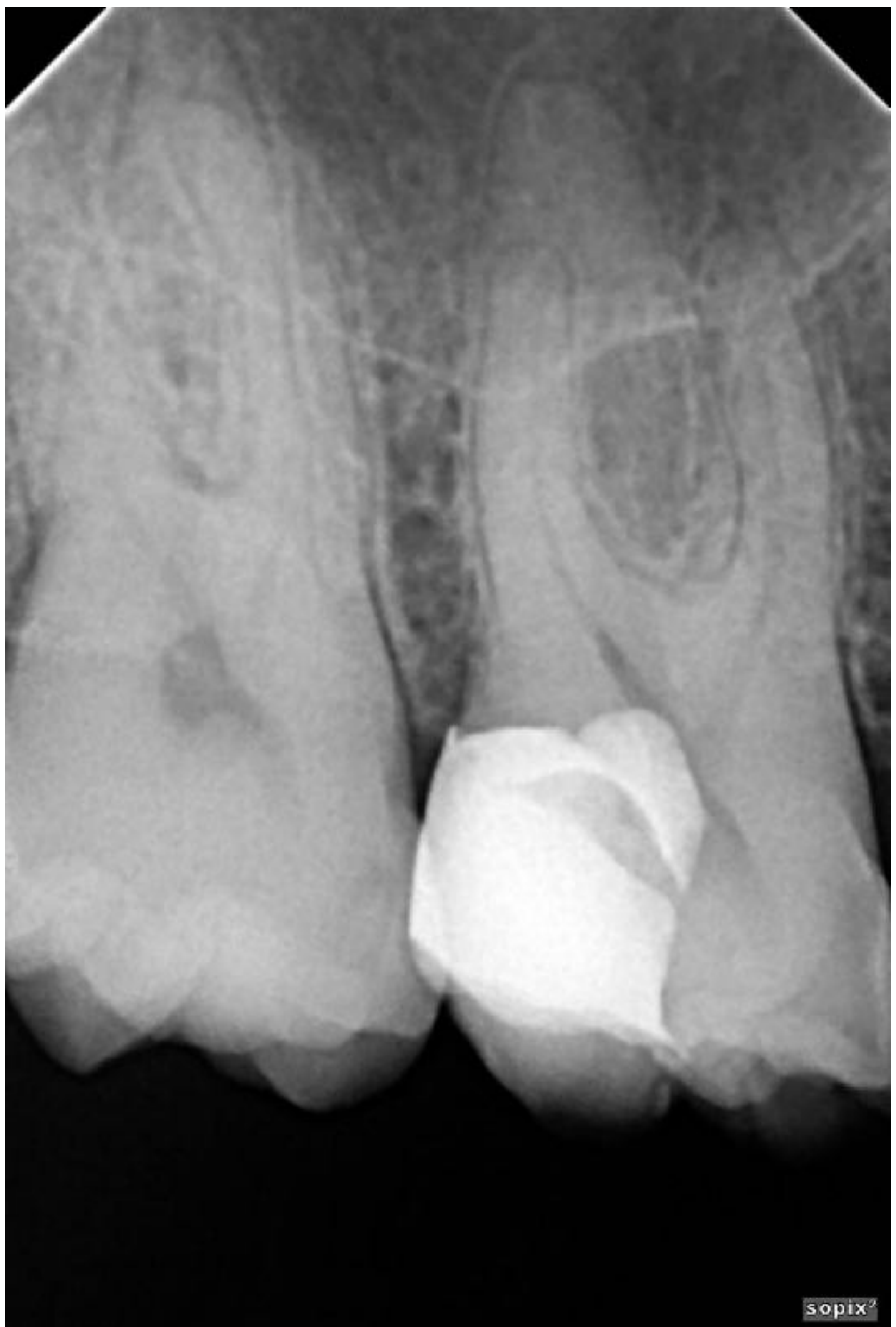
Clinical Case171819
32 yrs old
- Dull aching pain related to food impaction
- Cold & EP testing normal response
- No TTP
Courtesy: Dr Hytham Abdel-Aziz
Marginal acquisition using SDR and bulk fill composite, using saddle band
Courtesy: Dr Hytham Abdel-Aziz
Courtesy: Dr Hytham Abdel-Aziz
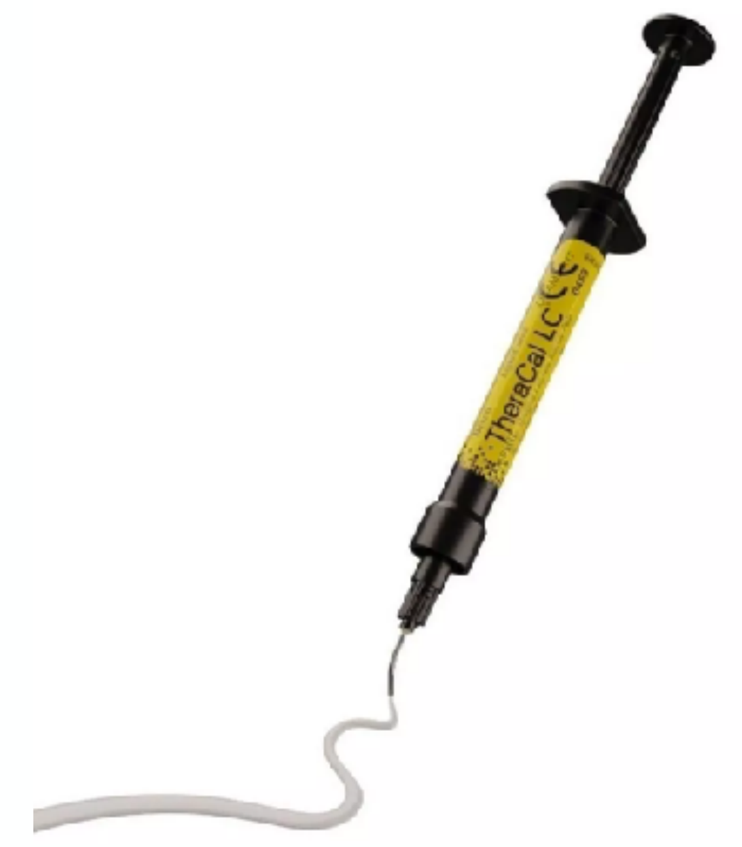
Procedure and Clinical Protocol20
Box 5.3 The Procedure and Clinical Protocol of Direct Pulp Capping21
-
After local anaesthesia, the tooth is isolated under a dental dam and the clinical crown is disinfected with NaOCl (2.5–5%), CHX (2%) or alcohol (70%). The primary preparation is completed with diamond burs in a high-speed contra-angle handpiece under constant water cooling.
-
A complete caries excavation is performed with bud burs in a low-speed handpiece supplemented by hand instruments if necessary. Optical magnification and illumination are strongly recommended for this purpose. Shortly before reaching the pulp chamber, a new, sterile instrument should be used in order to minimize the possible spread of microorganisms, e.g. infected dentine chips.
-
The cavity and the exposure area are cleaned with NaOCl 2.5–5%. Pulp bleeding can be staunched with a NaOCl-soaked cotton pellet under moderate pressure. (CHX 2% can be used as an alternative to NaOCl.) A pulp bleeding should be staunched within approx. five minutes. If pulp haemorrhage cannot be stopped after approx. five minutes, an attempt can be made to perform a partial or complete pulpotomy and, after successful haemostasis and assessment of the tissue condition, to cap the resected pulp or root pulp. All infected tissue must be removed. If sufficient haemostasis is not achieved even after a complete pulpotomy, a pulpectomy (vital extirpation) must be performed.
-
If no bleeding occurs after pulp exposure, the area must be examined for necrotic tissue. If present, the necrotic pulp tissue is removed with a diamond instrument at high speed under permanent water cooling until perfused tissue is exposed.
-
HCSC (or calcium hydroxide suspension) is prepared according to the manufacturer’s instructions.
-
Using a suitable instrument, the HCSC is applied to the exposed pulp in a thickness of approx. 1.5 mm, including part of the surrounding dentine. A sufficient dentine margin must be left free of cement for the dentine-adhesive restoration. Calcium hydroxide suspension, on the other hand, is only applied over a small area in the region of the pulp exposure and must be provided with a subbase (e.g. phosphate cement and glass ionomer cement) before application of a definitive restorative material.
-
Etching with a phosphoric acid gel and rinsing with water is not possible at this stage, as the HCSC would be washed away. Therefore, the setting time of HCSC (at least 15 minutes, depending on the preparation) should be observed.
-
Alternatively, the HCSC can be covered after application with a small amount of a light-curing flowable compomer, a resin-reinforced glass ionomer cement (RMGI), a self-etching, self-adhesive flowable composite resin or a flowable composite resin in combination with a self-etching dentine adhesive. The HCSC (or calcium hydroxide suspension) must be completely covered.
-
The cavity is then definitively restored adhesively with a dentine bonding agent and composite resin in the same appointment (use according to manufacturer’s instructions).
-
The sensibility of the pulp should be checked with a cold test at the next appointment (Figure 5.6a–j).
Source: Adapted from (8).
Pulpotomy22

Partial and Complete Pulpotomy232425
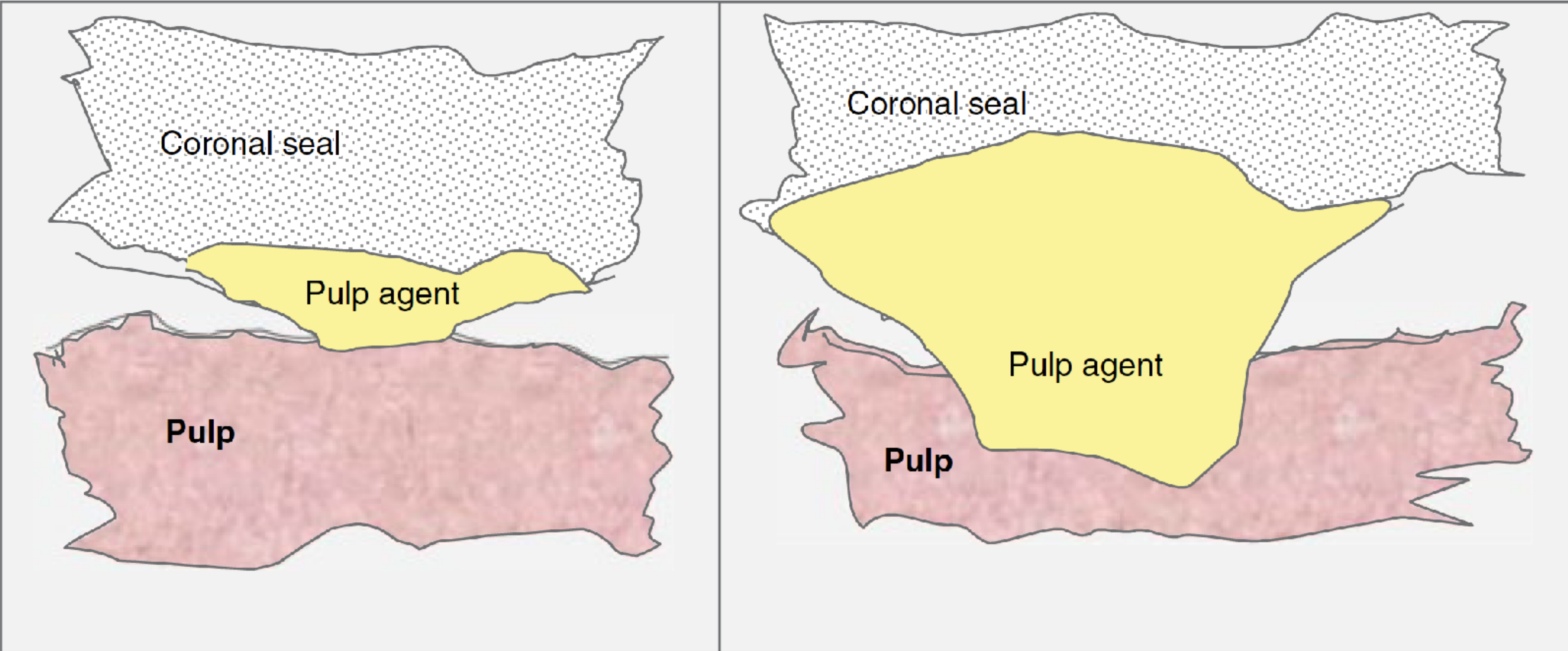
A partial pulpotomy is a clinical procedure in which a small portion of the coronal pulp is in direct contact with the affected area after pulp exposure is removed, followed by the application of a biomaterial directly onto the remaining pulp tissue prior to placement of a permanent restoration.
A full (or complete) pulpotomy involves the complete removal of the coronal pulp and the application of a biomaterial directly onto the pulp tissue at the level of the root canal orifice(s), prior to placement of a permanent restoration.
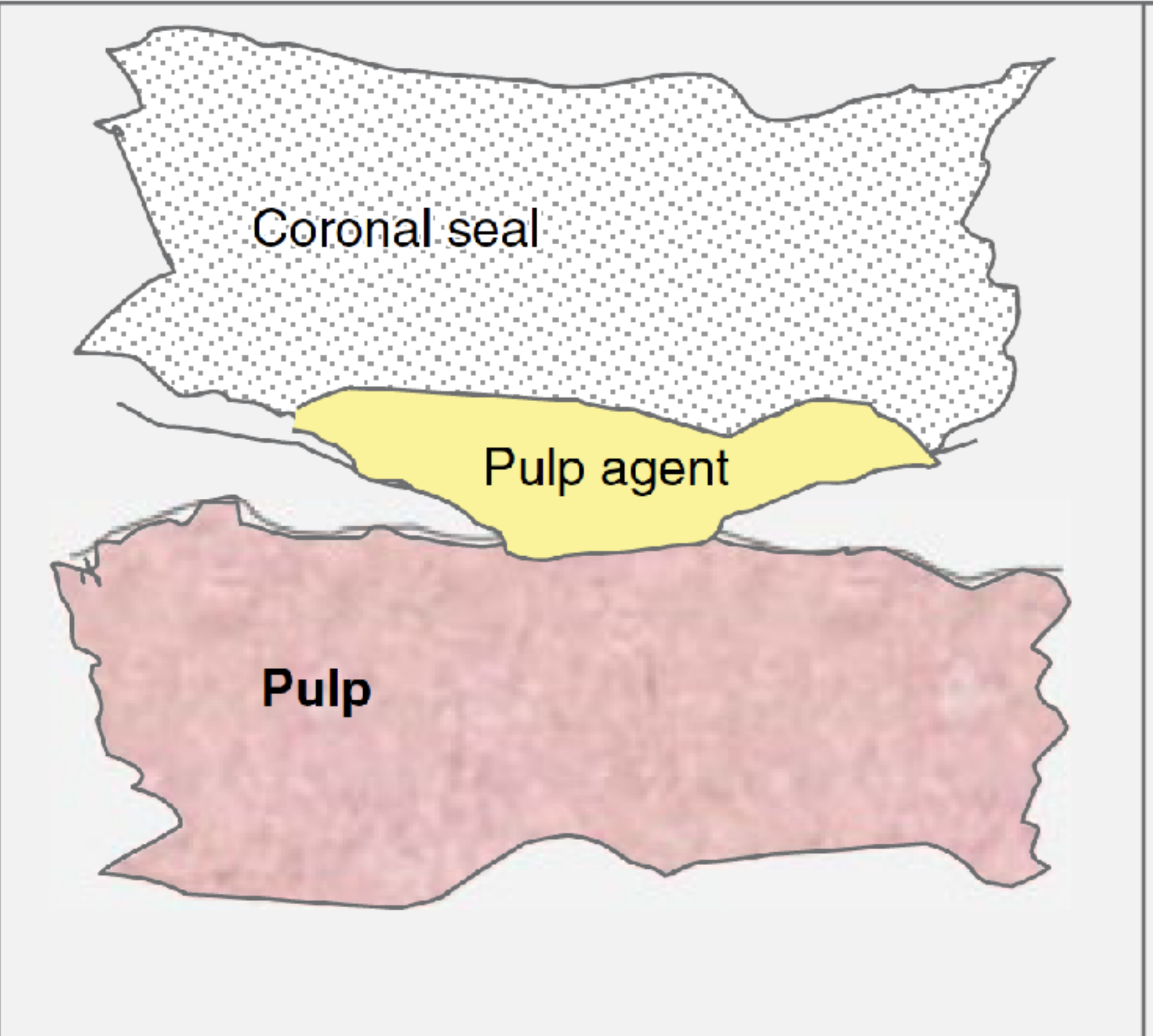
The decision to perform direct pulp capping or partial/complete pulpotomy depends on direct inspection and the ability to control pulpal bleeding during the operative procedure.
ESE position statement
 |  |
Indications26
- Definitive Treatment for Asymptomatic Cariously Exposed Pulp
- Definitive Treatment for Traumatically Exposed Pulp
- Other Indications
See - Traumatic crown-fracture management
Pulpotomy procedures have shown promising results in the management of severely furcation-involved molars requiring vital root resection.
J Clin Periodontol. 2020;47(8):970–9.
Pulpotomy has traditionally been used as a temporary or emergency treatment for pain relief for teeth with symptomatic irreversible pulpitis.
Contraindications
General Contraindications for Restorative Treatment27
- Poor oral hygiene
- Uncontrolled caries
- Periodontal disease
- Unrestorable tooth
- Inability to adequately isolate the tooth
Procedure-Specific Contraindications for Pulpotomy
- Inability to control pulpal bleeding
- Presence of necrotic pulp or lack of bleeding
- Need for post and core restoration
Specific Cases to Avoid Partial Pulpotomy
- Teeth with spontaneous pain
- Continuous uncontrollable pulp bleeding
- Presence of necrotic tissue in the pulp chamber
- Traumatic dental injuries affecting pulpal blood supply (considered negative prognostic factor for pulpotomy and a contraindication for VPT)
Clinical Technique
Assessment and Anaesthesia
Preoperative Assessment
- Detailed pain history and sensibility tests (heat, cold).
- Radiographic assessment (periapical and intraoral bitewing x-rays).
Anaesthesia
- Use adrenaline-containing solutions (2% lignocaine with adrenaline 1:80,000 or 4% articaine with adrenaline 1:100,000).
- Avoid intra-pulpal injection.
Pre-VPT Preparatory Procedures
Isolation and Aseptic Field
- Limit contamination and optimize marginal seal.
Preparation Scenarios
- Immediate pulpotomy if the tooth has ‘sound’ proximal walls.
- Pre-VPT restoration if interproximal defects are present.
Caries Removal
- Non-selective (complete) removal of all carious dentine.
- Use sharp, sterile hand excavators and water-cooled burs.
Pulp Tissue Removal
Visualize Exposed Pulp
- NaOCl rinsing for bleeding control and bacterial load reduction.
- Use fresh, sterile burs for tissue removal.
Partial vs. Full Pulpotomy
- Partial: Remove 2-3 mm of pulp.
- Full: Remove all pulp tissue to root canal orifices if bleeding persists.
Main reason for full pulpotomy & not RCT is immature apexes
See - Apexogenesis in immature teeth
Haemostasis
Control Bleeding
- Use NaOCl for haemostasis and antibacterial lavage.
- Apply soaked cotton pellets gently over the pulp.
Placement of Pulp Capping Material
Preferred Materials
- Hydraulic calcium silicate cements (HCSCs) over calcium hydroxide.
Application Protocol
- Apply a 2-3 mm layer of HCSC.
- Ensure surrounding dentine is clean for adhesion.
Handling of Different Materials
- MTA: Use carriers and compact with a moist paper point.
- Biodentine: Mix in an amalgamator, apply with a hand instrument, compact gently.
- Premixed materials: Introduce with a probe or plugger, adapt with a small brush.
Placement of Final Restoration
Coronal Seal
- Immediate permanent restoration preferred for long-term success.
- Delay should not exceed two days for temporary fillings.
Bonding Protocol
- Clean peripheral dentine for better bonding.
- Sandblast, selectively etch enamel, apply primer and bonding, cover HCSC with a flowable composite, then restore with bulk fill or incremental RBC.
Outcomes of Vital Pulp Treatment
The asymptomatic tooth is neither painful nor tender to bite on, but pain reporting varies across VPT studies, with some describing pain characteristics suggestive of irreversible pulpal changes, while others only note the presence or absence of pain, leading to uncertainty; proxy indicators like postoperative analgesic use and flare-ups requiring additional intervention should be interpreted cautiously.
Pain
Timing of Measurement of Pain28
The outcome of VPT must be measured at a consistent time point after treatment has been completed. This optimizes the likelihood that the effect intended for measurement is what is being measured and the true value of treatment can be assessed. Pain and the use of analgesics in the postoperative period should be measured as a minimum at seven days and for a maximum of three months post-treatment.
Sensibility and Vitality of Pulp
A key indicator of VPT success is pulpal health status. The gold standard in assessing pulpal health is histological assessment (28); however, as the pulp is encased in a rigid structure, histological assessment can only be carried out after commencing endodontic treatment or extraction of the tooth. In the absence of direct observation of tissue, proxy measures of pulpal status are frequently made based on neural responses to stimuli, the sensibility tests. Sensibility testing includes thermal, both cold and heat and electric pulp tests.
Timing of Sensibility Testing29
Based on the current ESE guidelines, pulp sensibility testing should be conducted at 6- and 12-months post-treatment, continuing at regular intervals if symptoms persist or when there is doubt about healing (15). The consensus group, however, have suggested that this follow-up reporting should continue for as long as possible beyond one year in all cases.
Absence of Apical Periodontitis and Root Resorption
Absence of apical periodontitis, root resorption and, in the case of immature teeth, continued root development.
Timing of Radiographic Assessment30
Early periapical changes cannot be visualized on radiographs and so sufficient time must elapse before a radiographic assessment can provide the information necessary to make a valid judgement about treatment outcome. The current recommendation is for radiographic assessment at one year, continuing annually if required for up to four years.
Future Directions31
Next-Generation Pulp Diagnostic Tools Using Blood and Biological Fluids32
- Sample taken using paper point, microcapillary tube or other collection vessel
- Chairside (or adjacent lab) analysis for biomarker of interest, e.g. MMP9
- Level of biomarker compared with established reference standard
- Objective measure of pulpitis obtained to inform clinician and patient of treatment options
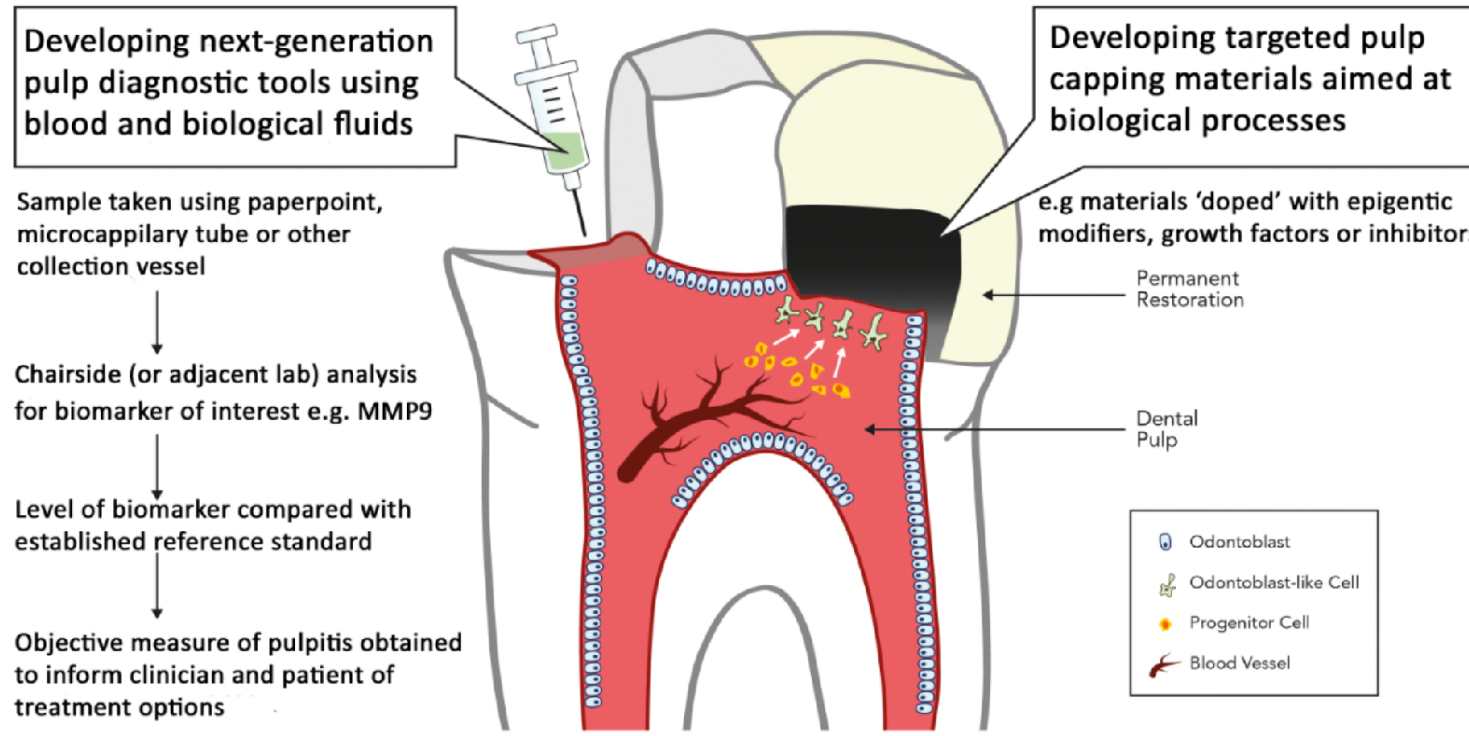
Targeted Pulp Capping Materials Aimed at Biological Processes
Next-Generation Pulp Diagnostic Tools
- e.g. materials ‘doped’ with epigenetic modifiers, growth factors or inhibitors
Diagram labels: Permanent Restoration — Dental Pulp — Odontoblast, Odontoblast-like Cell, Progenitor Cell, Blood Vessel.
Footnotes
-
Original PDF page 1: L8 Vital Pulp Treatment 2, p.1 ↩
-
Original PDF page 2: L8 Vital Pulp Treatment 2, p.2 ↩
-
Original PDF page 3: L8 Vital Pulp Treatment 2, p.3 ↩
-
Original PDF page 4: L8 Vital Pulp Treatment 2, p.4 ↩
-
Original PDF page 5: L8 Vital Pulp Treatment 2, p.5 ↩
-
Original PDF page 6: L8 Vital Pulp Treatment 2, p.6 ↩
-
Original PDF page 7: L8 Vital Pulp Treatment 2, p.7 ↩
-
Original PDF page 8: L8 Vital Pulp Treatment 2, p.8 ↩
-
Original PDF page 9: L8 Vital Pulp Treatment 2, p.9 ↩
-
Original PDF page 10: L8 Vital Pulp Treatment 2, p.10 ↩
-
Original PDF page 11: L8 Vital Pulp Treatment 2, p.11 ↩
-
Original PDF page 15: L8 Vital Pulp Treatment 2, p.15 ↩
-
Original PDF page 16: L8 Vital Pulp Treatment 2, p.16 ↩
-
Original PDF page 17: L8 Vital Pulp Treatment 2, p.17 ↩
-
Original PDF page 18: L8 Vital Pulp Treatment 2, p.18 ↩
-
Original PDF page 19: L8 Vital Pulp Treatment 2, p.19 ↩
-
Original PDF page 20: L8 Vital Pulp Treatment 2, p.20 ↩
-
Original PDF page 21: L8 Vital Pulp Treatment 2, p.21 ↩
-
Original PDF page 22: L8 Vital Pulp Treatment 2, p.22 ↩
-
Original PDF page 24: L8 Vital Pulp Treatment 2, p.24 ↩
-
Original PDF page 23: L8 Vital Pulp Treatment 2, p.23 ↩
-
Original PDF page 25: L8 Vital Pulp Treatment 2, p.25 ↩
-
Original PDF page 26: L8 Vital Pulp Treatment 2, p.26 ↩
-
Original PDF page 27: L8 Vital Pulp Treatment 2, p.27 ↩
-
Original PDF page 28: L8 Vital Pulp Treatment 2, p.28 ↩
-
Original PDF page 29: L8 Vital Pulp Treatment 2, p.29 ↩
-
Original PDF page 30: L8 Vital Pulp Treatment 2, p.30 ↩
-
Original PDF page 37: L8 Vital Pulp Treatment 2, p.37 ↩
-
Original PDF page 38: L8 Vital Pulp Treatment 2, p.38 ↩
-
Original PDF page 39: L8 Vital Pulp Treatment 2, p.39 ↩
-
Original PDF page 41: L8 Vital Pulp Treatment 2, p.41 ↩
-
Original PDF page 40: L8 Vital Pulp Treatment 2, p.40 ↩