L16 Management of Incompletely Developed Tooth 1
Management of Immature Teeth1
Dr Mostafa Elkholy, BDS, MSc (Endo), PhD (Endo)
Senior Lecturer and Discipline Lead in Endodontics
Dental School, UWA
Presenter Introduction23
Dr Mostafa Elkholy
Problems with Immature Roots
See also: Root Fractures and Developing Teeth.
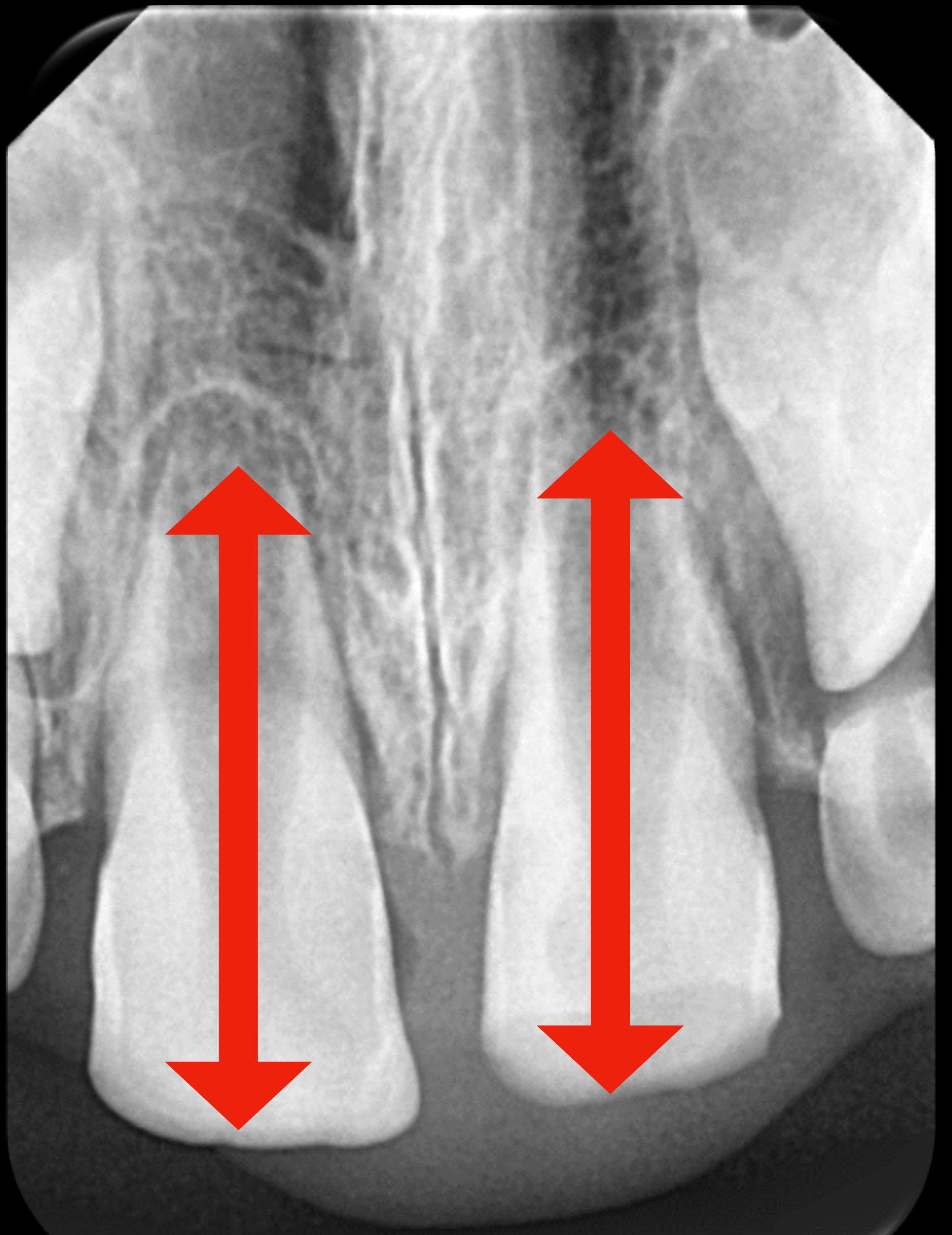
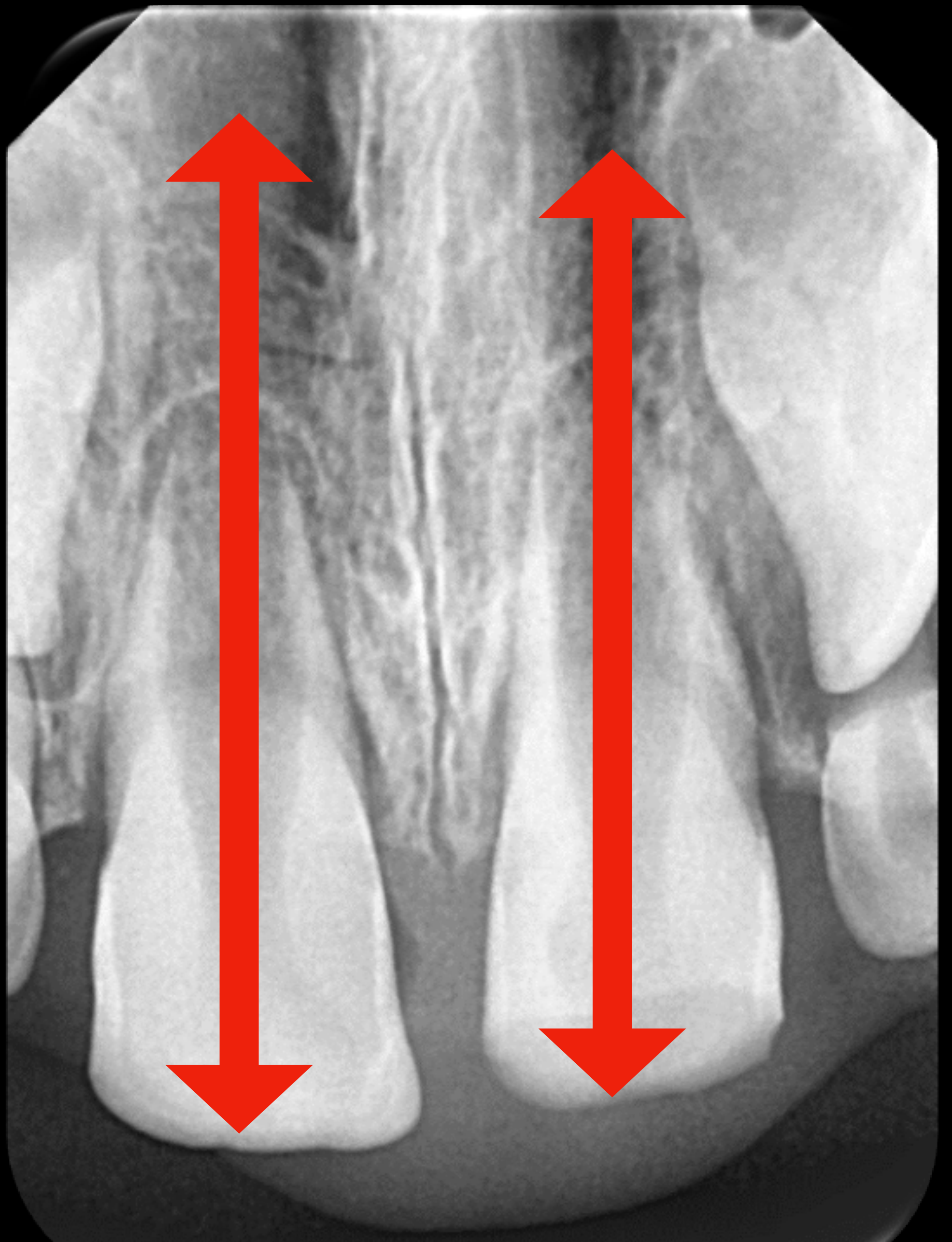
- Short root that will affect long-term retention of the tooth.
Short Root Length45
 |  |
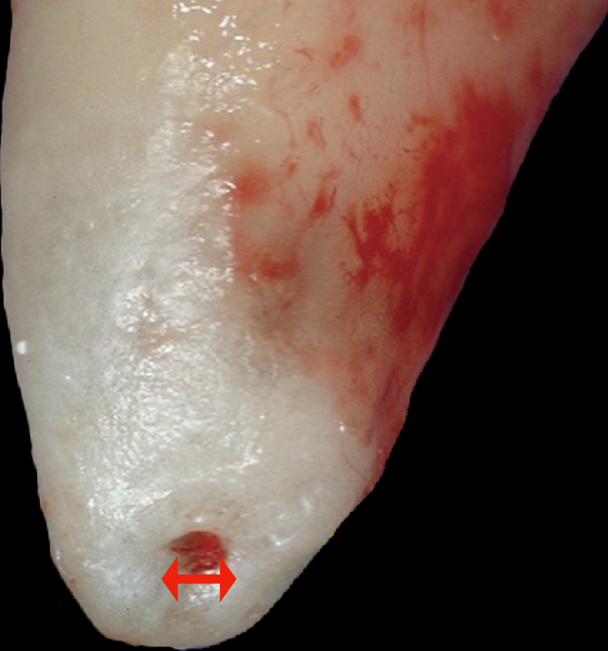
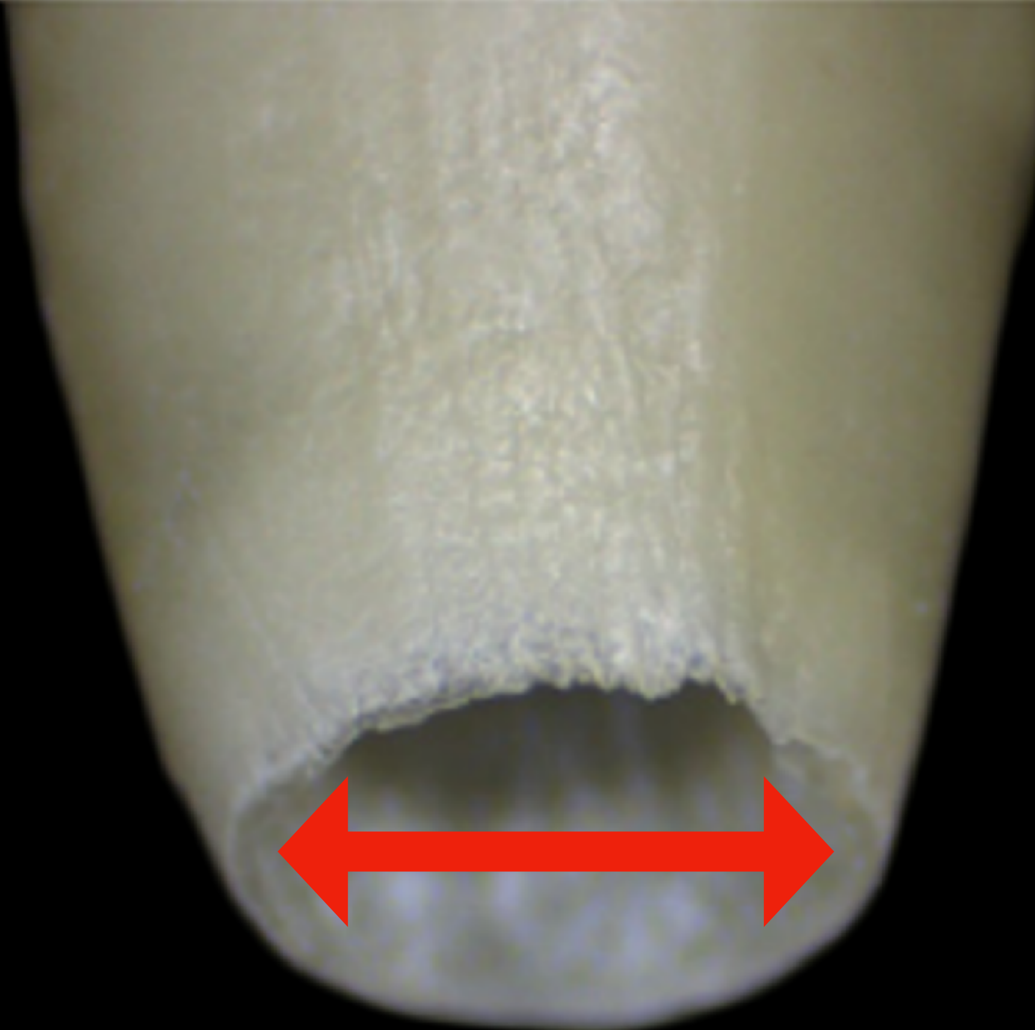
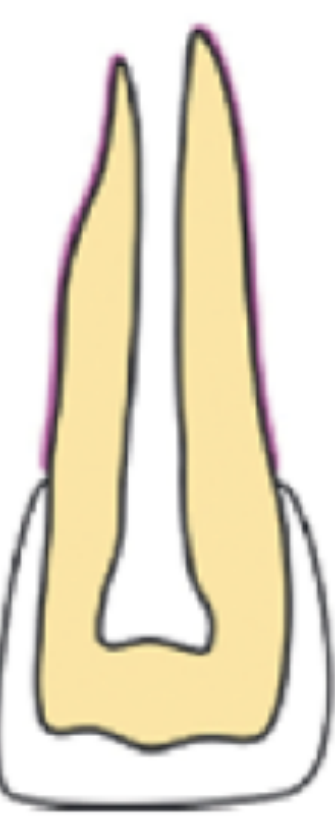
Thin Dentinal Walls and Cervical Fracture Risk67
- Thin dentinal walls that will make the tooth susceptible to cervical fracture.
 |  |
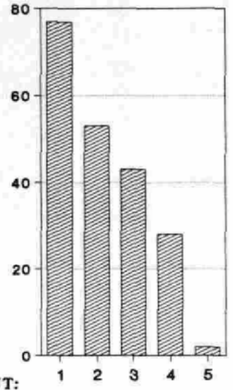
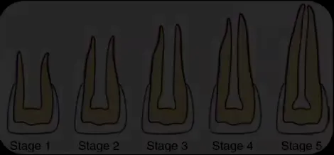
Stage of Root Development vs. Fracture Rate8
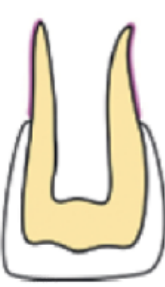
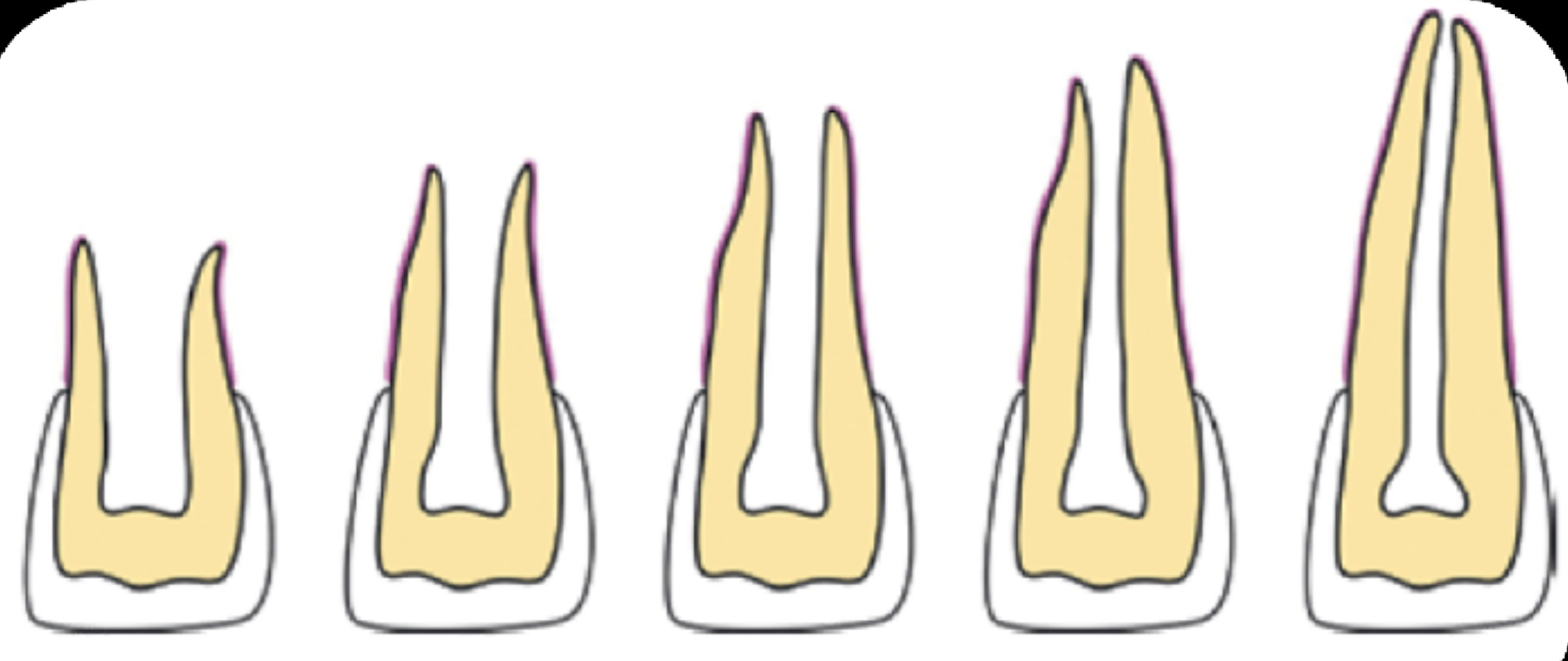
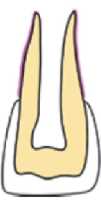
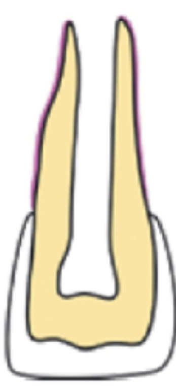
Stages 1-4 are immature and Stage 5 mature; in the retrospective cohort of luxated non-vital maxillary incisors treated with calcium hydroxide and gutta-percha, fracture proportions were 77% at Stage 1 and 2% at Stage 5. The full table and source context are in Cvek 1992 - Luxated Nonvital Incisors Apexification.
Lecturer — Root Fracture Risk
Immature roots are deficient not only in length but also in width, and their dentinal walls may be substantially thinner than those of fully developed roots.
- In traumatic dental injuries, loss of tooth structure is generally associated with the fracture itself rather than with caries or infection.
- Treatment should therefore preserve as much remaining tooth structure as possible.
- The lecturer described five stages of incomplete root development, from minimal root formation in Stage 1 to complete root formation without apical closure in Stage 5.
- Stage 1 teeth had the greatest number of fractures, possibly because of their very thin cervical dentinal walls.
 |  |  |
 |  |  |
 |
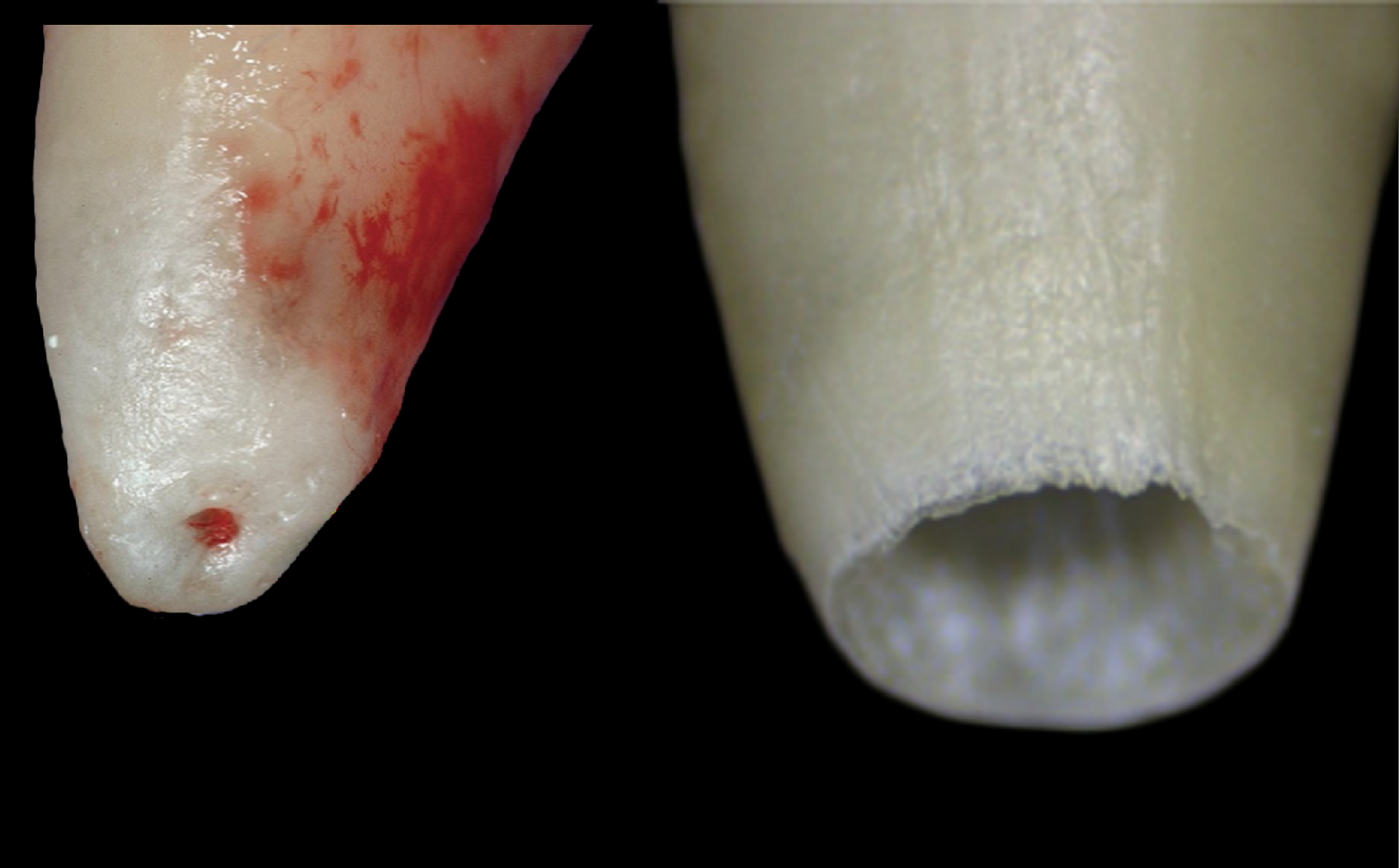
Large Apical Diameter and Leakage9
- Larger apical diameter potentially leading to more apical leakage and extrusion of the filling material.
Summary of Problems10
- Short root that will affect long-term retention of the tooth.
- Thin dentinal walls that will make the tooth susceptible to cervical fracture.
- Larger apical diameter potentially leading to more apical leakage and extrusion of the filling material.
Lecturer — Root Development
A longer root provides greater support and allows the tooth to better withstand functional forces in the oral cavity. The objective of treatment is therefore to promote continued root development wherever possible.
Lecturer — Apical Sealing
A large apical opening creates difficulties because there may be no adequate apical constriction or solid stop against which to condense the filling material. Irrigation solution or filling material may also be extruded, and the apical area can therefore be difficult to seal effectively; inadequate sealing increases the possibility of leakage.
- Continued root development is an important treatment objective when the pulp remains vital.
Supporting Evidence from IADT Guidelines
Received: 19 May 2020 | Accepted: 19 May 2020 DOI: 10.1111/edt.12578
International Association of Dental Traumatology guidelines for the management of traumatic dental injuries: 1. Fractures and luxations. Dental Traumatology (Wiley). Comprehensive review.
Stage of Root Development — Immature (Open Apex) vs Mature (Closed Apex) Permanent Teeth11
Every effort should be made to preserve the pulp, in both mature and immature teeth. In immature permanent teeth, this is of utmost importance in order to allow continued root development and apex formation.
Lecturer — Pulp Preservation
Preserving the pulp in immature permanent teeth permits more than continued root development and apex formation.
- It allows further root lengthening.
- It supports thickening of the dentinal walls.
- It permits formation or closure of the apex.
Lecturer — Treatment Selection
The treatment approach depends on the stage of root development, whether the pulp is vital or necrotic, and the extent of pulp exposure and inflammation. Immature teeth with vital pulp should generally receive vital pulp therapy, whereas immature teeth with necrotic pulp require a different approach because the odontoblasts and vital pulp tissue are no longer available to continue normal root development.
Lecturer — Apexogenesis Treatment
When the pulp is vital, the lecturer stated that treatment of choice is vital pulp therapy, usually by partial or complete pulpotomy depending on the level of inflammation and exposure.
- The treatment aims to preserve vital radicular pulp rather than remove the entire pulp.
Management Overview12
flowchart TD
    PulpStatus["Pulp Status"] --> VitalPulp["Vital Pulp"]
    VitalPulp --> SmallExposure["Small Exposure"]
    VitalPulp --> LargeExposure["Large Exposure"]
    SmallExposure --> PartialPulpotomy["Partial Pulpotomy"]
    LargeExposure --> Pulpotomy["Pulpotomy"]

    subgraph VitalPulpTreatment ["Vital Pulp Treatment = apexogenesis"]
        VitalPulp
        SmallExposure
        LargeExposure
        PartialPulpotomy
        Pulpotomy
    end
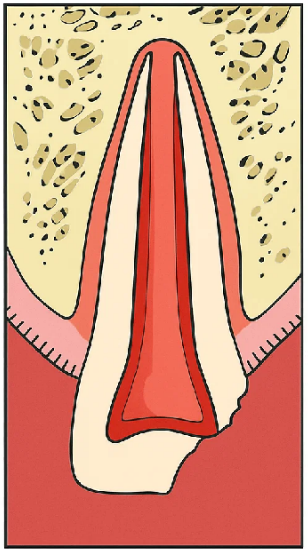
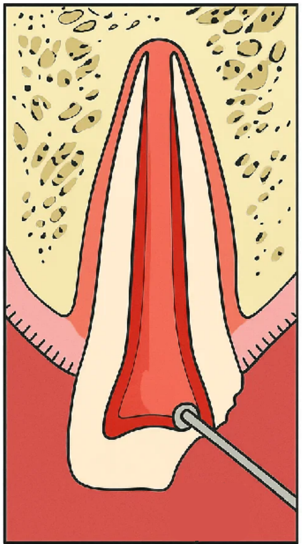
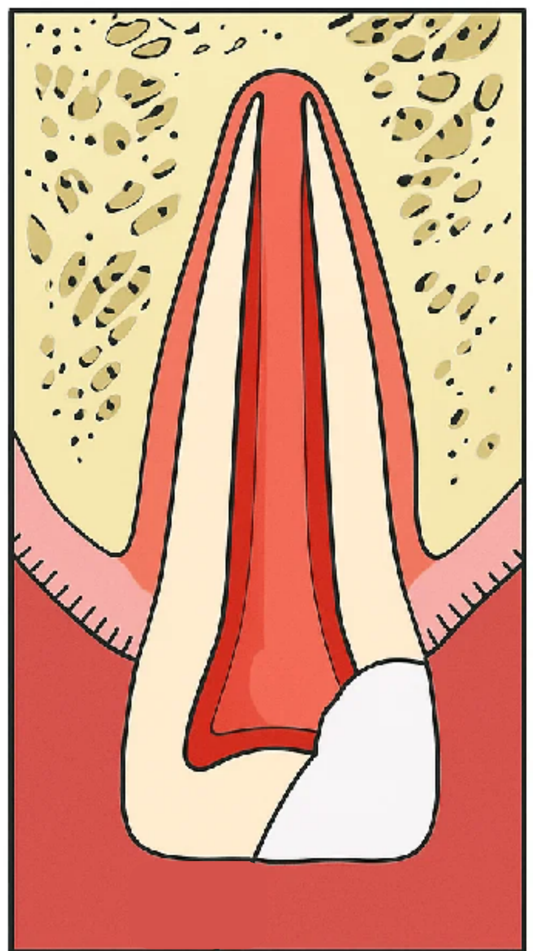
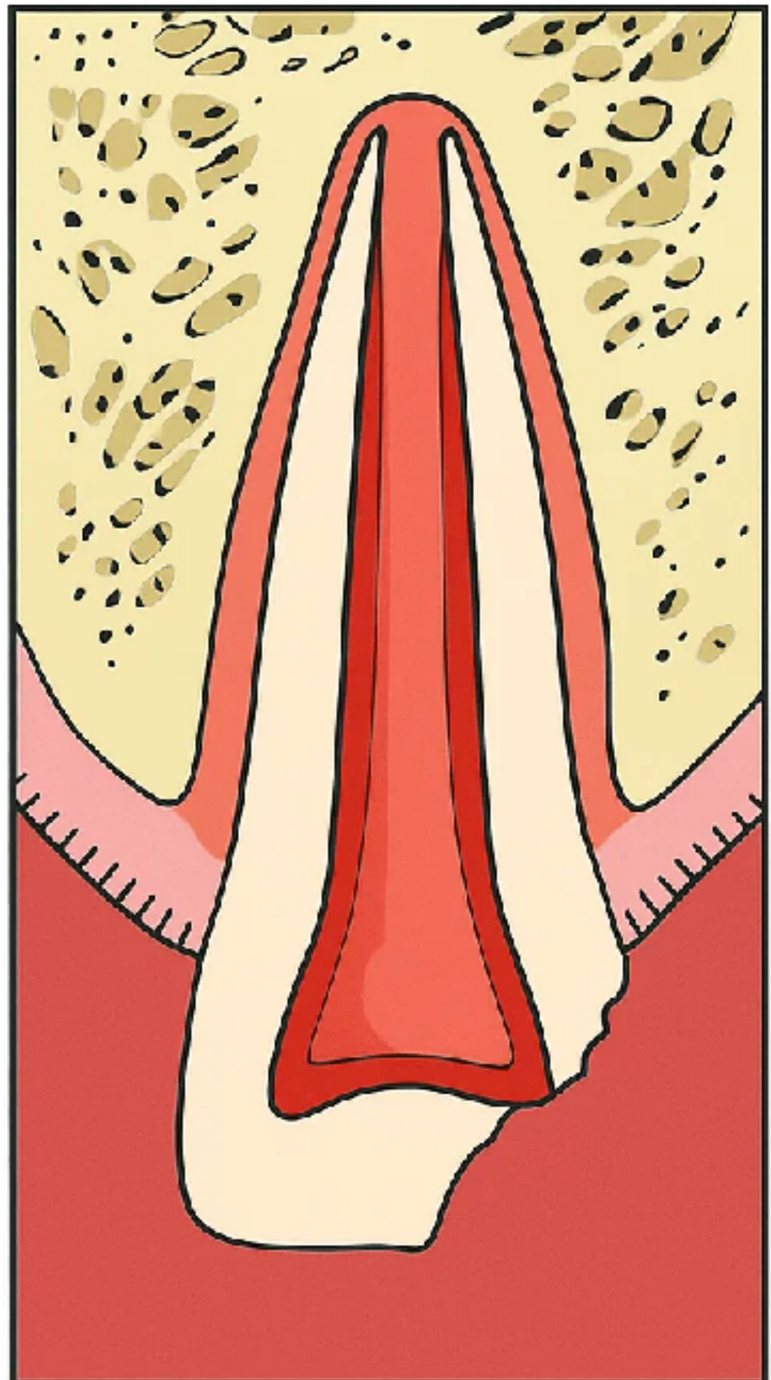
Objectives of Vital Pulp Treatment13
There are two essential objectives for treatment of the vital pulp that make the level of pulpal amputation important:
- A wound dressing should be placed on non-inflamed tissue.
- The loss of tooth structure should be kept to a minimum.
- Remove inflamed or infected pulp tissue while preserving non-inflamed radicular pulp tissue.
- Maintain pulp vitality and preserve the epithelial root sheath of Hertwig.
- Encourage further deposition of dentin by odontoblasts.
- Create conditions that would allow a future root canal treatment to be performed more predictably if it becomes necessary.
 |  |
 |
Apexogenesis
Apexogenesis refers to a vital pulp therapy procedure performed to encourage physiological development and formation of the root end.
S Goldstein, A Sedaghat-Zandi, M Greenberg, S Friedman. N Y State Dent J 1999 May;65(5):23-5.
Partial Pulpotomy versus Direct Pulp Cap14
-
The advantage of partial pulpotomy (removing 2 mm of coronal pulp) as compared with pulp capping lies in better control of the surgical wound and retention of the sealing material.
-
Capping of the pulp is recommended only when the exposure is small (less than 1 mm in size) and when it can be treated shortly after the accident. These indications apply to only a limited number of teeth and, in the majority of cases, partial pulpotomy is therefore performed.
-
For a larger exposure or more extensive inflammation, a complete pulpotomy may be required while preserving the radicular pulp.
-
In partial pulpotomy, removal continues until non-inflamed, vital pulp tissue is reached.
-
Suggested dressing materials include calcium hydroxide and hydraulic calcium silicate materials such as Biodentine, MTA, or bioceramic putty.
-
The most recent best practice described was to use a calcium silicate-based material after bleeding has been controlled.
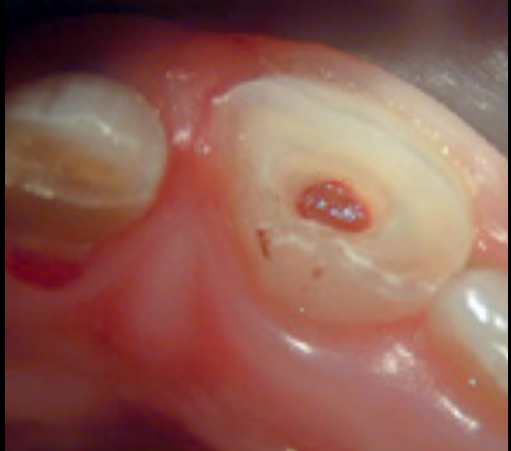
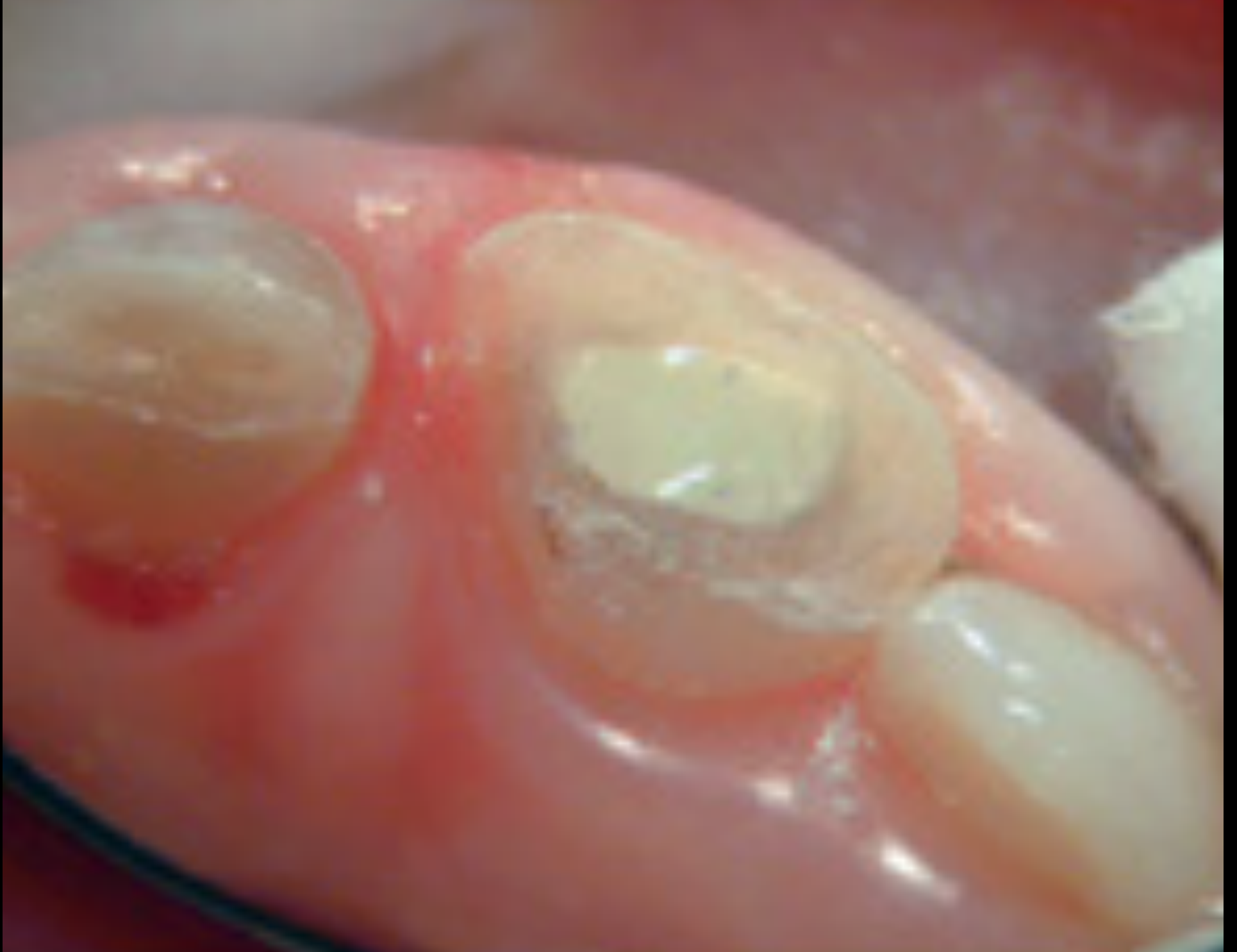
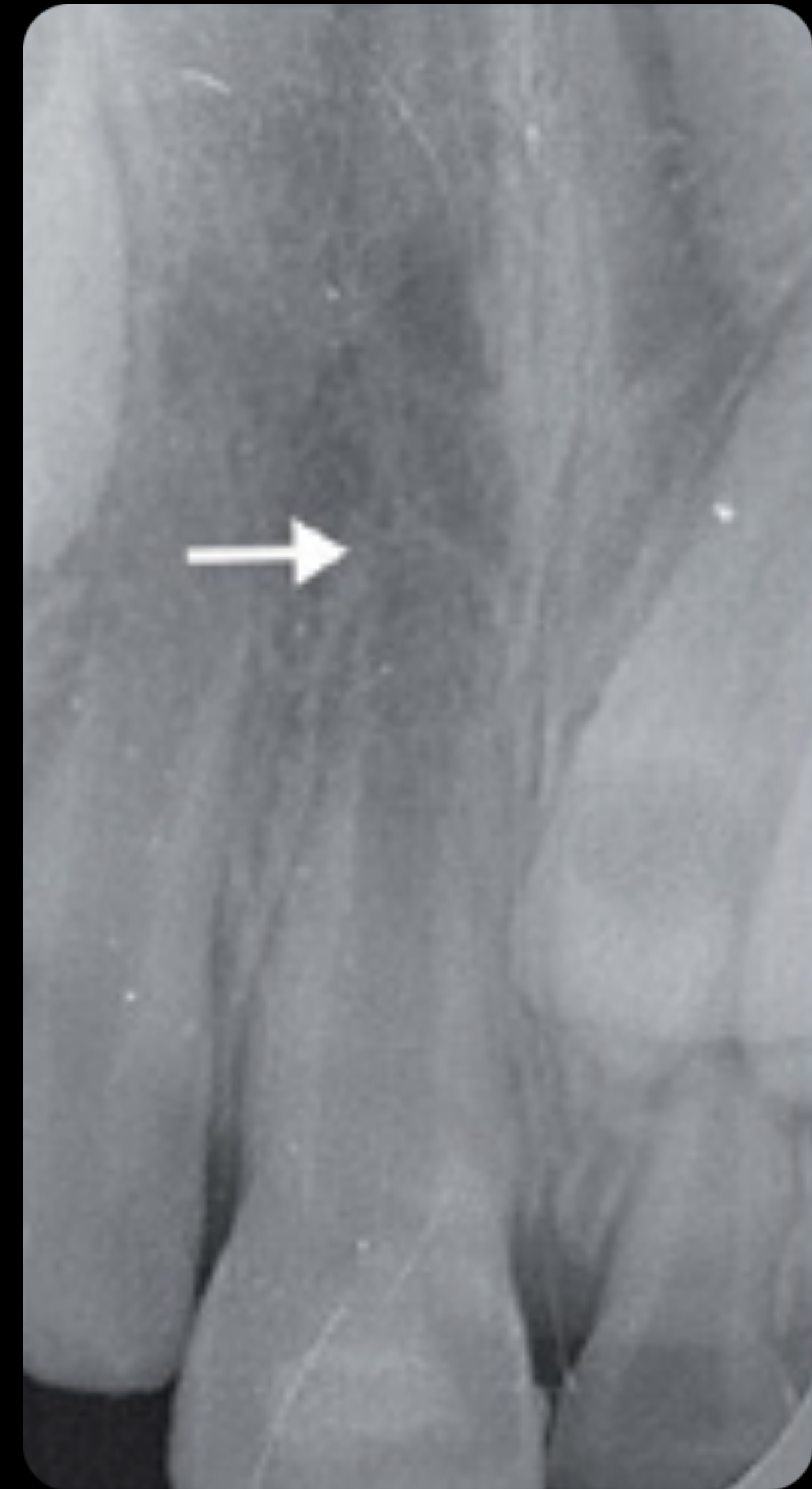
Clinical Examples of Vital Pulp Treatment1516
Vital Pulp Treatment
Courtesy of Dr Marga Ree. Courtesy of George Bogen. From Bogen G et al. Cohen’s Pathways of the Pulp, 12th ed.
Traumatized Immature Tooth
In a traumatized immature tooth with exposed but vital pulp, inflamed pulp tissue is removed and the remaining non-inflamed pulp is preserved.
- A calcium silicate cement may be placed over the pulp.
- If vitality is preserved, the root can continue to develop, become longer and thicker, and proceed toward apical closure.
- Root canal treatment and crown placement may still be required after root development is complete when the tooth has been fractured to a low gingival level.
 |  |
 |
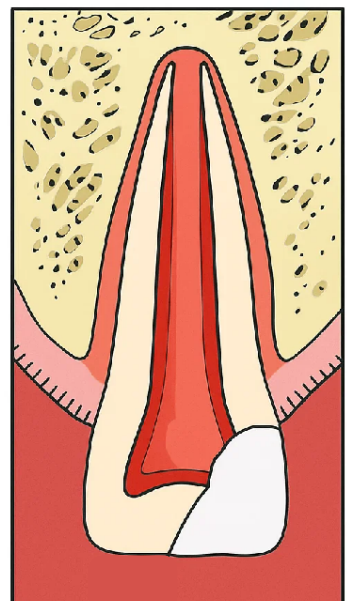
Goals of Apexogenesis17
- Sustaining a viable Hertwig’s sheath, thus allowing continued development of root length for a more favorable crown-to-root ratio.
- Maintaining pulpal vitality, thus allowing the remaining odontoblasts to lay down dentine, producing a thicker root and decreasing the chance of root fracture.
- Promoting root end closure, thus creating a natural apical constriction for root canal filling.
- Generating a dentinal bridge at the site of the pulpotomy.
- Allow odontoblasts to deposit additional dentin along the internal root walls, increasing dentinal wall thickness.
- Reduce the risk of extrusion during any future root canal filling. While the bridging is not essential for the success of the procedure, it does suggest that the pulp has maintained its vitality.
Webber RT. Apexogenesis versus apexification. Dent Clin N Am 1984;28:669–97.
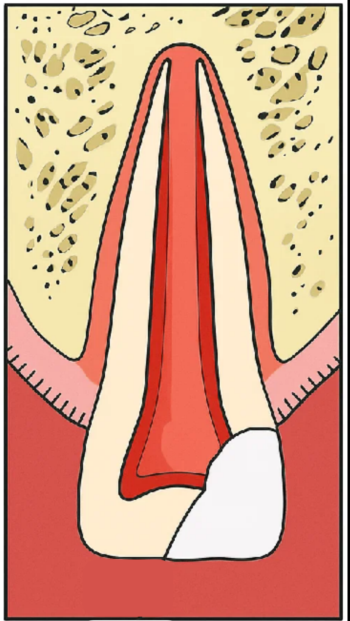
Outcomes of Apexogenesis18
A successful follow-up to vital pulpal treatment:
- Dentine bridge formation
- Continued root development
- Positive response to pulp vitality testing
- No symptoms
- No radiographic development of apical periodontitis or root resorption
Lecturer — Sensibility Testing
Sensibility testing may not always provide a positive result even when the radicular pulp has been preserved.
- Normal nerve stimulation may not be detected by cold, carbon dioxide, or electric testing.
Treatment Decision Flowchart19
See - Vital Pulp Pathway
flowchart TD
    PulpStatus["Pulp Status"] --> Necrotic["Pulpless and infected RCS"]
    Necrotic --> Regeneration["Regeneration"]
Lecturer — Regenerative Treatment
For an immature root with necrotic or infected pulp, the lecturer described revascularization or regenerative endodontic treatment as a possible initial trial in very immature roots.
- This was considered most appropriate particularly for Stage 1 or Stage 2 roots.
- Regenerative treatment is unpredictable and depends on careful case selection, infection control, patient consent, and compliance.
- For more advanced development, such as Stages 3, 4, and 5, apexification and apical plug procedures were identified as more appropriate topics for the following lecture.
Regenerative Endodontics Historical Foundation
Definition of Regenerative Endodontics
Regenerative endodontics is defined as biologically based procedures designed to replace damaged structures such as dentine, root structures, and cells of the pulp-dentin complex.
Lecturer — Regenerative Terminology
The objective is to restore tissue within the root canal after the original pulp has become necrotic and infected. The lecturer also used the terms pulp revascularization and repair, and emphasized that tissue formed clinically should not automatically be considered a true regenerated pulp.
Ostby 1961 — The Role of the Blood Clot in Endodontic Therapy20
Full citation, attribution and source-page wording are preserved in Nygaard-Östby 1961 - Blood Clot Experiment > Citation and attribution.
Ostby Blood Clot Experiment21
L16’s historical 17-patient experiment involved mature teeth: induced bleeding and clot formation were followed by reported tissue ingrowth/mineralization and clinical/radiographic resolution, but this is not the modern immature-tooth REP protocol. Full methods, outcomes, limitations, figure and source pages are in Nygaard-Östby 1961 - Blood Clot Experiment.
Ostby Treatment Protocol and Study Design22
Protocol, study-group and design details: Nygaard-Östby 1961 - Blood Clot Experiment > Method and Study Groups.
Treatment Protocol
See Nygaard-Östby 1961 - Blood Clot Experiment > Method and Study Groups.
Patient Groups
See Nygaard-Östby 1961 - Blood Clot Experiment > Method and Study Groups.
Hypothesis
See Nygaard-Östby 1961 - Blood Clot Experiment > Hypothesis and proposed mechanism.
Study Design
See Nygaard-Östby 1961 - Blood Clot Experiment > Method and Study Groups.
Patient Groups
See Nygaard-Östby 1961 - Blood Clot Experiment > Method and Study Groups.
Study Design
See Nygaard-Östby 1961 - Blood Clot Experiment > Method and Study Groups.
Treatment Protocol
See Nygaard-Östby 1961 - Blood Clot Experiment > Method and Study Groups.
Ostby Histology and Outcomes
Histology, outcomes and original source pages: Nygaard-Östby 1961 - Blood Clot Experiment > Histology and reported outcomes.
Treatment Protocol
See Nygaard-Östby 1961 - Blood Clot Experiment > Method and Study Groups.
Histology
See Nygaard-Östby 1961 - Blood Clot Experiment > Histology and reported outcomes.
Outcomes
See Nygaard-Östby 1961 - Blood Clot Experiment > Histology and reported outcomes.
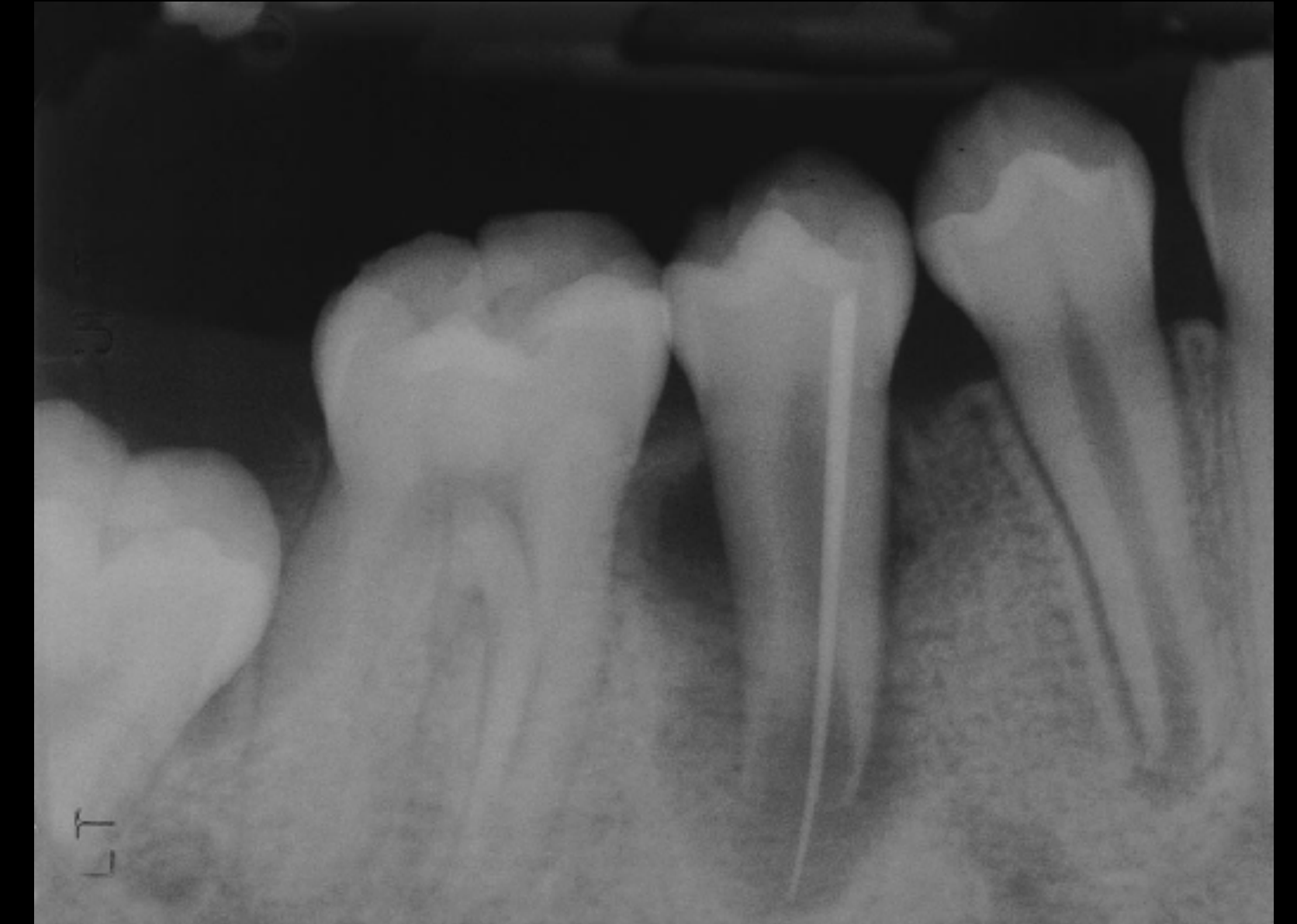
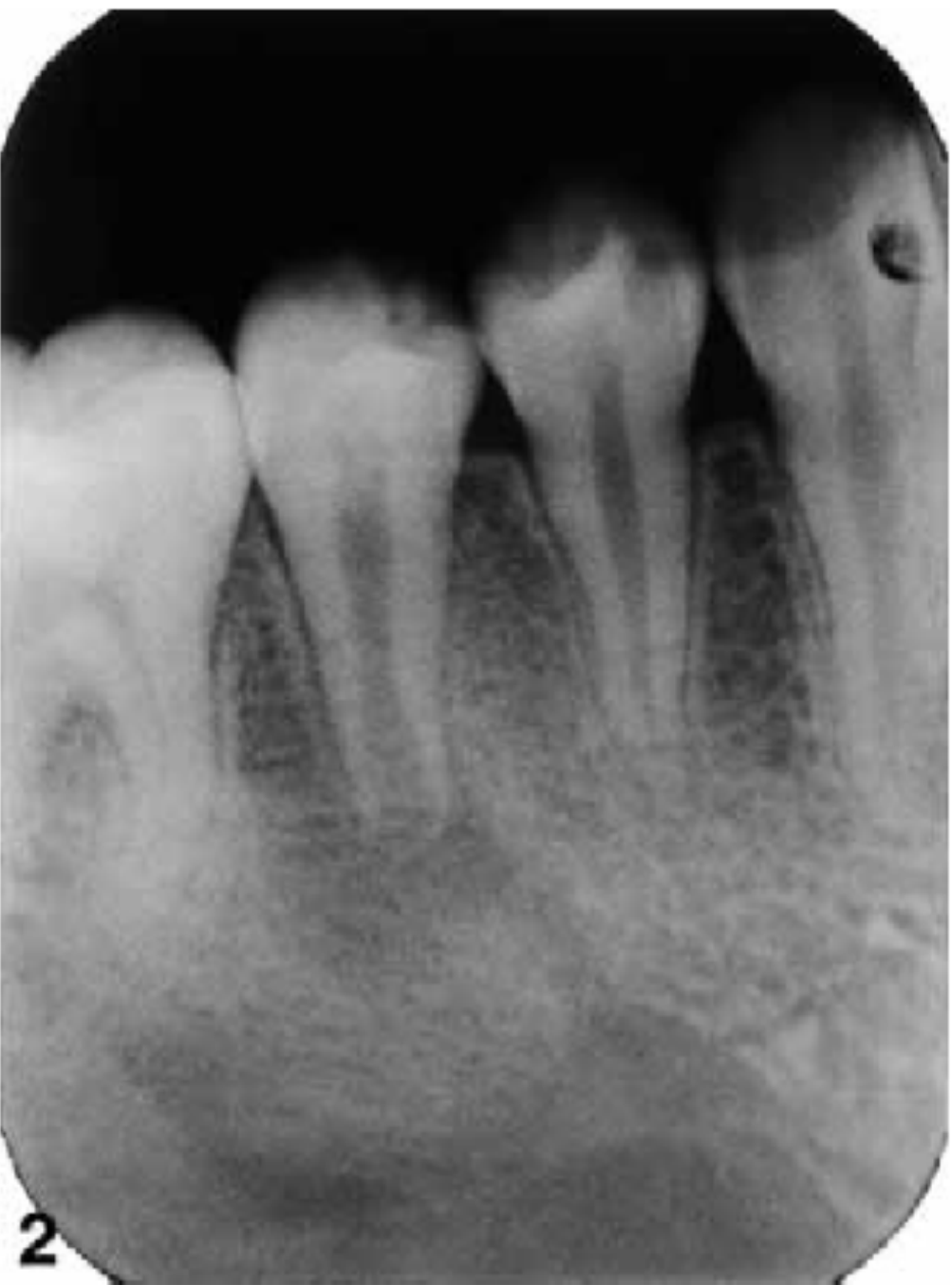
Iwaya Revascularization Case Report
In one immature mandibular premolar with apical periodontitis and a sinus tract, induced bleeding and a bioceramic/MTA seal were followed by continued root extension, pulp-space calcification and hard-tissue formation; the more regular-appearing result was not predictable. Full evidence and source page: Iwaya et al 2001 - Revascularization Case Report.
Revascularization of an Immature Permanent Tooth with Apical Periodontitis and Sinus Tract23
The associated clinical question and radiographs remain in the case below.
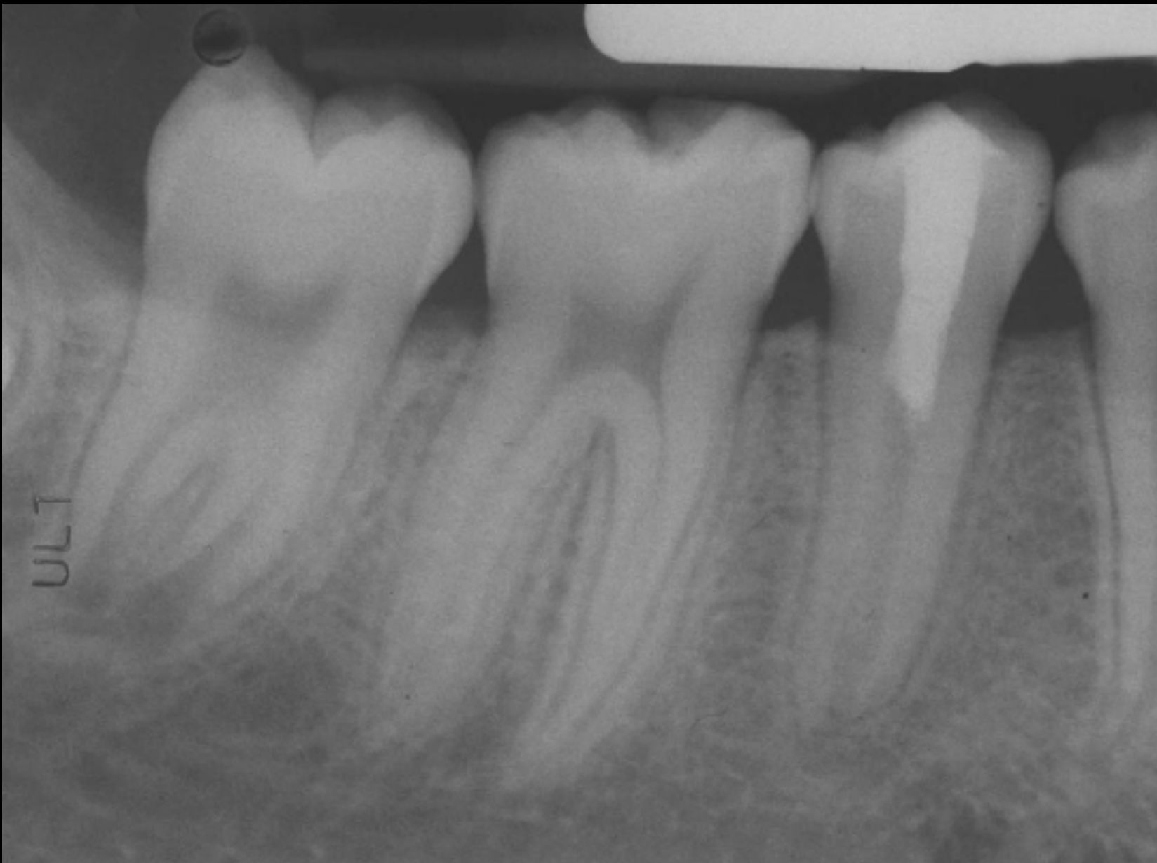
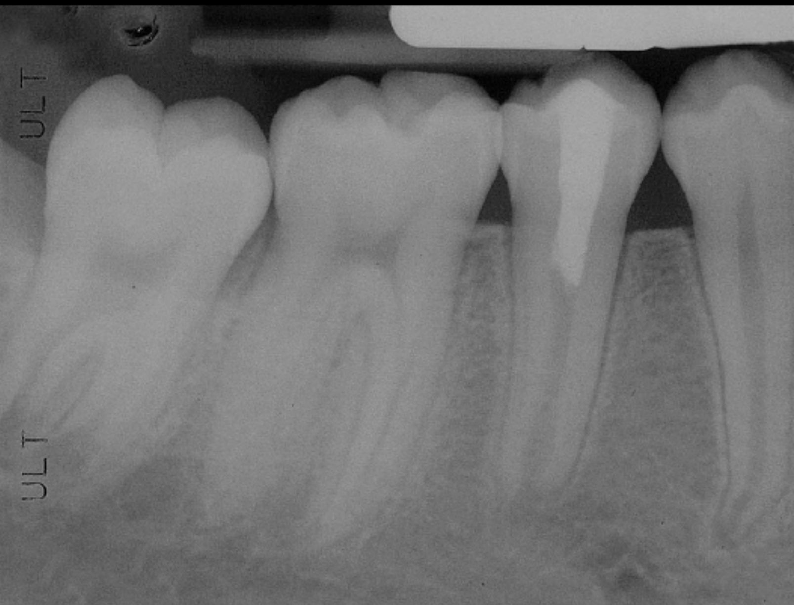
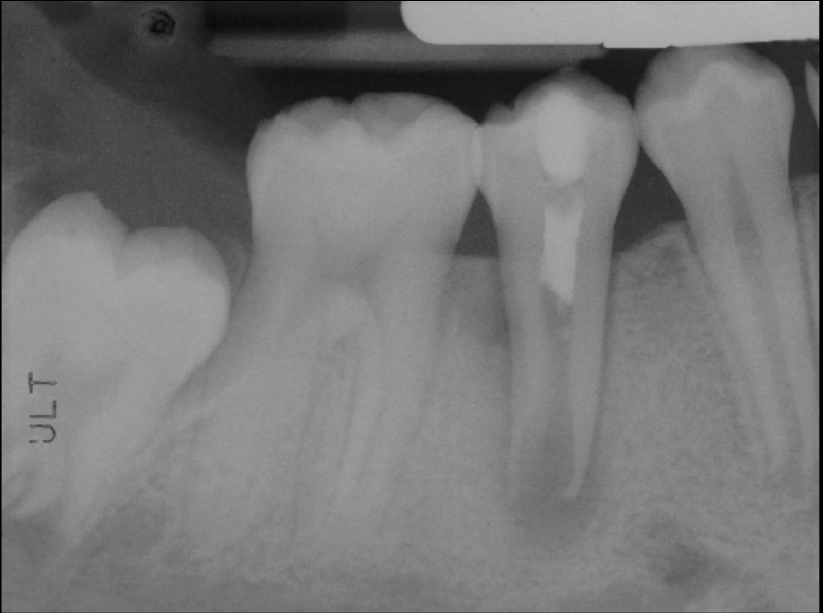
Banchs and Trope Revascularization Protocol2425
A single infected immature mandibular premolar showed continued root formation but irregular canal calcification and dentine after evoked bleeding and bioceramic sealing; this contrasted with the more regular-appearing Iwaya case and illustrates variable REP outcomes. Full evidence and source pages: Banchs and Trope 2004 - Revascularization Protocol.
 | 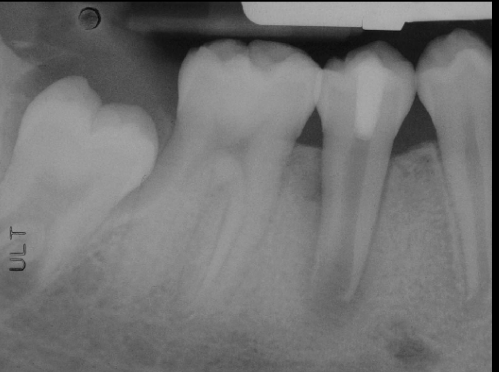 | 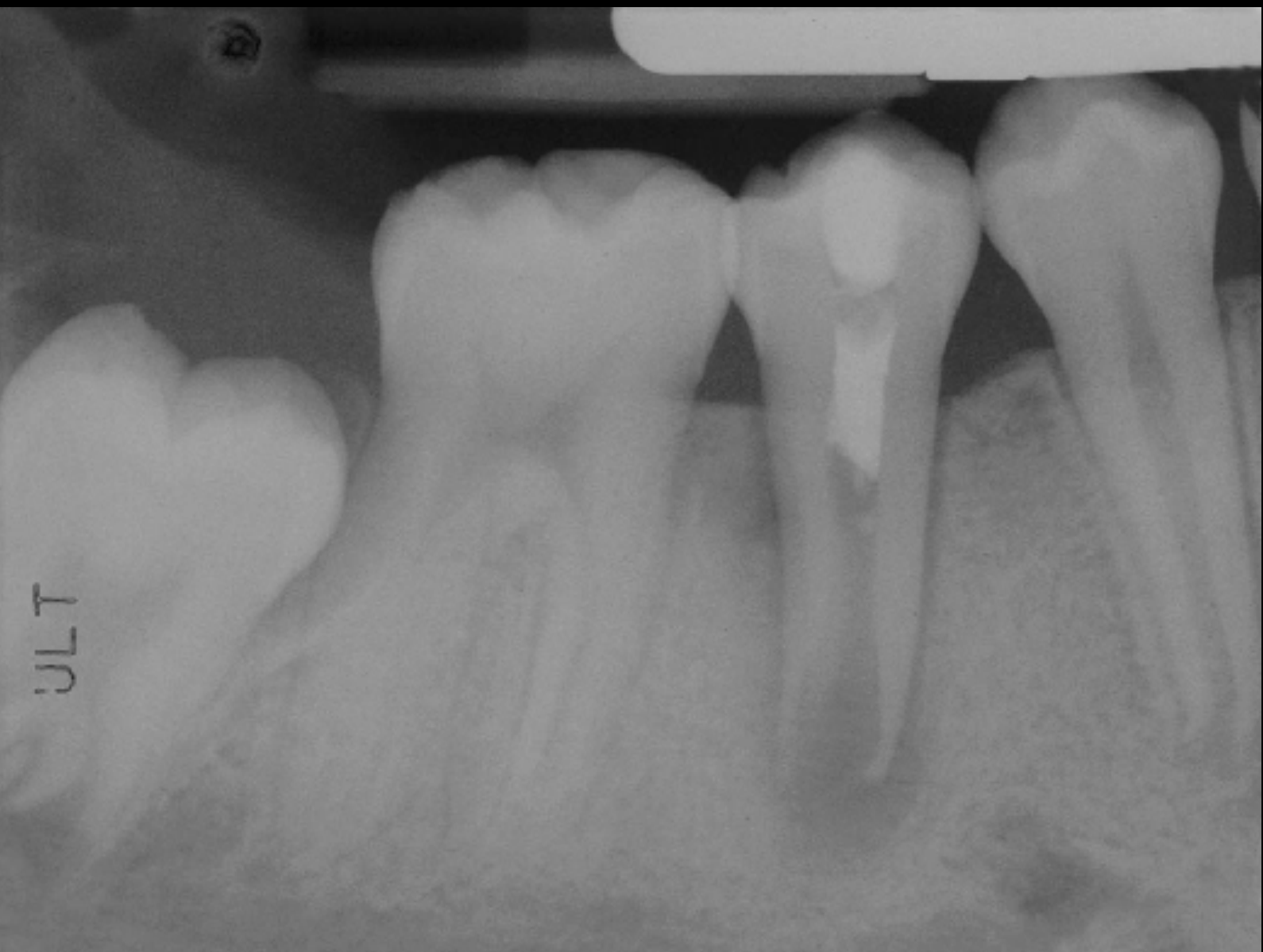 |
Revascularization of Immature Permanent Teeth with Apical Periodontitis: New Treatment Protocol?26
The distinct infected-premolar question and radiographs remain below.
Lecturer — Revascularization Outcomes
The lecturer observed that many published reports involved mandibular premolars, with variable regular dentine deposition or irregular pulp-space calcification; apparent success in those reports was not consistently seen in other tooth types.
Biology of Regenerative Endodontics
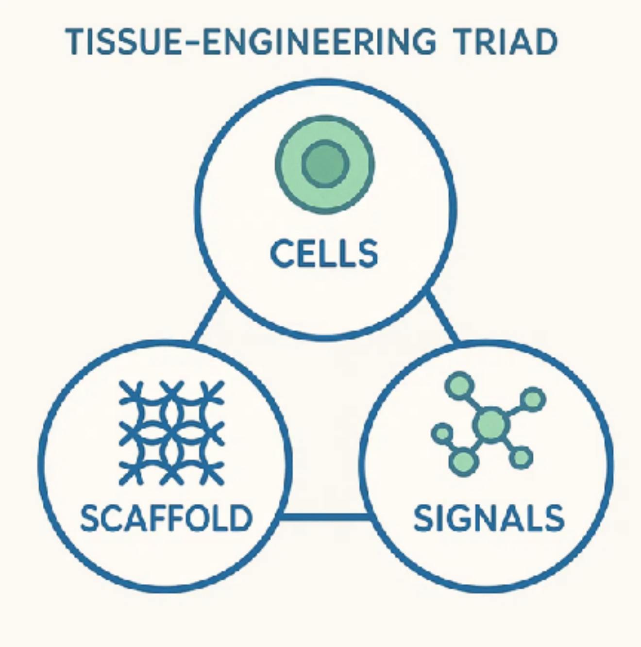
Cells, Scaffold, and Signals27
- CELLS — Viable progenitor/stem cells that can differentiate into pulp and dentin-forming cells
- SCAFFOLD — Physical matrix to host cells and provide structural support for tissue formation
- SIGNALS — Biochemical and mechanical cues that instruct cell proliferation and differentiation
These three components work together to rebuild a functional pulp-dentin complex
Key Cellular Players
SCAP — Stem Cells from Apical Papilla28
- Highly proliferative mesenchymal stem cells
- Main ‘workhorse’ for continued root development
- Produce dentine-forming cells and pulp-like tissue
Lecturer — SCAP Recruitment
SCAP may remain in the apical region as the root develops. When bleeding is induced from the periapical region, these cells may enter the root canal and contribute to the tissue formed there.
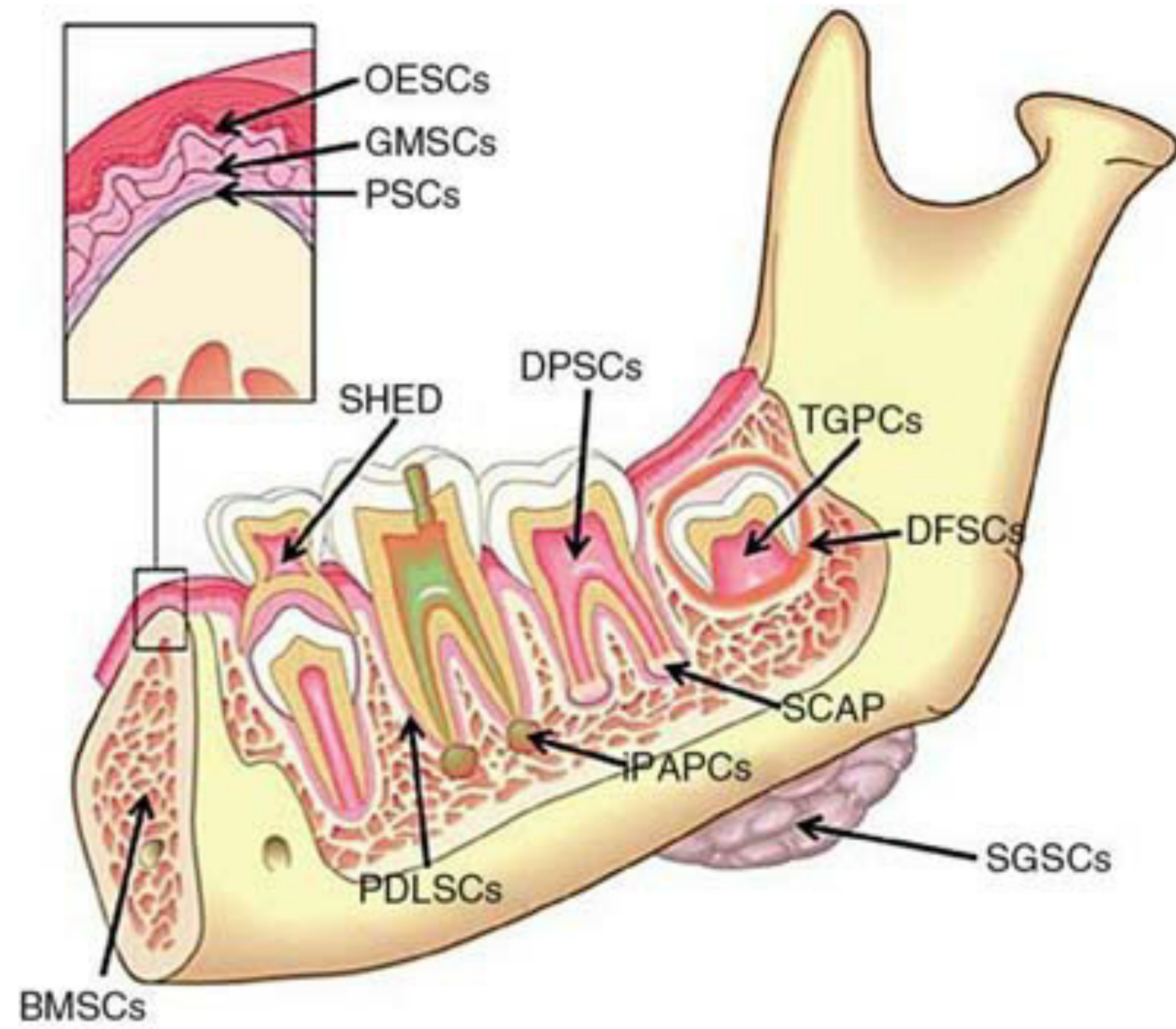
DPSCs — Dental Pulp Stem Cells
-
Found in residual pulp tissue
-
Contribute to regeneration when present
-
Support tissue repair processes
-
Residual dental pulp stem cells may remain within the root canal and contribute to repair or tissue formation
Other Contributors
- Periodontal ligament stem cells may differentiate into other cell types within the root canal environment
- Periodontal ligament stem cells
- Bone marrow-derived MSCs
- Creates mixed-origin regenerated tissue
Additional cell types identified: OESCs, GMSCs, PSCs, SHED, DPSCs, TGPCs, DFSCs, SCAP, iPAPCs, SGSCs, PDLSCs, BMSCs
Multiple cell types work together to regenerate pulp-dentin complex
Scaffold and Growth Factors
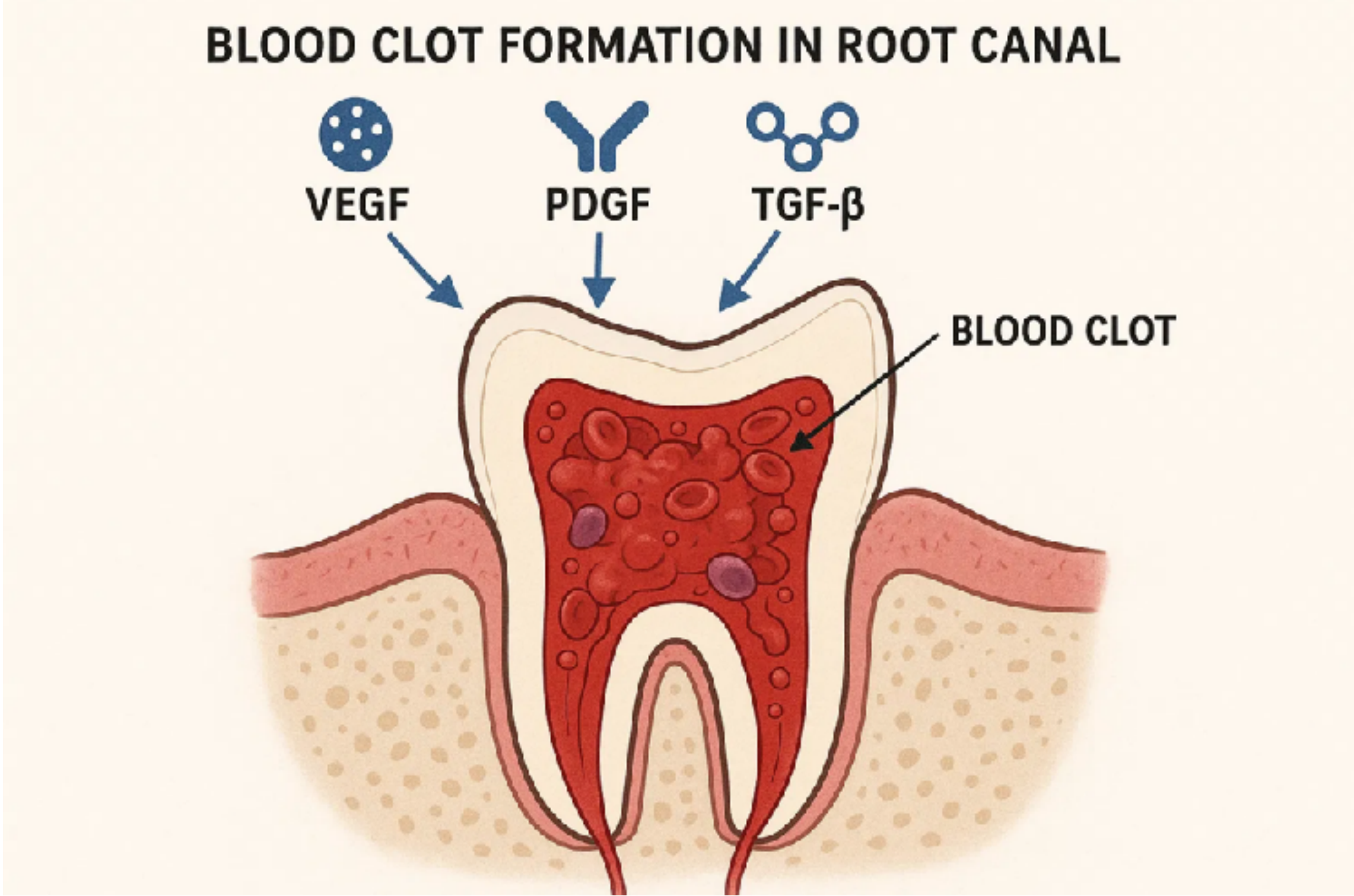
Blood Clot as Natural Scaffold29
- Provides fibrin matrix structure
- Contains platelets releasing growth factors
- Simplest clinical scaffold option
Lecturer — Blood Clot Scaffold
The blood clot is generated from apical or periradicular tissues and serves as a natural scaffold for cell homing.
- It provides fibrin and a matrix for cell attachment.
- It supports cell migration and population of the pulp space.
Alternative Scaffolds
- Autologous concentrates (PRF/PRP/CGF)
- Synthetic biomaterials (hydrogels, collagen)
- Polymer scaffolds (experimental)
Lecturer — Platelet Scaffolds
Platelet-rich fibrin and platelet-rich plasma can be prepared by drawing and centrifuging the patient’s blood.
- The resulting fibrin can be placed inside the root canal.
- These materials provide a scaffold and contain growth factors.
Blood clot formation in root canal involves: VEGF, PDGF, TGF-β → blood clot
Key Growth Factors
- VEGF — Promotes angiogenesis and vascular growth
- PDGF — Stimulates cell proliferation and migration
- TGF-β — Regulates cell differentiation and matrix formation
Lecturer — Growth Factor Release
Growth factors released from dentin may contribute to the regenerative process. The lecturer referred to tumor necrosis factor beta and platelet-derived growth factor as potentially relevant signaling factors, without requiring memorization of specific growth factors.
Microenvironment and Antimicrobial Balance
Infection Control30
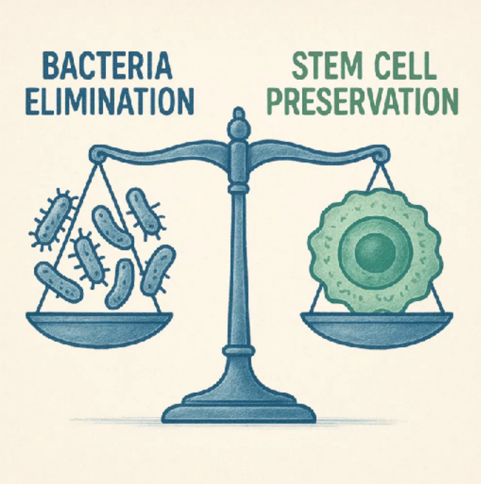
Elimination of bacteria is essential for successful regeneration, but harsh disinfection can damage stem cells.
Lecturer — Bleeding and Bacteria
Inducing bleeding may introduce additional bacteria into a canal that has already been disinfected.
- The article “The Trojan within the regeneration” described this as a possible contributor to failure of revascularization or regeneration.
- Regenerative treatment was therefore described as neither solid nor 100% predictable.
Stem Cell Viability
Preserving SCAP/DPSCs viability is crucial for tissue regeneration and continued root development.
Balanced Protocols
- Lower NaOCl concentrations
- EDTA irrigation to expose dentine-bound growth factors
- Gentle intracanal medicaments
Lecturer — Protocol Balance
The protocol must eliminate bacteria while preserving the viability of the cells intended to populate the canal. Strongly cytotoxic antimicrobial procedures may damage those cells, so antimicrobial effectiveness must be balanced against stem cell survival.
Cell Homing versus Cell Transplantation
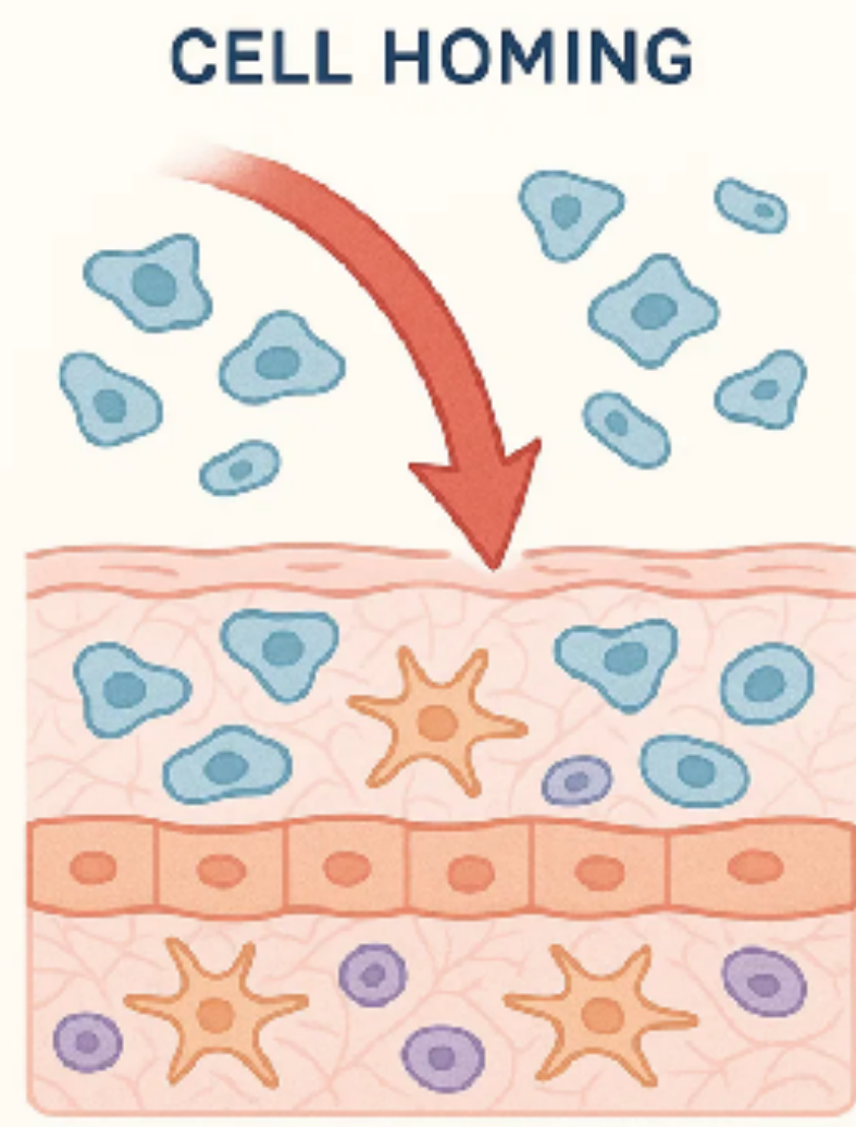
Cell Homing31
Recruiting the patient’s own stem/progenitor cells from periapical tissues into the canal using a scaffold and growth cues.
-
Common clinical approach
-
Simpler chairside procedure
-
No cell culture required
-
Less predictable cell types
-
Clinical cell homing involves inducing bleeding so cells from the periapical region can enter and populate the canal
 |  |
 |
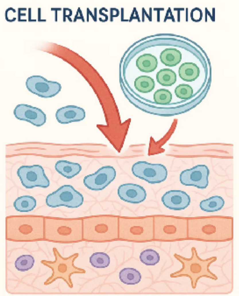
Cell Transplantation
Lab-grown stem cells (DPSCs/SCAP) seeded into engineered scaffolds and implanted into the root canal system.
- Controlled cell selection
- Potentially more predictable outcomes
- Complex laboratory procedures
- Regulatory and logistic challenges
Lecturer — Transplantation Status
Cell transplantation remains largely a research approach and has not yet been implemented clinically in routine treatment. The intended result is differentiation into odontoblast-like or pulp-like cells, followed by possible reinnervation.
Tissue Formed Inside the Canal
Fibrovascular Connective Tissue32
Most common finding in regenerated canals. Contains collagen fibers, blood vessels, and various cell types but lacks the organized structure of normal pulp.
Lecturer — Healing Response
The body may respond to induced bleeding as though it were healing an extraction site rather than regenerating an organized pulp-dentin complex. A blood clot is followed by the normal sequence of healing, producing fibrovascular connective tissue rather than a true regenerated pulp.
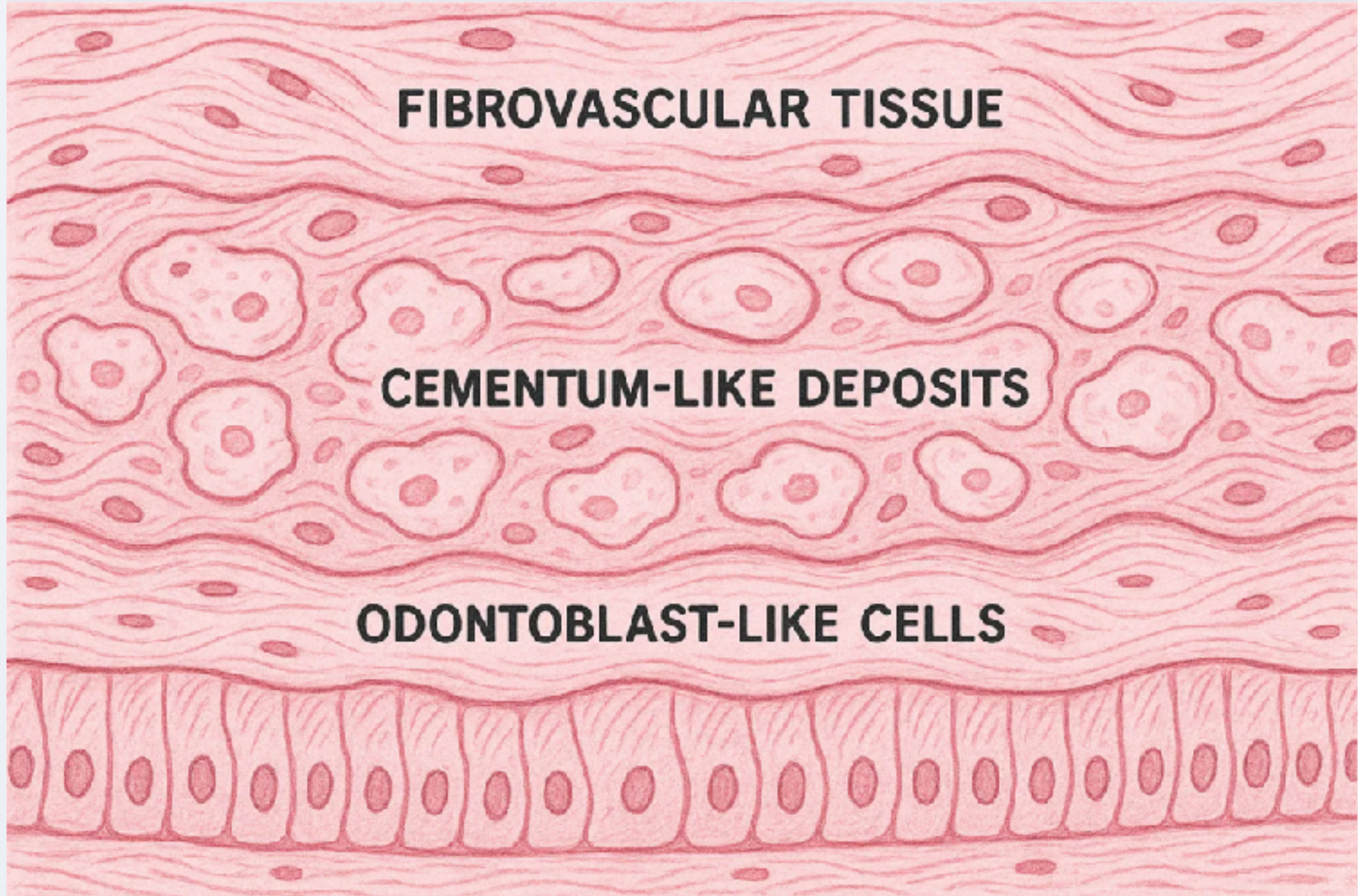
Cementum-like Deposits
Hard tissue formation along canal walls resembling cementum or bone-like tissue. Contributes to root strengthening but differs from normal dentine.
Odontoblast-like Cells
Occasionally observed cells resembling odontoblasts that may produce dentine-like tissue. True odontoblast layer with tubular dentine is uncommon.
- A true pulp and true odontoblast formation should not be assumed after regenerative treatment
Histological outcomes are variable and often differ from normal pulp architecture
Clinical Success ≠ Complete Biological Restoration — Root maturation and infection resolution can occur even without perfect pulp regeneration
Current Challenges and Future Directions
Current Challenges33
- Organised Pulp Formation — Directing stem cells to form organised pulp with proper odontoblast layer
- Predictable Re-innervation — Achieving consistent restoration of pulp sensibility
- Disinfection Protocols — Standardizing protocols that preserve cell viability
Lecturer — Additional Limitations
The lecturer also described variable root development and unpredictable outcomes between different teeth and cases.
- Possible findings include cementum-like tissue, water absorption, or ankylosis.
- The irregular calcification or cementum-like formation may not represent organized regeneration.
- There is uncertainty about whether regenerative treatment is ethical in teeth with more advanced root development when more predictable options are available.
Future Research Directions
- Advanced Scaffolds — Development of bioactive scaffolds with controlled release of growth factors
- Exosome Therapy — Utilising stem cell-derived exosomes for targeted regeneration
- Gene Therapy — Genetic modification to enhance regenerative potential
“Full pulp regeneration with organized tissue architecture remains an active research goal”
AAE Clinical Considerations for Regenerative Procedures34
AAE Clinical Considerations for a Regenerative Endodontic Procedure
Revised 5/18/2021
Case Selection Criteria35
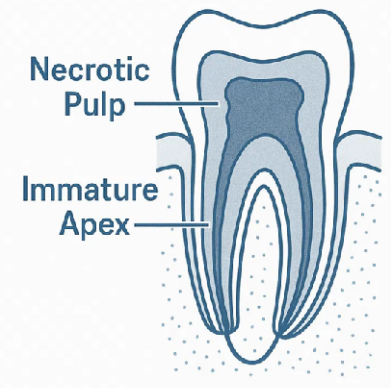
- Tooth Condition: Necrotic pulp with immature apex
- Pulp Space: Not needed for post/core or final restoration
- Patient Compliance: Compliant patient/parent
- Medical History: No allergies to medications and antibiotics necessary for procedure (ASA 1 or 2)
- Contraindications: Allergies to required medications, non-compliant patients
Lecturer — Root Maturity
The lecturer recommended regenerative treatment principally for very immature roots, particularly Stage 1 or Stage 2.
- In a Stage 1 or Stage 2 tooth with necrotic pulp, very little root may be present.
- Regeneration may be reasonable if the patient understands its risks, benefits, and unpredictable nature.
- If regeneration fails, apexification or another treatment modality may be attempted.
- For Stage 3, Stage 4, and Stage 5 roots, the lecturer preferred approaches such as apexification or an apical plug.
Informed Consent Requirements36
- Multiple Appointments: Two (or more) appointments required
- Medications: Use of antimicrobial(s) during treatment
- Possible Adverse Effects: Staining of crown/root, lack of response to treatment, pain/infection
- Alternatives: MTA apexification, no treatment, extraction (when deemed nonsalvageable)
Lecturer — Consent and Compliance
Patients must understand that root formation may not occur and that another treatment, such as apexification, may subsequently be required. Compliance with appointments and follow-up is necessary.
 |  |
 |
First Appointment Protocol
Anesthesia & Access37
Local anesthesia, dental dam isolation and access
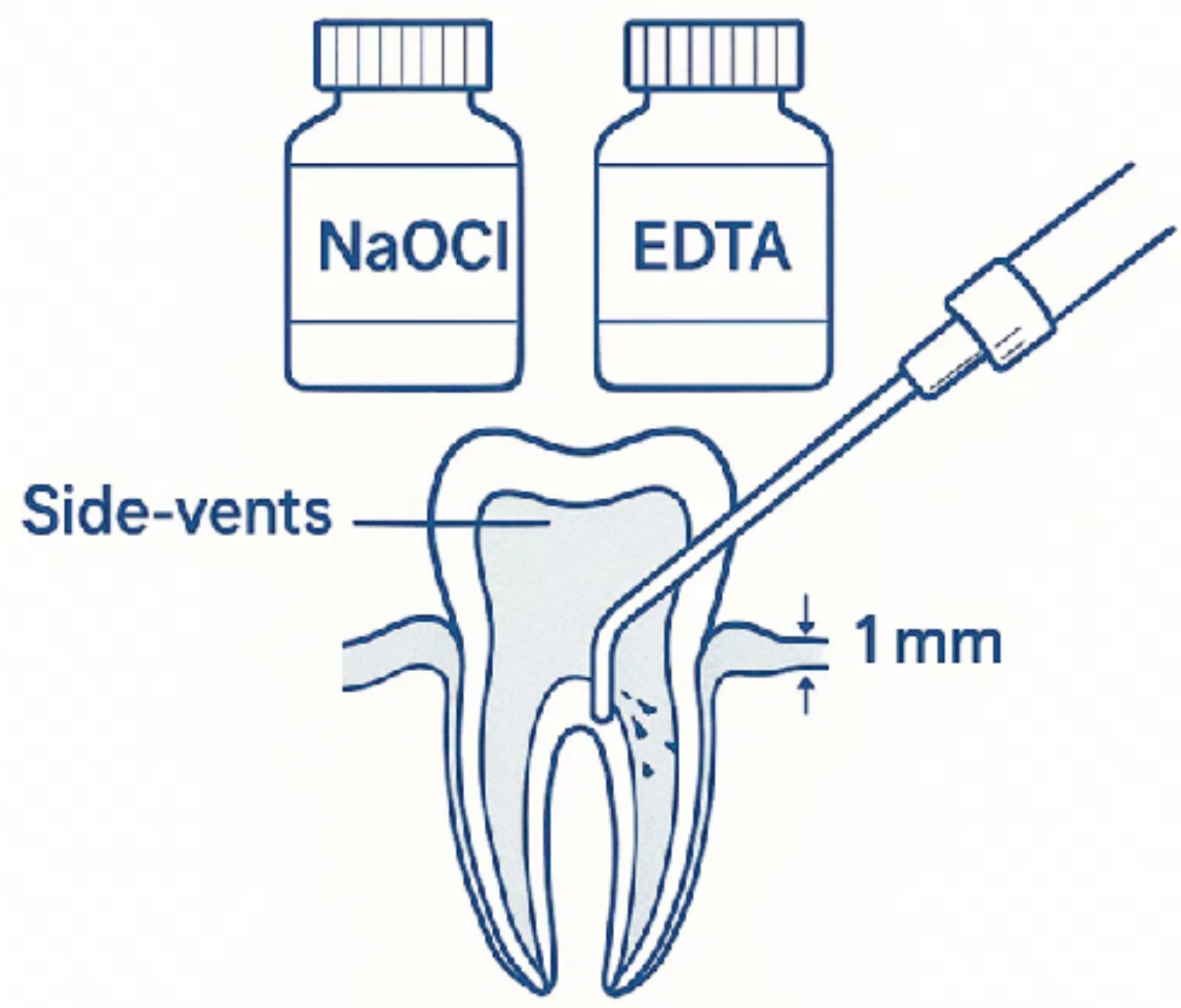
Irrigation Protocol
- Copious, gentle irrigation with 20 ml NaOCl
- Lower concentration: 1.5%–3% NaOCl
- Secondary irrigation with saline or EDTA
- Needle positioned ~1 mm from root end
Medication Placement
- Dry canals with paper points
- Place calcium hydroxide or triple antibiotic paste
- Keep below the CEJ to minimize staining
Lecturer — Canal Medicaments
Calcium hydroxide is now favored because the lecturer considered it less cytotoxic than triple antibiotic paste.
- Historically, triple antibiotic paste consisted of ciprofloxacin, minocycline, and metronidazole.
- EDTA helps expose dentin and release growth factors from the dentinal walls.
Temporary Restoration
Seal with 3–4 mm temporary material. Dismiss patient for 1–4 weeks.
Proper irrigation technique for regenerative endodontic procedure
Second Appointment Protocol
(1–4 weeks after first visit)
Assessment and Preparation38
- Assess response to initial treatment; if persistent infection, consider additional antimicrobial treatment
- Anesthesia with 3% mepivacaine without vasoconstrictor
- Copious, gentle irrigation with 20 ml of 17% EDTA
Lecturer — Second Appointment Assessment
Before proceeding, the tooth should be asymptomatic, with any swelling or draining sinus resolved. The tooth is then isolated and gently irrigated with EDTA.
 |  |
Blood Clot Formation
- Create bleeding by over-instrumenting with pre-curved K-file 2 mm past apical foramen
- Goal: Fill entire canal with blood to the level of the cemento-enamel junction
Lecturer — Blood Clot Material
A small K-file is extended slightly beyond the canal to initiate bleeding from the periapical area. Once the clot is established, a calcium silicate-based material, such as bioceramic material or MTA, may be placed over it in approximately two or three layers.
Restoration Placement
- Place resorbable matrix over blood clot with white MTA as capping material
- Apply 3–4 mm layer of glass ionomer, followed by final restoration
Follow-Up Protocol
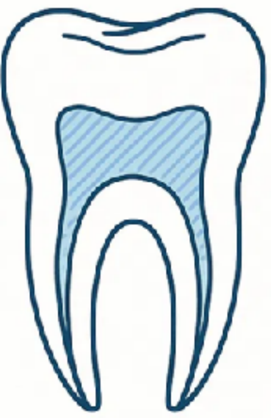
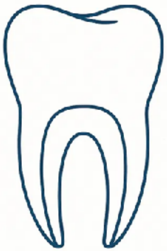
Clinical Examination39
No pain, soft tissue swelling or sinus tract
 |  |  |
 |  |
Radiographic Examination
Resolution of apical radiolucency (6–12 months)
Root Development
- Root width: Increased width of root walls (12–24 months)
- Root length: Increased root length (12–24 months)
Pulp Testing
Positive response to vitality testing
Lecturer — Pulp Sensibility
A positive sensibility response may suggest preservation or development of viable tissue, but it often does not occur because the tissue formed is not a normally innervated pulp.
Long-Term Follow-Up
Yearly follow-up after the first 2 years
Lecturer — Long-Term Outcomes
Long-term review is needed because regenerative treatment is unpredictable and infection may recur.
- Continued root development must be assessed at follow-up visits, including approximately 6, 12, and 24 months.
- Radiographs may show irregular calcification rather than organized dentin formation.
- Other possible outcomes include cementum-like tissue, water absorption, or ankylosis.
- If the procedure fails, alternative treatment such as apexification may be required.
Success Criteria for Regenerative Endodontic Procedure
- Primary goal: Elimination of symptoms and bony healing
- Secondary goal: Increased root wall thickness
- Tertiary goal: Positive vitality testing
Clinical Cases
Case: Right floor of mouth SCC with altered anatomy post-surgery
Question
Scenario: A lady diagnosed with right floor of mouth SCC in June 2024.
What’s shown: Significant alterations to the anatomy following surgery and chemo-radiotherapy, with a history of full dental clearance due to poor dentition.
Consider: What are the challenges in her rehabilitation and how is it being managed?
Answer
Observations:
- Significant alterations to the anatomy following partial glossectomy, marginal mandibulectomy, bilateral neck dissection, free flap, and post-op chemo-radiotherapy.
- Full dental clearance was performed at the same time as ENT surgery due to very poor dentition.
Reasoning: The significant anatomical changes make making a denture much trickier.
Outcome: Referred to prosthodontic colleagues who are currently in the process of making her a conventional full upper and full lower denture.
Takeaway: Complex anatomical alterations following head and neck cancer surgery and radiotherapy require specialized prosthodontic rehabilitation for denture fabrication.
Case: Facial structure rehabilitation with silicone prosthesis
Question
Scenario: A case of facial rehabilitation involving entire facial structures beyond just teeth.
What’s shown: A silicone prosthesis with natural hairs individually threaded into it.
Consider: What specialized skills are required for this type of rehabilitation and what details are notable in the prosthesis?
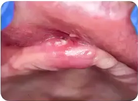
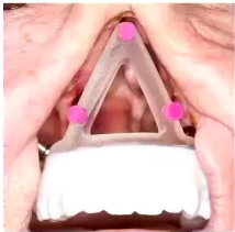
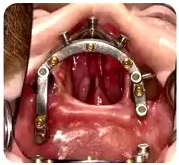
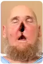
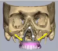

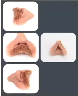
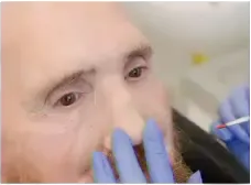
Answer
Observations:
- Rehabilitation of entire facial structures (eyes, ears, noses) rather than just teeth.
- Natural hairs are individually threaded into the silicone.
Reasoning: Maxillofacial prosthetists have the specialized experience and art form required to make these prostheses look and mimic the natural situation.
Outcome: The case demonstrates the successful involvement of a maxillofacial prosthetist in the multidisciplinary team to rehabilitate complex facial defects.
Takeaway: Rehabilitation after head and neck cancer can extend beyond teeth to entire facial structures, requiring the specialized artistry of a maxillofacial prosthetist.
Case: Periapical radiograph of immature maxillary central incisors
Question
Scenario: A clinical scenario involving an immature or incompletely formed root in upper anterior teeth following traumatic dental injury.
What’s shown: A periapical radiograph showing two maxillary central incisors.
Consider: What specific problem related to the root structure is visible on this radiograph?
Answer
Observations:
- Very minimum dentine thickness in one tooth as compared to the other.
Reasoning: The immature or incompletely formed root is not only short in length but also narrow in width, resulting in thinner dentinal walls which make the tooth susceptible to cervical fracture.
Outcome: None stated for this specific exhibit.
Takeaway: Immature roots have thin dentinal walls in addition to short length, increasing the risk of cervical fracture.
Case: Immature tooth with exposed pulp due to trauma
Question
Scenario: A clinical scenario of a tooth with exposed pulp following trauma.
What’s shown: Close-up clinical photographs of the tooth showing the exposure and the placed restoration, alongside a periapical radiograph.
Consider: What material was used for the treatment, and what does the follow-up radiograph demonstrate about the root?
Answer
Observations:
- Clinical photographs show exposed pulp due to trauma and the placement of calcium silicate cement.
- The periapical radiograph shows the root at a follow-up review.
Reasoning: The goal of placing the calcium silicate cement is to preserve the odontoblasts and vital pulp tissue inside the radicular root. Maintaining this vitality sustains the epithelial root sheath of Hertwig, which is necessary for the root to continue its physiological development.
Outcome: The root maintained its vitality and continued its formation, as demonstrated on the follow-up radiograph.
Takeaway: Preserving vital pulp tissue in a traumatized immature tooth (apexogenesis) allows the root to continue its physiological development and formation.
Case: Permanent mandibular premolar, revascularization case report
Question
Scenario: A published case report from the literature describing the revascularization of an immature permanent mandibular premolar.
What’s shown: Periapical radiographs of the treated premolar following the procedure.
Consider: What treatment was performed, and what radiographic outcomes are observed in this case?
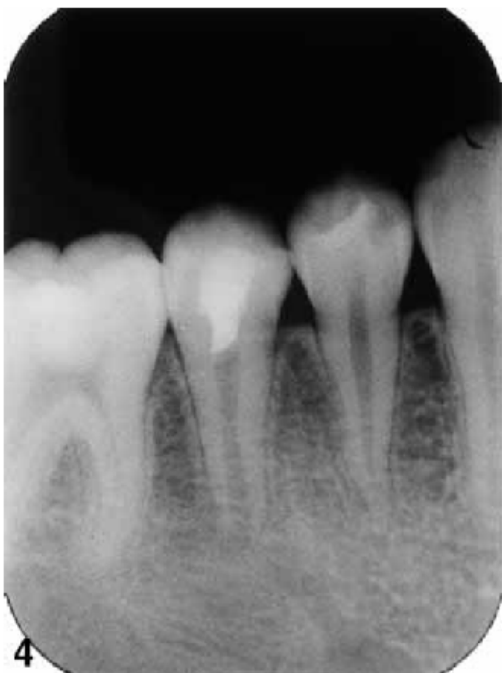
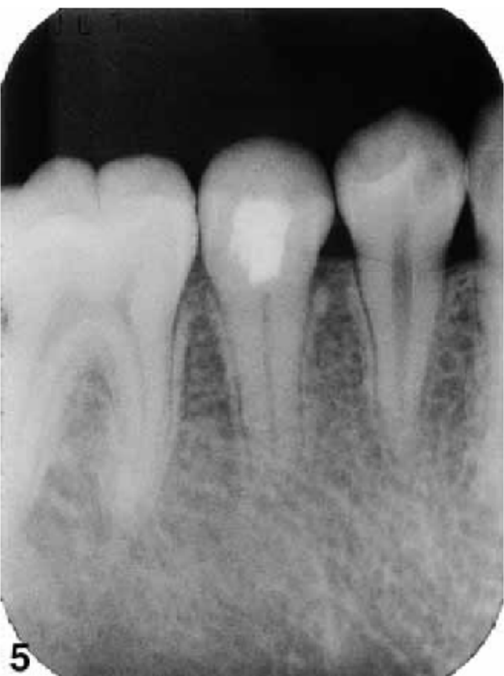
Answer
Answer: Evoked bleeding and a bioceramic/MTA seal were followed by root extension and calcification, with more regular-appearing dentine than in the infected Banchs case; this single result does not make REP outcomes predictable. Full study evidence: Iwaya et al 2001 - Revascularization Case Report.
Case: Infected immature mandibular premolar, revascularization case report
Question
Scenario: A published case report detailing the revascularization of an infected, immature permanent mandibular premolar.
What’s shown: Periapical radiographs of the treated infected premolar.
Consider: How was the infected tooth managed, and what does the radiographic outcome reveal about the tissue formed inside the canal?
Answer
Answer: Bleeding was induced in the root-canal system and sealed with bioceramic material. Root formation continued, but canal calcification and dentine were irregular compared with Iwaya’s case, illustrating variable REP outcomes in a single case. Full study evidence: Banchs and Trope 2004 - Revascularization Protocol.
Footnotes
-
Original PDF page 1: L16 Management of Incompletely Developed Tooth 1, p.1 ↩
-
Original PDF page 2: L16 Management of Incompletely Developed Tooth 1, p.2 ↩
-
Original PDF page 3: L16 Management of Incompletely Developed Tooth 1, p.3 ↩
-
Original PDF page 4: L16 Management of Incompletely Developed Tooth 1, p.4 ↩
-
Original PDF page 5: L16 Management of Incompletely Developed Tooth 1, p.5 ↩
-
Original PDF page 6: L16 Management of Incompletely Developed Tooth 1, p.6 ↩
-
Original PDF page 8: L16 Management of Incompletely Developed Tooth 1, p.8 ↩
-
Original PDF page 7: L16 Management of Incompletely Developed Tooth 1, p.7 ↩
-
Original PDF page 9: L16 Management of Incompletely Developed Tooth 1, p.9 ↩
-
Original PDF page 10: L16 Management of Incompletely Developed Tooth 1, p.10 ↩
-
Original PDF page 11: L16 Management of Incompletely Developed Tooth 1, p.11 ↩
-
Original PDF page 12: L16 Management of Incompletely Developed Tooth 1, p.12 ↩
-
Original PDF page 13: L16 Management of Incompletely Developed Tooth 1, p.13 ↩
-
Original PDF page 14: L16 Management of Incompletely Developed Tooth 1, p.14 ↩
-
Original PDF page 15: L16 Management of Incompletely Developed Tooth 1, p.15 ↩
-
Original PDF page 16: L16 Management of Incompletely Developed Tooth 1, p.16 ↩
-
Original PDF page 17: L16 Management of Incompletely Developed Tooth 1, p.17 ↩
-
Original PDF page 18: L16 Management of Incompletely Developed Tooth 1, p.18 ↩
-
Original PDF page 19: L16 Management of Incompletely Developed Tooth 1, p.19 ↩
-
Original PDF page 20: L16 Management of Incompletely Developed Tooth 1, p.20 ↩
-
Original PDF page 21: L16 Management of Incompletely Developed Tooth 1, p.21 ↩
-
Original PDF page 23: L16 Management of Incompletely Developed Tooth 1, p.23 ↩
-
Original PDF page 25: L16 Management of Incompletely Developed Tooth 1, p.25 ↩
-
Original PDF page 27: L16 Management of Incompletely Developed Tooth 1, p.27 ↩
-
Original PDF page 28: L16 Management of Incompletely Developed Tooth 1, p.28 ↩
-
Original PDF page 26: L16 Management of Incompletely Developed Tooth 1, p.26 ↩
-
Original PDF page 29: L16 Management of Incompletely Developed Tooth 1, p.29 ↩
-
Original PDF page 30: L16 Management of Incompletely Developed Tooth 1, p.30 ↩
-
Original PDF page 31: L16 Management of Incompletely Developed Tooth 1, p.31 ↩
-
Original PDF page 32: L16 Management of Incompletely Developed Tooth 1, p.32 ↩
-
Original PDF page 33: L16 Management of Incompletely Developed Tooth 1, p.33 ↩
-
Original PDF page 34: L16 Management of Incompletely Developed Tooth 1, p.34 ↩
-
Original PDF page 35: L16 Management of Incompletely Developed Tooth 1, p.35 ↩
-
Original PDF page 36: L16 Management of Incompletely Developed Tooth 1, p.36 ↩
-
Original PDF page 37: L16 Management of Incompletely Developed Tooth 1, p.37 ↩
-
Original PDF page 38: L16 Management of Incompletely Developed Tooth 1, p.38 ↩
-
Original PDF page 39: L16 Management of Incompletely Developed Tooth 1, p.39 ↩
-
Original PDF page 40: L16 Management of Incompletely Developed Tooth 1, p.40 ↩
-
Original PDF page 41: L16 Management of Incompletely Developed Tooth 1, p.41 ↩