L13 Dento-Alveolar Trauma 6 - Managing Root Fractures
Management of Transverse Root Fractures12
W/Prof. Paul V. Abbott AO
BDSc, MDS, FRACDS(Endo), FPFA, FADI, FICD, FACD, FIADT
- Specialist Endodontist
- Winthrop Professor of Clinical Dentistry
- UWA Dental School, The University of Western Australia
Dent Traumatol 2019; 35: 333-347.
J Endod 2019; 45: S13-S27.
 | 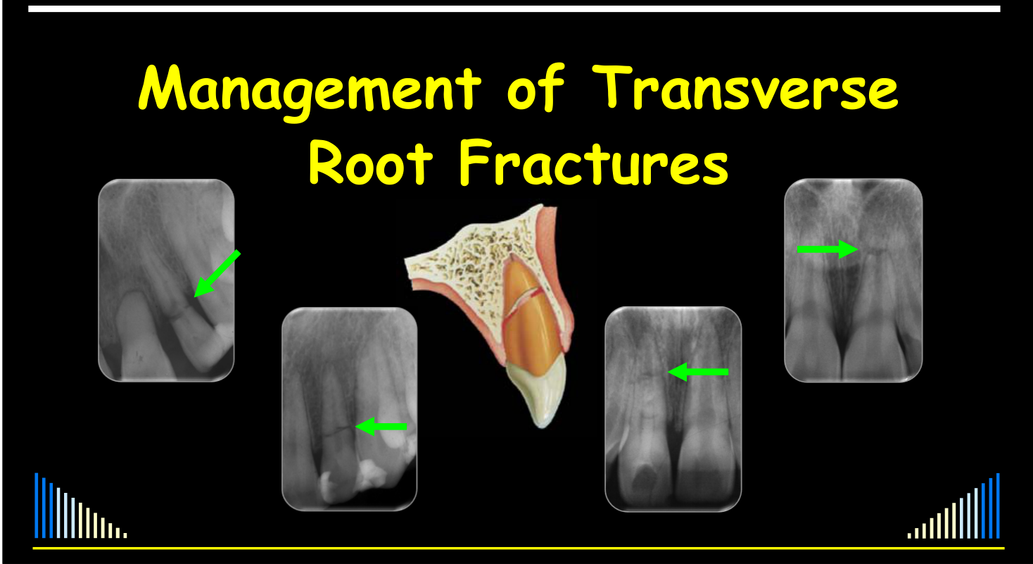 |
 |  |
Introduction and Scope3
Related topic index: Root Fractures and Developing Teeth.
W/Prof. Paul V. Abbott AO
Outline:
- Background information
- Classification of Root Fractures
- Diagnosis of Root Fractures
- Responses to Root Fractures
- Prognosis
- Management
Lecturer — Lecture Scope
Transverse root fractures are an intriguing and relatively poorly understood traumatic injury.
- The lecture draws extensively on Andreasen’s textbook, the Dental Trauma Guide, and articles published in the Dental Traumatology Journal and the Journal of Endodontics after the 2018 World Congress on Dental Trauma.
- Crown-root fractures are mentioned later because some management principles are similar.
Transverse Root Fractures
-
What I will NOT be discussing:
- Crown:root fractures
- Vertical fractures
-
A crown fracture may also occur as a concurrent injury
Background
W/Prof. Paul V. Abbott AO
See the lecture outline above.
Root Fractures4
-
Root Fractures
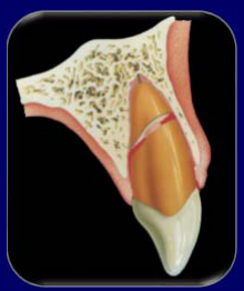
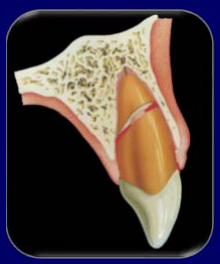
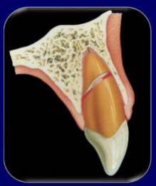
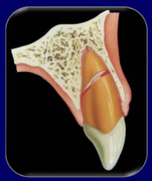
- Defined as fractures involving dentine, cementum and pulp
-
A relatively uncommon injury
- Andreasen & Andreasen (2007)
- 0.5 – 7.0% of injuries to permanent teeth
- 2.0 – 4.0% of injuries in primary teeth
- Stockwell (1988) – WA School Dental Service — study note
- 0.4% of the injuries (6–12 year old children)
- Lam, Abbott et al (2008) – Bunbury, WA — study note
- 3.0% of the injuries (children and adults)
- Andreasen & Andreasen (2007)
 |  |
Mechanism of Injury
- Usually a frontal impact
- Typically caused by fights and objects striking the teeth
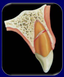
- Fracture can occur in different planes
- Depends on the direction and the distribution of the force
- More common to be oblique
- Even though many say “horizontal” !!
Lecturer — Force Direction
A force directed from a more coronal direction is more likely to produce a crown fracture or crown-root fracture.
Concurrent Injuries
- The coronal fragment:
- Concussion
- Subluxation
- Extrusion
- Lateral luxation
- Avulsion
Tissue Damage and Effects
Apical Fragment
- Neurovascular supply intact
- Pulp is not damaged
- PDL is not damaged
Coronal Fragment
- Pulp is stretched or lacerated at the fracture line
- Rupture and/or compression of the PDL
- Root surface may be exposed if coronal fragment is displaced
- Amount of damage will depend on whether the coronal fragment is displaced and the extent of any displacement
Lecturer — Tissue Damage
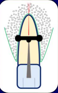
The apical and coronal fragments can be considered almost as two separate tooth segments.
- In the apical fragment, the neurovascular supply usually remains intact because the apical portion has not moved from its normal position, making pulp damage unlikely.
- In the coronal fragment, displacement may completely sever the pulp at the fracture line; periodontal ligament rupture may occur with avulsion or extrusion, whereas compression may occur with lateral luxation, particularly on the palatal aspect.
- The extent of damage also depends on the direction of displacement and the associated concurrent injury.
Classification of Root Fractures
See the lecture outline above.
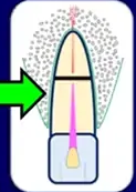
Classified According to Position5
- Position:
- Apical third
- Middle third
- Coronal third
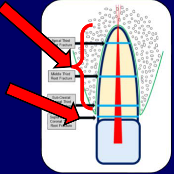
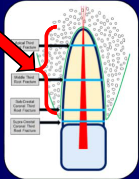
Figure labels: Apical Third Root Fracture; Middle Third Root Fracture; Sub-Crestal Coronal Third Root Fracture
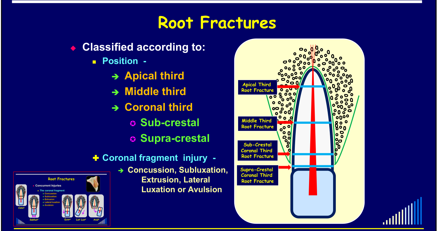
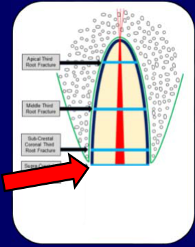
Classified According to Position and Coronal Fragment Injury6
- Position:
- Apical third
- Middle third
- Coronal third
- Sub-crestal
- Supra-crestal
- Plus coronal fragment injury:
- Concussion, Subluxation, Extrusion, Lateral Luxation or Avulsion
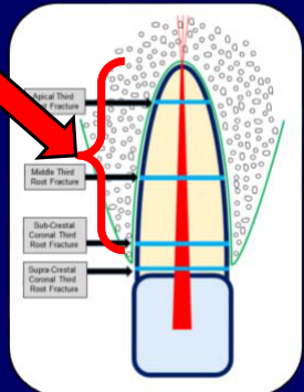
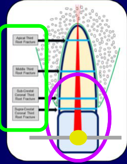
Figure labels: Apical Third Root Fracture; Middle Third Root Fracture; Sub-Crestal Coronal Third Root Fracture; Supra-Crestal Coronal Third Root Fracture
Inset: The coronal fragment — Concussion, Subluxation, Extrusion, Lateral luxation, Avulsion
Lecturer — Coronal Fracture Levels
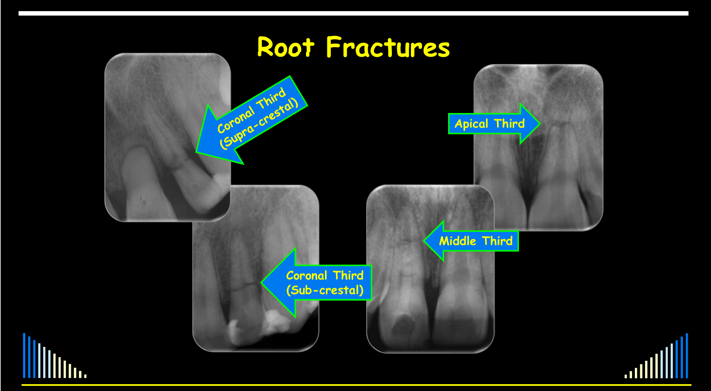
The coronal third group is further divided according to the relationship of the fracture to the alveolar crest.
- Subcrestal: the fracture is below the level of the bone.
- Supracrestal: the fracture is above the level of the bone and is more coronal.
- This distinction is important because supracrestal coronal third fractures require substantially different management from apical, middle, and subcrestal coronal fractures.
- Clinical assessment must also identify injuries to the coronal fragment, including concussion, subluxation, extrusion, lateral luxation, and avulsion.
Radiograph labels: Coronal Third (Supra-crestal); Apical Third; Coronal Third (Sub-crestal); Middle Third
 |  |
Diagnosis of Root Fractures
See the lecture outline above.
Clinical Examination7
- History - as for all trauma cases
- Clinical - as for all trauma cases
- Special things to note:
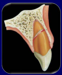
- Coronal segment may be mobile or displaced
- May be bleeding from the gingival sulcus
- May have crown discolouration (red or grey)
- May be tender to percussion
- Special things to note:
Lecturer — Clinical Interpretation
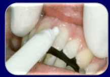
It may initially be difficult to determine whether mobility involves only the coronal fragment or the entire tooth.
- The clinical findings must be interpreted together with pulp tests and radiographs because the same signs may occur with other traumatic injuries.
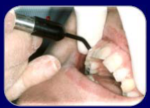
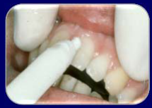
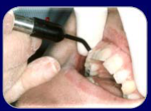
Pulp Sensibility Tests
- Usually initially negative for root fractures
- Except when there is no, or only minor, displacement of the coronal fragment
- Pulp tests are important to help assess the risk of healing complications
- Positive initial pulp test responses indicate a significantly reduced risk of pulp necrosis later
- Essential to perform at the emergency visit
- Time is NO EXCUSE for not doing these tests !!!
- Lack of response at the emergency appointment is not an indication to begin root canal treatment
- Time is NO EXCUSE for not doing these tests !!!
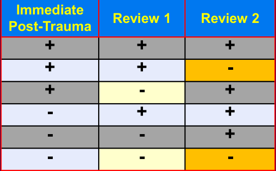
Initial Pulp Test Results at Review Appointments8
Initial pulp test results are necessary to assess the pulp status at subsequent review appointments, based on changes in how the pulp responds.
Lecturer — Pulp Recovery Patterns
Pulp responses can fluctuate after trauma, so decisions should not be made too quickly.
- The tooth may respond normally at all reviews, lose an initially positive response, regain a response after initially failing to respond, or alternate between positive and negative responses.
- Pulp testing helps determine whether the pulp is recovering or whether pulp necrosis is developing.
| Immediate Post-Trauma | Review 1 | Review 2 |
|---|---|---|
| + | + | + |
| + | + | - |
| + | - | + |
| - | + | + |
| - | - | + |
| - | - | - |
 |  |  |
 |  |
Radiographic Examination91011
- Radiographs are essential to reveal root fractures
- Minimum radiographs for typical trauma incident:
- 3 x periapical radiographs
- 1 x occlusal radiograph
- 1 - 2 PA’s of the opposing arch
- Consider other views - vertical tube shifts
→ To diagnose all injuries and to assess the healing at reviews
Lecturer — Radiographic Baseline
A complete initial radiographic series establishes a baseline for later assessment.
- It helps identify associated injuries and assess healing or non-healing, including the type of healing, at review appointments.
- Additional views may include an occlusal radiograph of the opposing arch when required.
- Occlusal radiograph
- Best for apical third and middle third root fractures
- Bisecting angle periapical radiograph
- Best for coronal third root fractures
- NOTE: Some root fractures not evident initially
- But become more obvious at review appointments - due to haemorrhage or granulation tissue in the fracture line
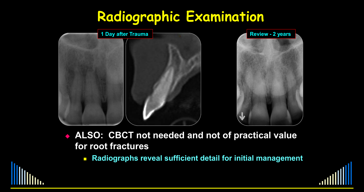
- ALSO: CBCT not needed and not of practical value for root fractures
- Radiographs reveal sufficient detail for initial management
Lecturer — Fracture Appearance
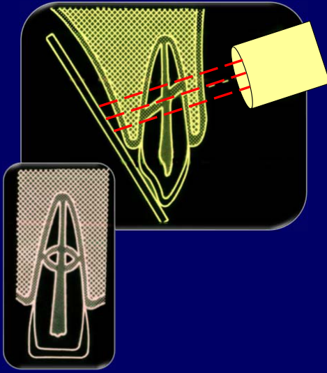
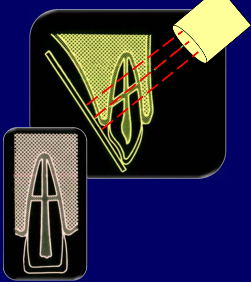
Radiographic appearance depends on the relationship between the fracture angle, the X-ray beam angle, and the position of the film or sensor.
- Unfavourable beam geometry can make one fracture appear as two lines.
- Changing the angulation may make it appear as a single line.
- Fractures may be especially difficult to see immediately when the coronal fragment has not been displaced.
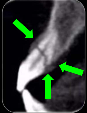
CBCT images - have revealed the likelihood of fractures of the alveolar socket wall being associated with root fractures
Lecturer — Associated Bone Fracture
An associated bone fracture is particularly likely when the coronal fragment has been displaced.
- Knowing about the bone fracture does not usually change management because repositioning and splinting the tooth also stabilizes the bone.
Lecturer — Connective Tissue Healing
Displacement may occur through lateral luxation, extrusion, or avulsion. Radiographs may show a space occupied by connective tissue, likely periodontal ligament tissue, along with rounding and remodelling of the fracture edges.
Responses to Root Fractures
Four typical responses:
- Healing with calcified tissue
- Interposition of connective tissue
- Interposition of bone and connective tissue
- Interposition of granulation tissue
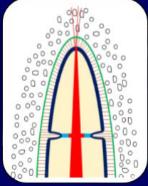
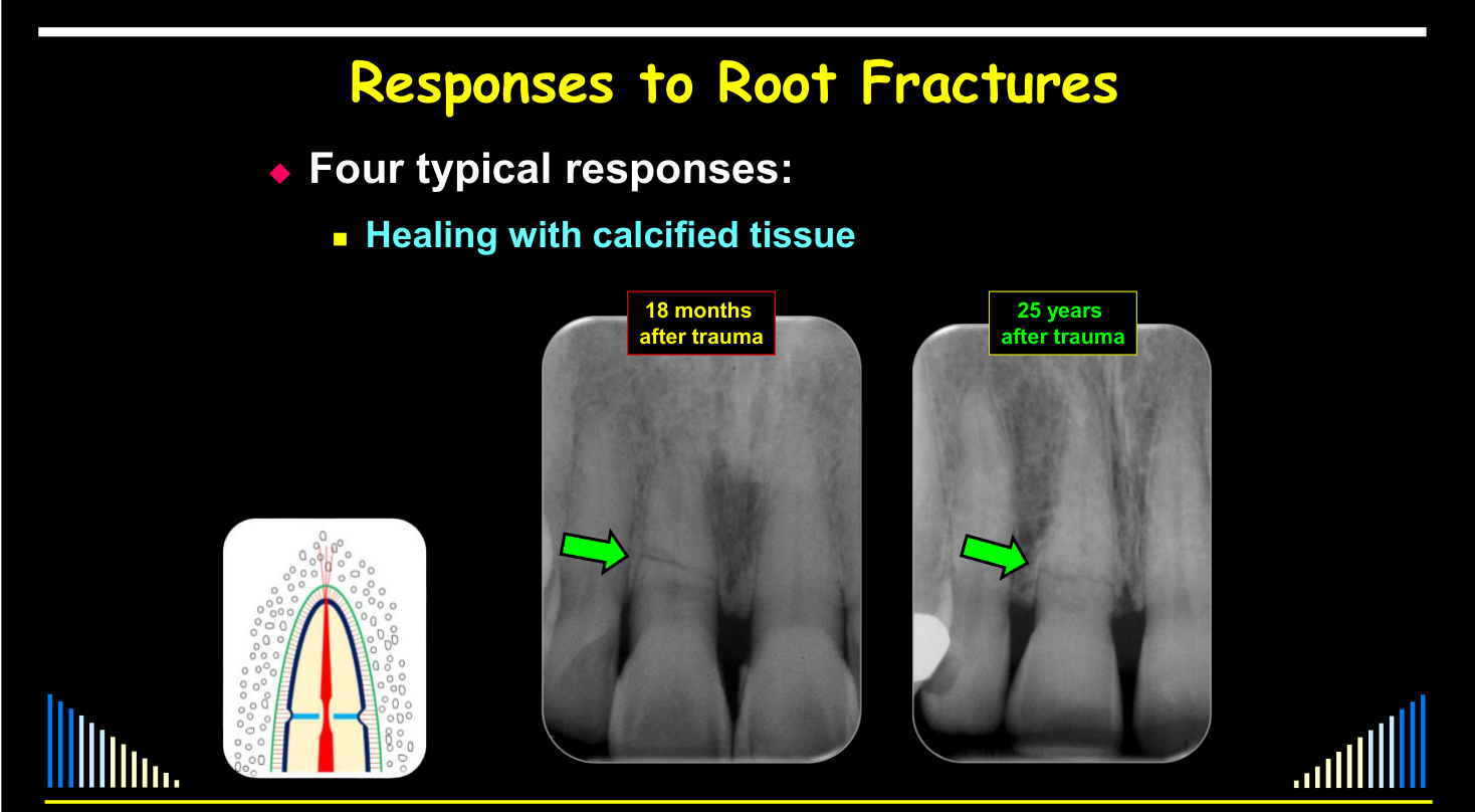
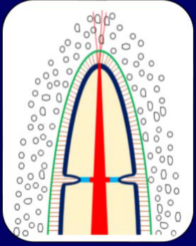
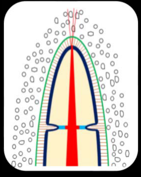
Healing with Calcified Tissue121314
- Most likely when no, or little, displacement of the coronal fragment
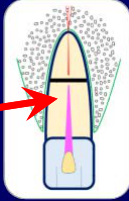
- “Internal repair” — dentine
- May be some osteodentine or cementum
- Usually have rounding of peripheral fracture edges
- Usually get Pulp Canal Calcification over time
Lecturer — Calcified Tissue Repair
Calcified-tissue healing is most favourable when the fragment is rapidly repositioned and the tooth is well stabilized.
- Dentine is laid down along the root-canal walls, narrowing the pulp canal.
- Cementum may repair the external fracture edges, and osteodentine may also be present.
- This tissue is not true dentine because odontoblasts are not present externally to produce it.
- Radiographs usually continue to show evidence of the original fracture, and calcification may occur in both fragments.
See the four response types above.
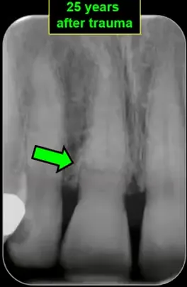
Radiographs: 18 months after trauma; 25 years after trauma.
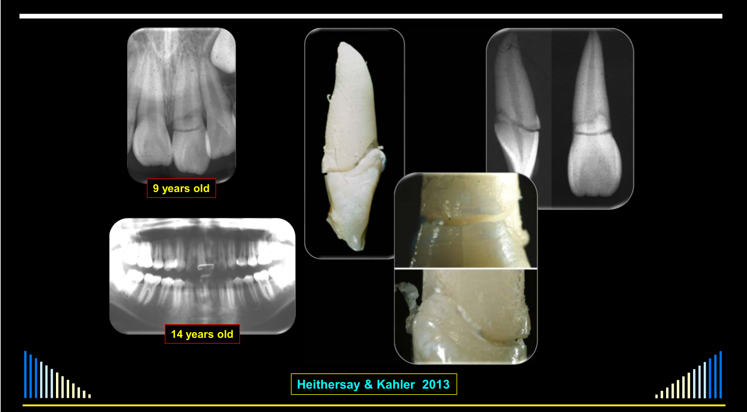
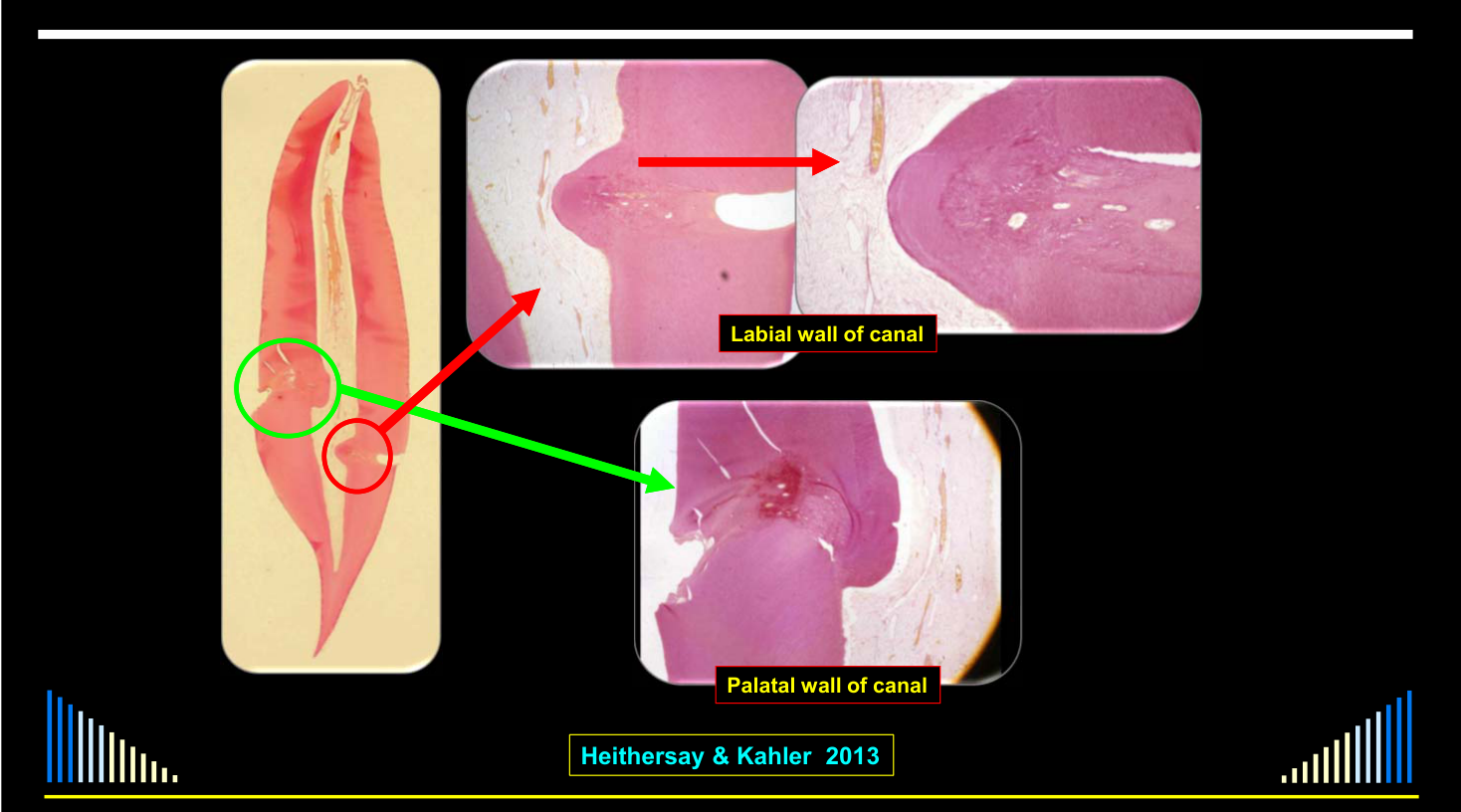
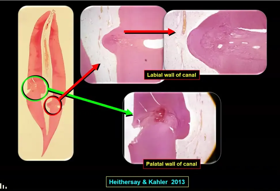
Heithersay, G. S., and Kahler, B. (2013). Healing responses following transverse root fracture: a historical review and case reports showing healing with (a) calcified tissue and (b) dense fibrous connective tissue, Dent Traumatol, 29, 253-265.
Radiographs: 9 years old; 14 years old.
Labels: palatal wall of canal; labial wall of canal.
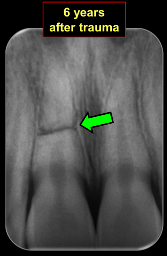
Interposition of Connective Tissue15
See the four response types above.
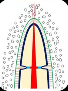
- Most likely when the coronal fragment is displaced
- Pulp is severely stretched or severed completely
- May repair / revascularize if no bacteria present
- PDL cells dominate healing in the fracture line
- Hence, connective tissue repair
- Usually get Pulp Canal Calcification over time
Radiographs: 6 years after trauma; 42 years after trauma.
 |  |
 |
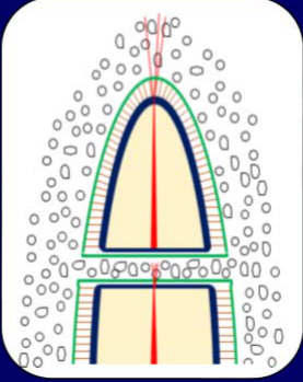
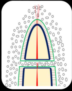
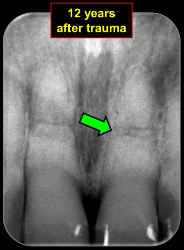
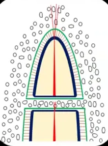
Interposition of Bone and Connective Tissue16
See the four response types above.
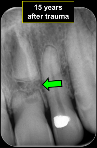
- If fracture occurs before completion of alveolar growth
- No union between fragments for this to occur
- The coronal fragment erupts normally
- The apical fragment remains in the same position
- Usually have rounding of peripheral fracture edges
- Usually get Pulp Canal Calcification over time
Lecturer — Bone and Tissue Interposition
In young patients, alveolar growth may allow the coronal fragment to erupt while the apical fragment remains in its original position, producing a gap between them.
- Bone may form between the fragments, sometimes together with connective tissue.
Radiographs: 12 years after trauma; 15 years after trauma.
 |  |
 |  |
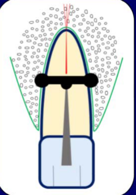
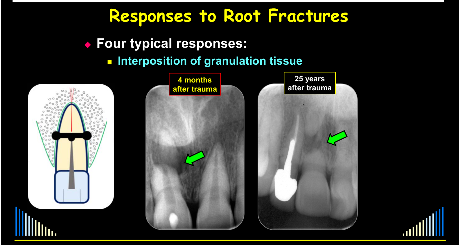
Interposition of Granulation Tissue17
See the four response types above.
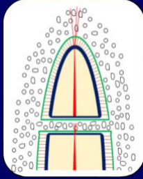
- If the pulp in the coronal fragment necroses and becomes infected
- Granulation tissue forms in the fracture line in response to the infection
- Similar to apical periodontitis from an infected root canal system
- No union between fragments
- May be some inflammatory resorption leading to widening of the foramen at the fracture line
- Granulation tissue forms in the fracture line in response to the infection
- The pulp in the apical fragment remains normal and usually has Pulp Canal Calcification over time
- Usually have rounding of peripheral fracture edges
Lecturer — Prognosis of Responses
The first three responses are considered favourable, whereas granulation tissue formation is considered unfavourable.
Lecturer — Granulation Tissue Features
The fracture line functions similarly to an apical foramen for the coronal fragment when its infected root canal system produces granulation tissue.
- Pulp necrosis and infection may occur within the first three or four months after trauma or many years later.
- Late infection may be associated with crown infractions that permit bacterial penetration or with breakdown of a restoration.
- Radiographs may show radiolucency on either side of the fracture.
Radiographs: 4 months after trauma; 25 years after trauma.
 |  |
Pulp Responses After Root Fractures1819
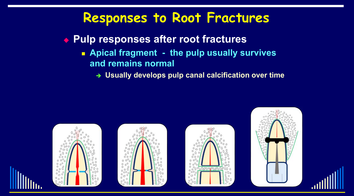
- Apical fragment — the pulp usually survives and remains normal
- Usually develops pulp canal calcification
- Coronal fragment
- Pulp may recover / revascularise
- Typically then get pulp canal calcification
- OR may get pulp necrosis and infection of the root canal system
- Inflammation in the fracture line
- i.e. granulation tissue
- Inflammation in the fracture line
- Pulp may recover / revascularise
Lecturer — Pulp Healing Prognosis
Pulp canal calcification is a normal physiological response and is not, by itself, an indication of a poor prognosis. A wide pulp space in an incompletely developed tooth provides a greater opportunity for revascularization and healing.
 | 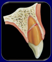 |
Studies of Pulp Necrosis After Root Fractures20
- 15 studies from 1930–2004
- No. of teeth in each study ranged from 18–400
- Pulp necrosis in each study ranged from 4%–55%
- Total: 1017 teeth
- 274 had pulp necrosis = 26.9%
Hence — the prognosis for pulp healing after root fracture is very good!
Lecturer — Overall Pulp Outcome
Overall, approximately three out of four teeth with root fractures have pulp healing. Pulp necrosis is therefore not the expected outcome in most cases.
- Many factors affect the pulp response
- Age
- Stage of root development
- Size of pulp lumen at the fracture line
- Mobility of the coronal fragment
- Displacement of the coronal fragment
- Diastasis between fragments
- i.e. whether the pulp was stretched or ruptured at the fracture line
- The treatment provided
Lecturer — Pulp Response Factors
The pulp response depends on the exact injury and its management, so it is difficult to generalize about pulp healing.
Hence, it is difficult / impossible to generalise!
Outline: Background information; Classification of Root Fractures; Diagnosis of Root Fractures; Responses to Root Fractures; Prognosis; Management.
Lecturer — Incompletely Developed Teeth
Root fractures are very rare in incompletely developed teeth; the Dental Trauma Guide data included only 13 such teeth over approximately 50 years.
- Pulp canal calcification is a normal physiological response and does not indicate a poor prognosis.
Lecturer — Fully Developed Teeth
Bone loss refers specifically to loss of crestal bone height.
- Ankylosis and replacement resorption may be associated with concurrent injuries such as avulsion or lateral luxation.
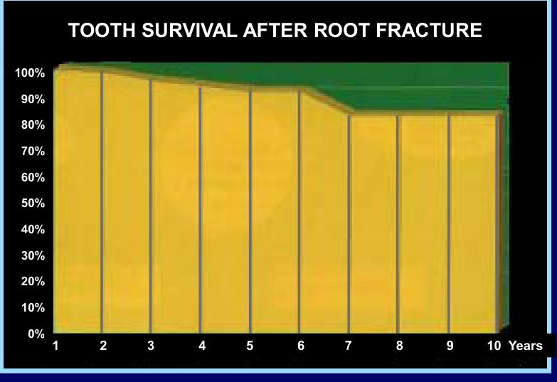
Lecturer — Tooth Survival
Tooth survival decreases progressively over time, but most teeth remain in place. Pulp necrosis and infection are uncommon overall, although the risk varies according to the severity and position of the injury.
Prognosis After Root Fracture
Incompletely developed teeth — 13 teeth:
- NO teeth were lost
- NO cases with external resorption or ankylosis
- NO cases with bone loss
- NO cases of pulp necrosis over 10 years
- Pulp canal calcification (note: cumulative numbers)
- 38.5% (4 teeth) after 1 year
- 55.1% (6 teeth) after 3 years
- 82.1% (9 teeth) after 10 years
Hence — the prognosis for incompletely developed teeth after root fracture is very good!
Source: www.dentaltraumaguide.com
Incompletely Developed Teeth21
For the related thin-wall and cervical-fracture risk in immature teeth, see L16.
Uncommon problems after root fracture:
- Bone loss
- Coronal third fractures — 18.8% after 10 years
- Middle third fractures — 6% after 10 years
- Apical third fractures — NIL
- Surface resorption — NIL
- Inflammatory root resorption — NIL
- Ankylosis and replacement resorption
- 2.5% of middle third fractures after 10 years
- NIL for apical and coronal third fractures
Source: www.dentaltraumaguide.com
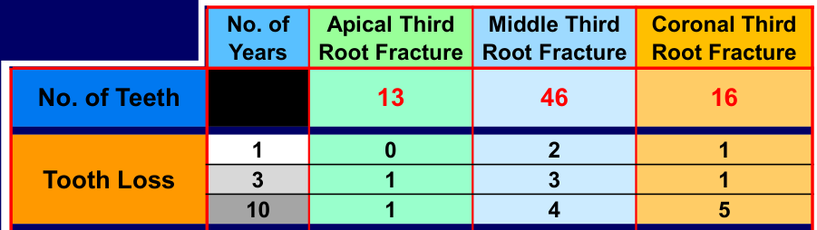
Fully Developed Teeth222324
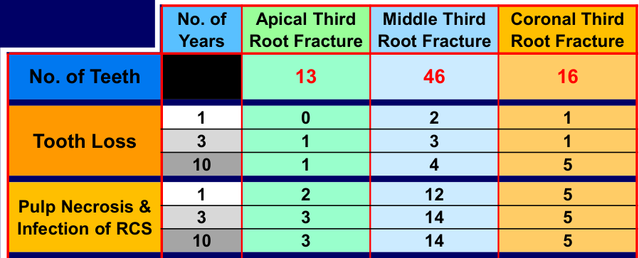
tooth loss:
| No. of Years | Apical Third Root Fracture | Middle Third Root Fracture | Coronal Third Root Fracture | |
|---|---|---|---|---|
| No. of Teeth | 13 | 46 | 16 | |
| Tooth Loss | 1 | 0 | 2 | 1 |
| 3 | 1 | 3 | 1 | |
| 10 | 1 | 4 | 5 |
Source: www.dentaltraumaguide.com
Andreasen et al 2007.
tooth loss and pulp necrosis:
| No. of Years | Apical Third Root Fracture | Middle Third Root Fracture | Coronal Third Root Fracture | |
|---|---|---|---|---|
| No. of Teeth | 13 | 46 | 16 | |
| Tooth Loss | 1 | 0 | 2 | 1 |
| 3 | 1 | 3 | 1 | |
| 10 | 1 | 4 | 5 | |
| Pulp Necrosis & Infection of RCS | 1 | 2 | 12 | 5 |
| 3 | 3 | 14 | 5 | |
| 10 | 3 | 14 | 5 |
Source: www.dentaltraumaguide.com
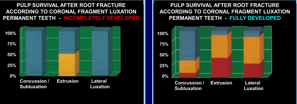
Graphs: Pulp survival after root fracture according to coronal fragment luxation (permanent teeth — incompletely developed; permanent teeth — fully developed), with categories Concussion/Subluxation, Extrusion, Lateral Luxation (Y-axis 0%–100%).
Tissue responses: Hard tissue; Connective tissue; Granulation tissue.
Andreasen et al 1989.
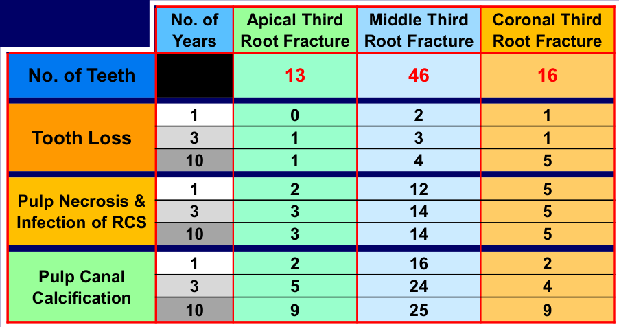
tooth loss, pulp necrosis and pulp canal calcification:
| No. of Years | Apical Third Root Fracture | Middle Third Root Fracture | Coronal Third Root Fracture | |
|---|---|---|---|---|
| No. of Teeth | 13 | 46 | 16 | |
| Tooth Loss | 1 | 0 | 2 | 1 |
| 3 | 1 | 3 | 1 | |
| 10 | 1 | 4 | 5 | |
| Pulp Necrosis & Infection of RCS | 1 | 2 | 12 | 5 |
| 3 | 3 | 14 | 5 | |
| 10 | 3 | 14 | 5 | |
| Pulp Canal Calcification | 1 | 2 | 16 | 2 |
| 3 | 5 | 24 | 4 | |
| 10 | 9 | 25 | 9 |
Source: www.dentaltraumaguide.com
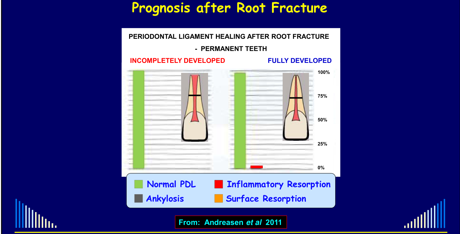
Graph: Periodontal ligament healing after root fracture — permanent teeth (incompletely developed vs fully developed); Y-axis 0%–100%; categories: Normal PDL, Inflammatory Resorption, Ankylosis, Surface Resorption.
Management of Root Fractures
Outline25
See the lecture outline above.
|  |
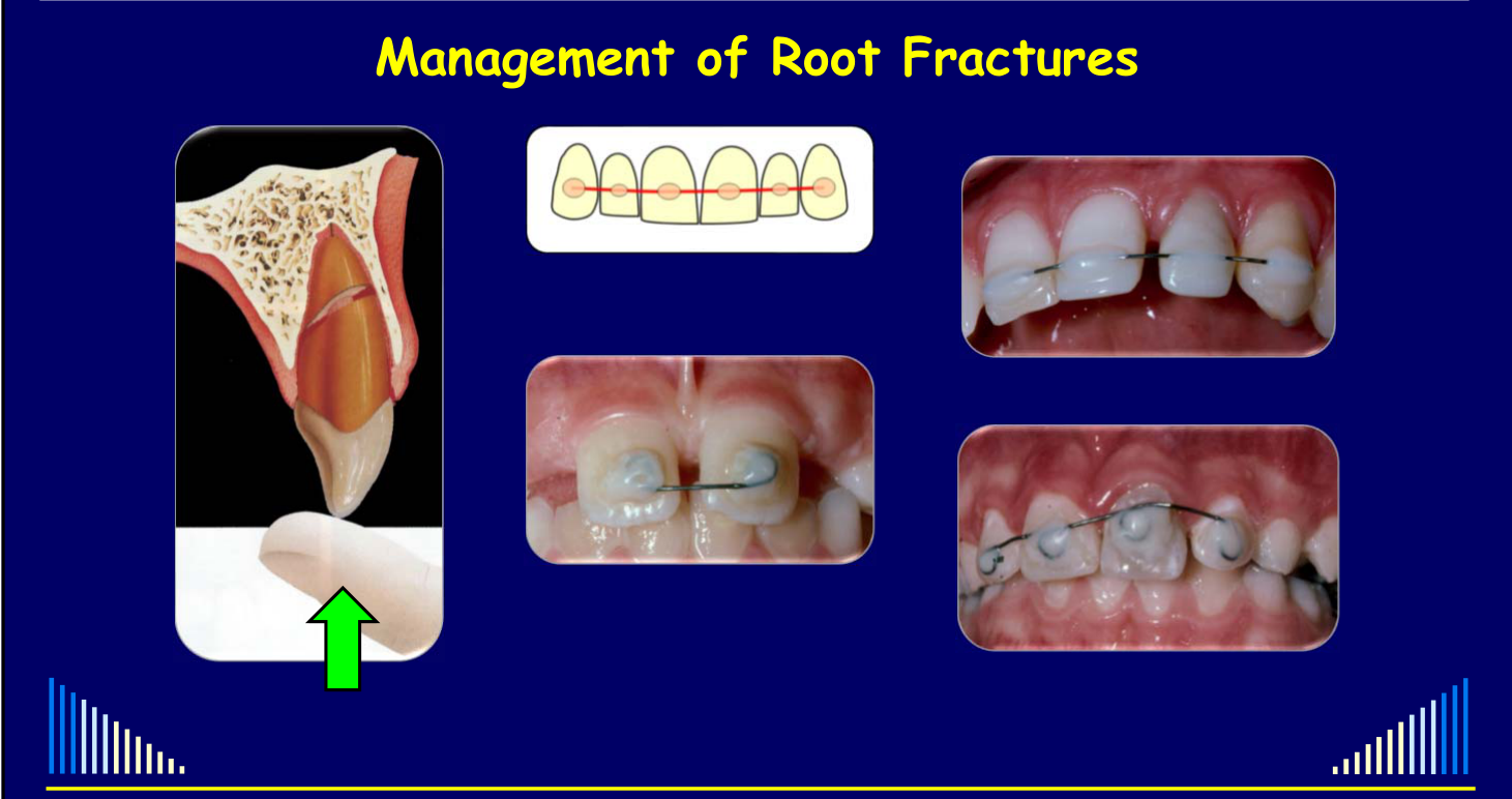
Initial Management by Fracture Position2627
- Initial management depends on fracture position
- Apical, middle, & coronal sub-crestal fractures — manage conservatively
- Reposition coronal fragment — if displaced
- Stabilise with wire/composite splint
- For 1 – 3 months
- The more coronal the fracture, the longer the splinting period
- Review — PDL and pulp healing
- Critical case — supra-crestal fracture in the coronal third
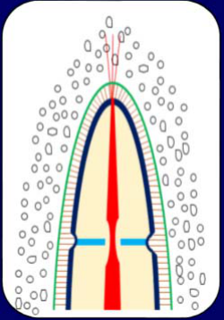
- AIM: to stabilise the fragments to provide the ideal environment for healing with calcified tissue
- i.e. internal dentine repair
- Healing is less likely if the fragments are moving
- Only one time / opportunity for this repair
- If repair does not occur, then cannot try again with further splinting
- There is no harm in splinting for longer periods compared to short periods
- Apical, middle, & coronal sub-crestal fractures — manage conservatively
Lecturer — Splint Stabilization
The splint should prevent movement of the coronal fragment without being excessively rigid.
- Reposition an extruded or laterally luxated tooth with the fingers before splint placement.
- Periodontal ligament healing takes at least approximately three months, while pulp healing and internal hard-tissue repair may take several months.
- If there are no signs of pulp necrosis, continue reviewing the tooth for as long as possible.
- Do not begin root canal treatment at the emergency appointment solely because the tooth does not respond to pulp testing.
 |  |
 |  |
Coronal and Middle Third Case Examples
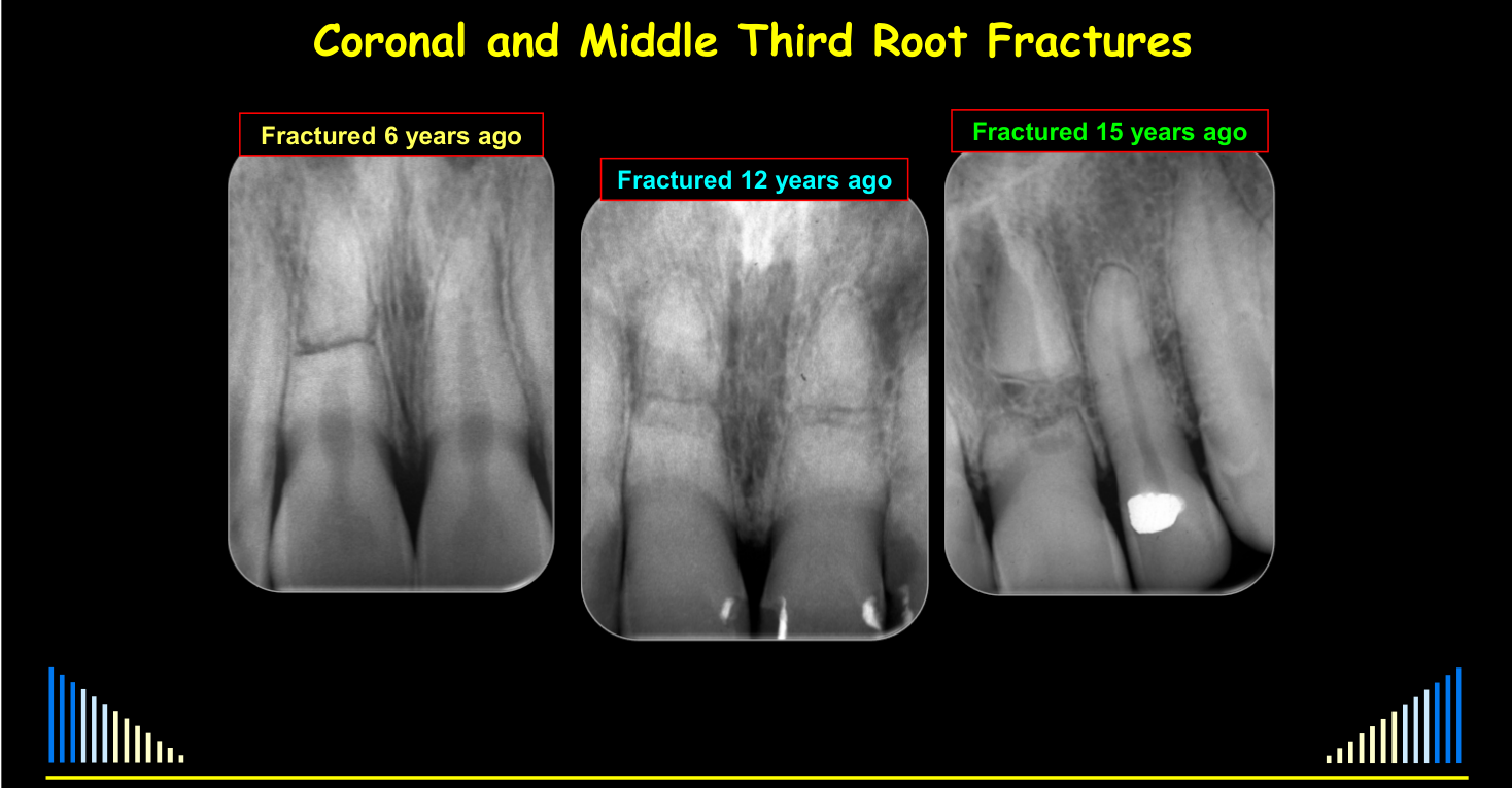
Coronal and Middle Third Root Fractures28
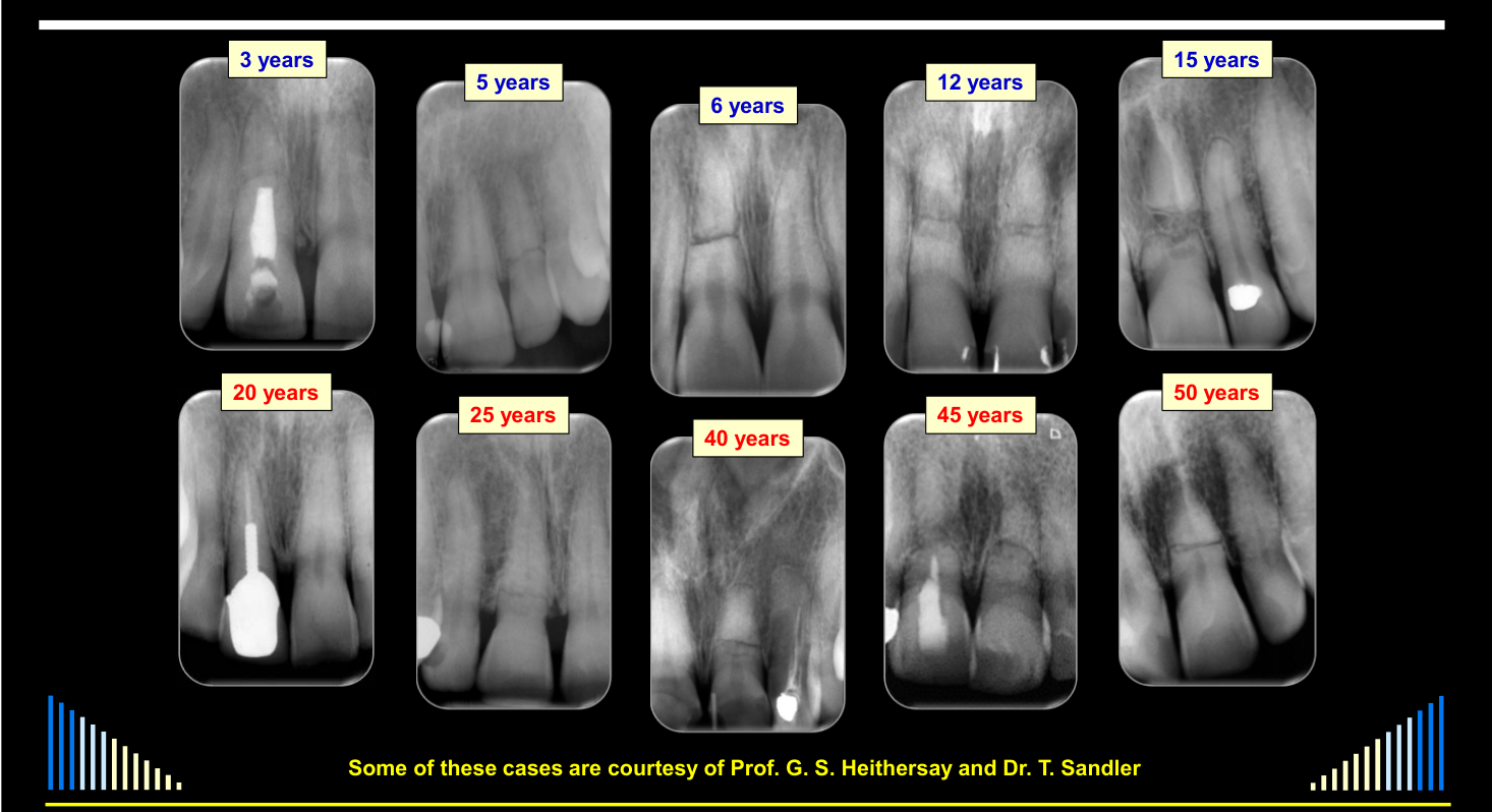
- Fractured 6 years ago
- Fractured 12 years ago
- Fractured 15 years ago
 | 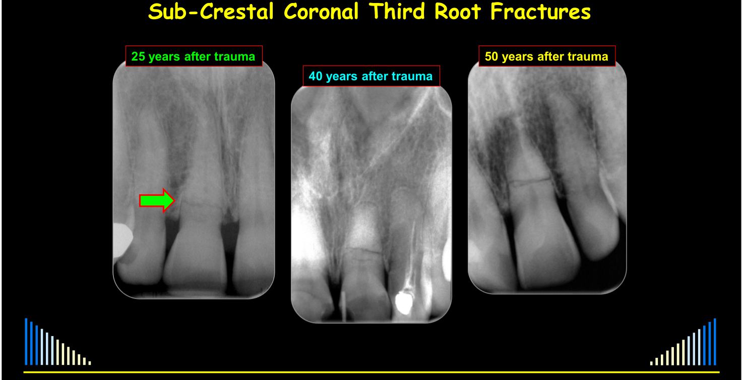 |
Sub-Crestal Coronal Third Root Fractures
- 25 years after trauma
- 40 years after trauma
- 50 years after trauma
Lecturer — Long-Term Outcomes
The teeth were managed by repositioning and stabilization, and pulp canal calcification occurred while the teeth remained stable.
- Clinically, the teeth could appear normal with respect to mobility, percussion, and general appearance.
- Some fractures were identified only radiographically, and some cases developed bone and connective tissue between the fragments.
Managing Pulp Necrosis and Infection
- Root Canal Treatment — coronal fragment only
- Preferred / simplest method with predictably favourable outcomes
- If necrosis + infection occurs early → hence, the foramen at the fracture line is usually wide open
- Need “apexification” — e.g. with Ca(OH)₂
- Or place a “plug” — e.g. with MTA
- If necrosis and infection occur later → PCC has usually narrowed the canal and opening
- Can do routine root canal treatment
- Related calcium-hydroxide apexification method for immature roots: L17.
Root Canal Treatment of the Coronal Fragment29
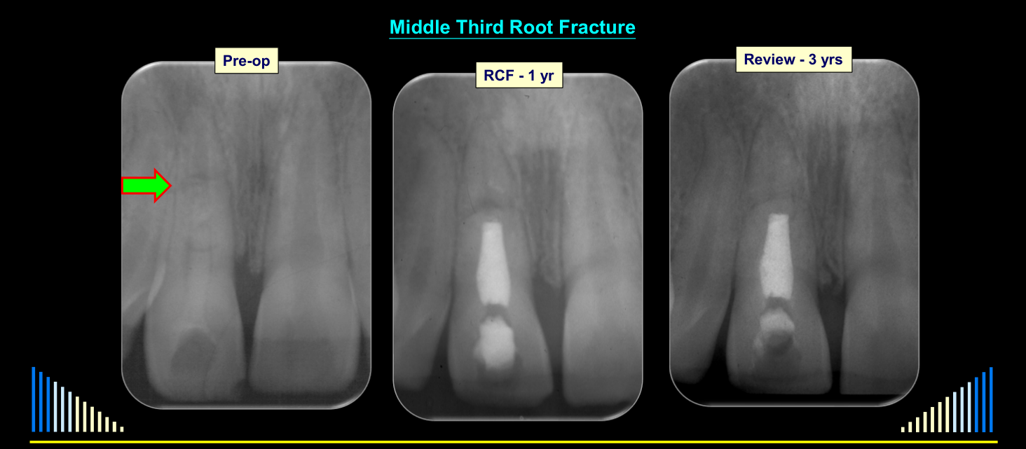
Middle Third Root Fracture
- 3 Months Ca(OH)₂
- 9 Months Ca(OH)₂
- RCF – 15 months
| Pre-op | RCF - 1 yr | Review - 3 yrs |
|---|---|---|
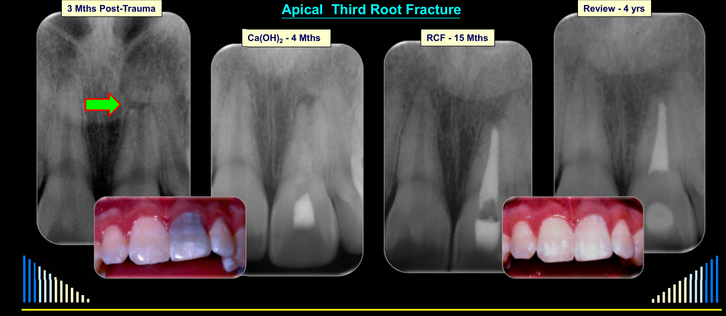
Apical Third Root Fracture
| 3 Mths Post-Trauma | Ca(OH)₂ - 4 Mths | RCF - 15 Mths | Review - 4 yrs |
|---|---|---|---|
See the coronal-fragment treatment summary above.
Lecturer — Early Pulp Necrosis
When early pulp necrosis occurs, the end of the coronal fragment is open and resembles an immature tooth.
- Long-term calcium hydroxide is generally preferred because placement of an apical plug is technically difficult.
- Calcium hydroxide is changed approximately every three months and placed only to the fracture line, not beyond it.
- Once a hard-tissue barrier has formed, root canal filling can be completed; examples showed root filling after approximately twelve to fifteen months.
- In an apical third fracture, the apical fragment is generally left untreated.
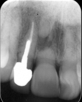
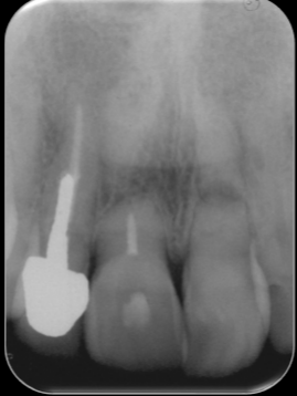
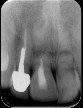
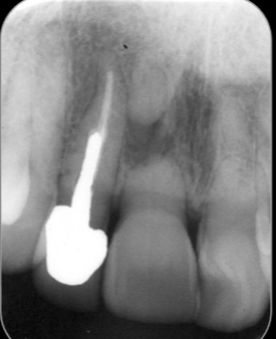
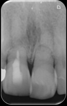
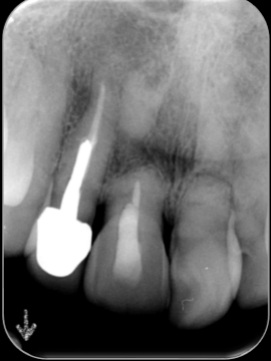
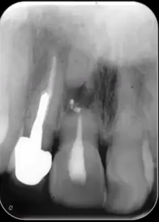
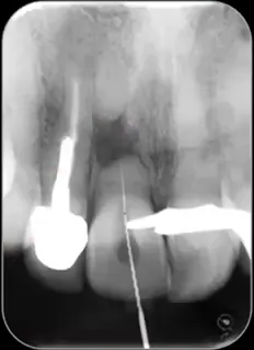
Case: 11, 21 — Root Fractures when 8 years old — 1969
- 11: Infected RCS + “apical” periodontitis in fracture line — 1994 (i.e. 25 years later)
- 11: WL — 1994
- 11: RCF — 1994
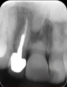
Case: 11, 21 — Root Fractures when 8 years old — 1969
11: Infected RCS + “apical” periodontitis in fracture line — 1994 (i.e. 25 years later)
- Review: 2003 (34 yrs post-trauma; 9 yrs since 11 RCF)
- Review: 2011 (42 yrs post-trauma; 17 yrs since 11 RCF)
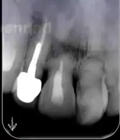
Case: 11, 21 — Root Fractures when 8 years old — 1969
11: Infected RCS + “apical” periodontitis in fracture line — 1994 (i.e. 25 years later)
- Review: 2014 (45 yrs post-trauma; 20 yrs since 11 RCF)
- Review: 2016 (47 yrs post-trauma; 22 yrs since 11 RCF)
 |  |  |
 |  |  |
Root Canal Treatment of Both Fragments
- Root Canal Treatment — coronal and apical fragments
- Not usually needed / recommended
- Only when the entire root canal is infected or develops irreversible pulpitis
- Difficulties controlling debris, bacteria, tissue, materials, etc. at the fracture line
- May be extruded → can cause inflammation, delayed healing, foreign body reaction, etc.
- Generally has a poor prognosis
- Surgery may be needed to remove apical fragment and excess materials
Case Series — From: M. Cvek - Fig. 22.23 - Andreasen 4th Edⁿ 2007
| 3 yr | 5 yr | 5 yr | 5 yr | 9 yr |
|---|---|---|---|---|
The 21 root fracture healed initially but the tooth developed Irreversible Pulpitis due to caries 5 years after the injury
Lecturer — Treating Both Fragments
The absence of extrusion through the fracture line suggested that the fragments had already healed internally with hard tissue.
- Treatment through the fracture line can displace debris, bacteria, tissue, or materials into the fracture site.
- Inflammation, delayed healing, or a foreign-body reaction may result, so treatment to the fracture line is generally preferred.
See the criteria and treatment risks above.
Case Series — From: M. Cvek - Fig. 22.22 - Andreasen 4th Edn 2007
| 3 d | 4 mo | 5 mo | 8 mo | 4 yr |
|---|---|---|---|---|
Surgical Removal of the Apical Fragment
- Root Canal Treatment — coronal fragment PLUS surgical removal — apical fragment
- Rarely required
- Since the apical fragment is not usually affected by the trauma and rarely develops pulp necrosis
- Most cases are due to treatment problems after extrusion of materials into fracture site
Extruded Root Filling Material
In one case, rapid root canal filling without prior formation of an apical barrier caused extensive extrusion of material into the fracture line.
- Granulation tissue and the radiolucency enlarged, and the root filling became effectively displaced from the apical fragment.
- Surgery was required to remove the apical fragment and excess material.
- The resulting root was short, but the short root itself was not considered the main problem.
- The complication might have been avoided by using long-term calcium hydroxide before root filling.
Case Series — From: M. Cvek - Fig. 22.20 – Andreasen 4th Edn 2007
0 d | 4 mo | 4 mo | 6 mo | 4 yr
Supra-Crestal Coronal Third Fractures
See initial management by fracture position.
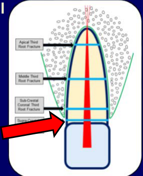
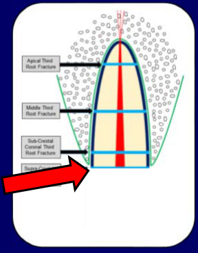
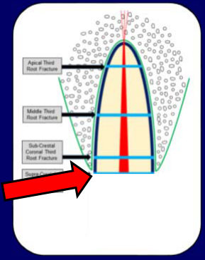
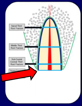
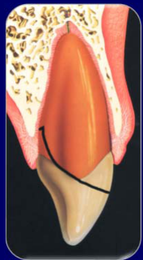
- Must decide whether to retain or remove the coronal fragment or even the entire tooth
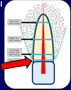
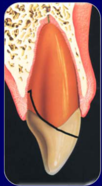
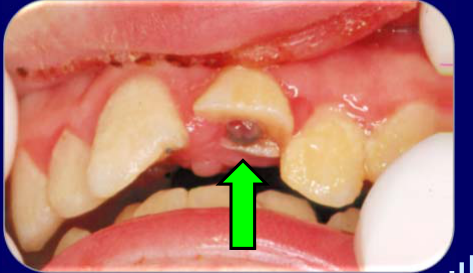
Complicated Crown:Root Fractures30
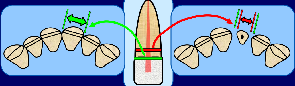
L12 covers crown-root classification and vertical crown-root management: L12 Crown-Root Fractures. This section applies the related supracrestal root-fracture management principles.
- Initial management is very similar to managing a supra-crestal root fracture in the coronal third
- Since the fracture extends into the coronal third of the root
- Margins are sub-gingival
- Likely to have lost a significant amount (or all) of the tooth’s crown
-
Or the crown will be loose / detached so it cannot be retained
-
 |  |
 |
Treatment Options3132
See the supra-crestal management decision above.
- Options:
- Remove coronal fragment and do Root Canal Treatment — followed by:
- Post, crown
- Usually difficult or not feasible
- Depends on whether an adequate restoration can be placed without compromising the gingivae
- Depends largely on the position of the fracture
- Usually difficult or not feasible
- Crown lengthening surgery, post, crown
- Is possible in some cases
- Depends on whether an adequate restoration can be placed without compromising the gingivae
- Can surgery be done without compromising the adjacent teeth and the aesthetics
- Depends largely on the position of the fracture → especially depth on the labial -v- palatal
- Is possible in some cases
- Orthodontic extrusion ± periodontal surgery, post, crown
- Several factors to consider
- Post, crown
- Extract entire tooth and replace with prosthesis
- Remove coronal fragment and do Root Canal Treatment — followed by:
 |  |
 |  |
Orthodontic Extrusion333435
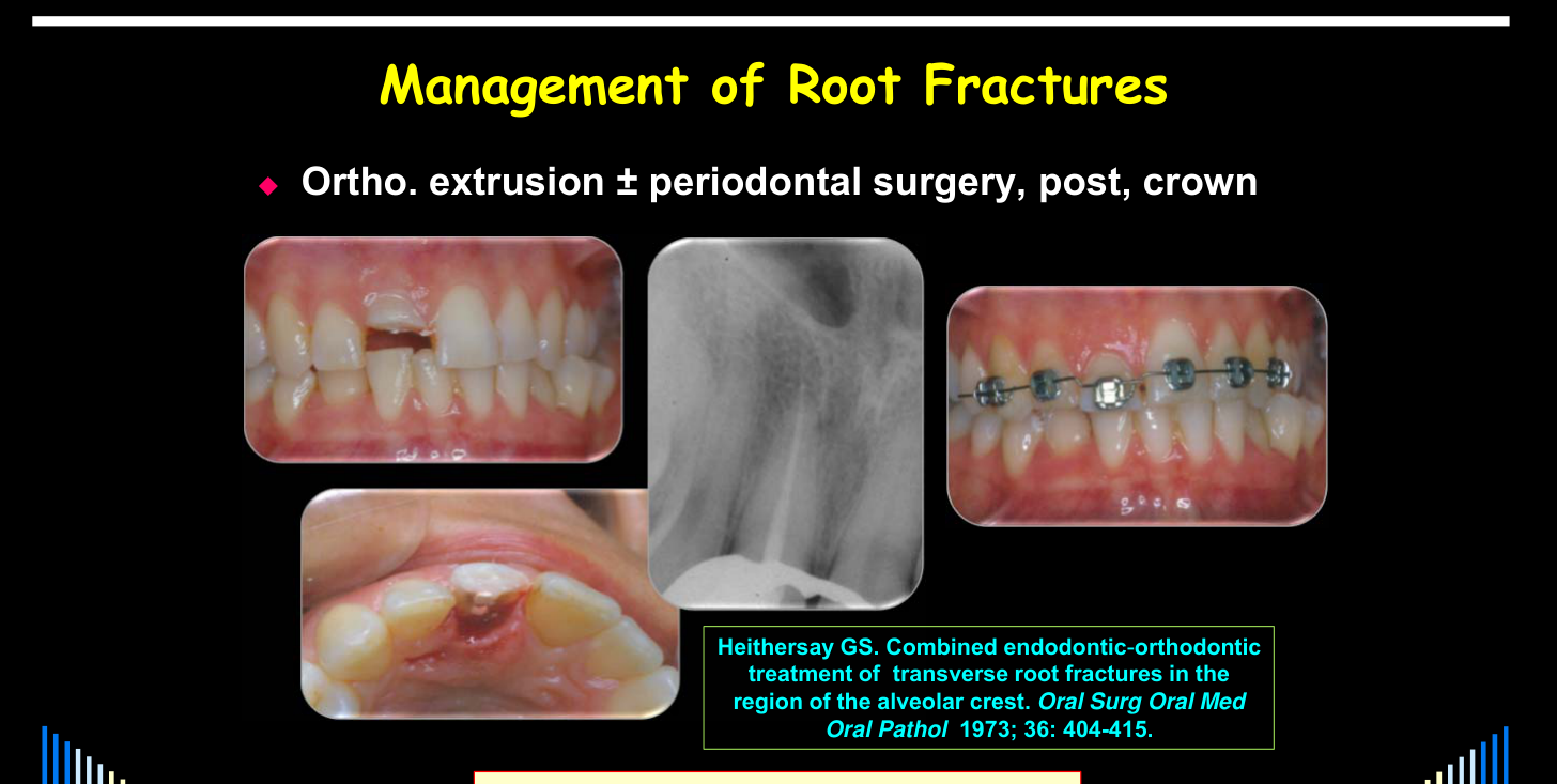
- Ortho. extrusion ± periodontal surgery, post, crown
- Several factors to consider - esp:
- Length of root / Length of crown
- Emergence profile
- Width of root / Width of crown
- Amount & strength of remaining tooth structure
- Ferrule
- Occlusion
- Aesthetics
- Periodontal condition
- Parafunctional habits
- etc, etc.
- Several factors to consider - esp:
Orthodontic Root Extrusion
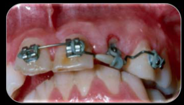
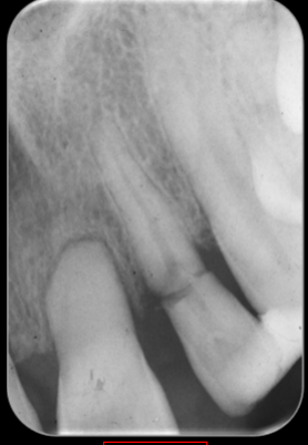
In the described supracrestal root fracture, gingival recession and bone loss were already present, and natural spacing made the emergence profile less critical.
- The coronal fragment was removed and root canal treatment was completed in the remaining root, leaving space for a post.
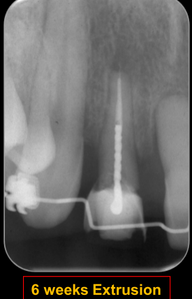
- A temporary post with a hook, orthodontic brackets, a bent wire, and an elastic chain were used to extrude the root; composite prevented the wire from cutting the elastic.
- Extrusion took approximately six weeks, followed by retention for approximately three months while the periodontal ligament reorganized and bone repaired at the socket entrance.
- A composite crown served as an interim restoration and retainer before removal of the retainer and periodontal recontouring.
- Extrusion is more favourable when root width at the fracture level resembles the space between adjacent teeth; a narrow, tapered root may require an over-contoured crown with an unfavourable emergence profile.
- Teeth are preferably extruded relatively quickly, over approximately six weeks, and periodontal recontouring is often required because soft tissue follows the extruded tooth.
See the extrusion considerations above.
Heithersay GS. Combined endodontic-orthodontic treatment of transverse root fractures in the region of the alveolar crest. Oral Surg Oral Med Oral Pathol 1973; 36: 404-415.
Courtesy of Prof. Geoffrey Heithersay AO
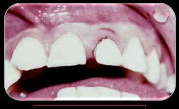
Restoration following Extrusion — 20 year Review
Courtesy of Prof. Geoffrey Heithersay AO
Summary36
- Root fractures have a good prognosis
- Depends on whether coronal fragment is displaced
- Depends on level of fracture — supra-crestal fractures in the coronal third of the root have the least predictable prognosis
- Prognosis also depends on concurrent injuries, the pulp response, and the quality and duration of stabilization.
- Apical third, middle third, and subcrestal coronal third fractures are usually managed conservatively.
- Emergency management is simple
- Reposition / Stabilise (splint) / Review
- A tooth should not receive root canal treatment at the emergency appointment solely because it does not respond to pulp testing.
- Reposition / Stabilise (splint) / Review
- Healing responses:
- Hard tissue, or connective tissue, or bone & connective tissue
- Most pulps survive
- Do NOT commence RCT at emergency appointment
- Wait and reassess over time
- RCT only if the pulp in the coronal fragment necroses and becomes infected — only about 27% of cases
- Pulp necrosis may become evident within the first few months or many years after trauma.
- Treatment of both fragments and surgical removal of the apical fragment are rarely required.
- Long-term follow-up is important because many fractured teeth remain functional for decades.
- Then only do RCT to the fracture line
- Do NOT commence RCT at emergency appointment
 |  |
 |  |
Case Illustrations37
W/Prof. Paul V. Abbott AO
Some Cases to Illustrate …
93 — 94
DMD 3rd Year - 2020
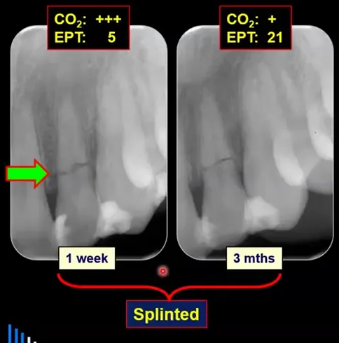
Root Fracture After a Squash-Racket Injury
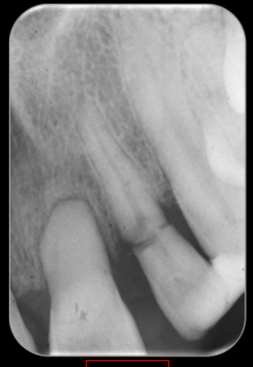
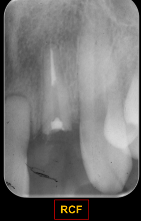
A squash-racket injury caused a root fracture that was stabilized with composite attached to the two adjacent teeth.
- The tooth remained sensitive to hot and cold at the one-week assessment, consistent with reversible pulpitis after trauma.
- No endodontic treatment was performed initially, and the splint was removed after three months.
- Pulp testing remained stable for approximately five years; although canal calcification eventually prevented a carbon-dioxide response, the tooth continued to respond normally to electric pulp testing.
- The tooth remained clinically stable.
 |  |
Clinical Cases
Case: Root fracture with associated bone fractures
Question
Scenario: A patient who fell and sustained a dental injury, resulting in a root fracture.
What’s shown: Radiographic images, including a CBCT, showing the root fracture and surrounding structures.
Consider: The utility of CBCT in this scenario and the overall management plan.
Answer
Observations:
- A root fracture is visible.
- Bone fractures are present on the labial and lingual aspects.
Reasoning: The CBCT clearly reveals the root fracture and concurrent bone fractures. However, the management of the bone fractures is the same as the root fracture (e.g., stabilization with a splint), so the advanced imaging does not change the treatment plan.
Outcome: The patient was managed with regular review. After two years, the tooth remained intact with no need for root canal treatment.
Takeaway: Periapical radiographs are usually sufficient to diagnose and manage transverse root fractures; CBCT may reveal concurrent bone fractures but typically does not alter the management.
Case: Tooth 18 months post-trauma
Question
Scenario: A tooth evaluated 18 months after sustaining a root fracture.
What’s shown: Radiographic images of the tooth.
Consider: The radiographic signs of healing and tissue response.
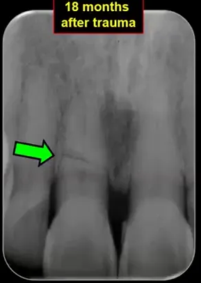
Answer
Observations:
- Internal repair with hard tissue.
- Rounding off of the peripheral fracture edges.
- Pulp canal calcification in both the apical and coronal fragments.
Reasoning: These radiographic features indicate the most favorable healing response, where calcified tissue forms between the fragments and the pulp canal calcifies over time.
Outcome: Successful healing with hard tissue repair.
Takeaway: Healing with calcified tissue is the most ideal response to a root fracture, characterized by internal repair, rounding of edges, and pulp canal calcification.
Case: Tooth 25 years post-trauma
Question
Scenario: A tooth evaluated 25 years after sustaining a root fracture.
What’s shown: Radiographic images of the tooth.
Consider: The long-term radiographic signs of healing.
Answer
Observations:
- Internal repair with hard tissue.
- Rounding off of the peripheral fracture edges.
- Pulp canal calcification in both the apical and coronal fragments.
Reasoning: Even after 25 years, the radiographic features demonstrate sustained, favorable healing with calcified tissue and pulp canal calcification.
Outcome: Successful long-term healing with hard tissue repair.
Takeaway: Favorable healing responses, such as calcified tissue repair and pulp canal calcification, can be maintained over decades.
Case: Extracted tooth with coronal third root fracture
Question
Scenario: A patient with a coronal third root fracture that was initially stabilized and later extracted for orthodontic reasons.
What’s shown: Panoramic radiograph, close-up clinical views of the extracted tooth, and histological slides.
Consider: The histological and radiographic evidence of healing in the extracted fragments.
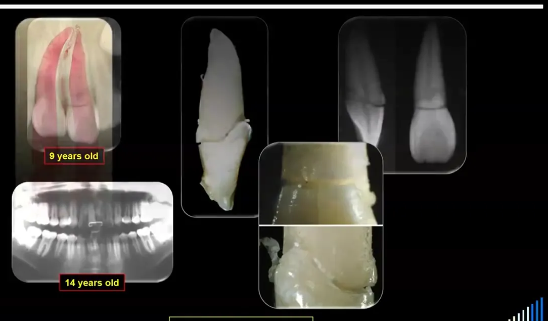
Answer
Observations:
- The tooth was extracted in one piece, indicating the fragments were joined.
- Close-up and radiographic views show repair inside the tooth on the labial and palatal sides of the canal.
- Histology shows laying down of dentine and cementum on the outside.
Reasoning: The fragments healed with hard tissue between them, demonstrating internal repair and external cementum repair, which is the most favorable response to a root fracture.
Outcome: The tooth was extracted but histological examination confirmed complete hard tissue healing between the fragments.
Takeaway: Root fractures can heal with hard tissue (dentine and cementum) joining the fragments, which can be confirmed histologically when the tooth is extracted.
Case: Root fracture, 6 years after trauma
Question
Scenario: A radiograph taken 6 years after a root fracture.
What’s shown: A radiograph of a tooth root.
Consider: Identify the tissue response and the radiographic features of the fracture line.
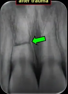
Answer
Observations:
- Remodeling and rounding off of the edges at the fracture line.
Reasoning: The remodeling and rounding off of the edges indicate that connective tissue, in the form of periodontal ligament, has formed between the fragments, representing a favorable connective tissue healing response.
Takeaway: Connective tissue repair between root fracture fragments presents radiographically as remodeling and rounding off of the fracture edges.
Case: Root fracture, 42 years after trauma
Question
Scenario: A radiograph taken 42 years after a root fracture.
What’s shown: A radiograph of a tooth root.
Consider: Identify the tissue response and the radiographic features of the fracture line.
Answer
Observations:
- Remodeling and rounding off of the edges at the fracture line.
Reasoning: Similar to the 6-year example, the rounding off of the edges indicates connective tissue (periodontal ligament) repair between the fragments, showing this favorable response can persist long-term.
Takeaway: Connective tissue repair between root fracture fragments can remain stable and show remodeling over decades.
Case: Root fracture in a young patient, 12 years after trauma
Question
Scenario: A radiograph taken 12 years after a root fracture in a young patient.
What’s shown: A periapical radiograph of upper anterior teeth.
Consider: Identify the healing response and the radiographic signs of the fragments.
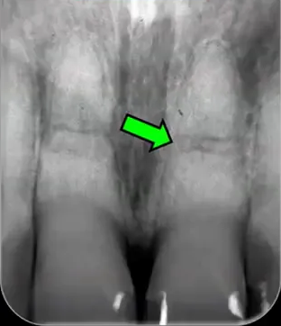
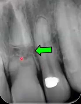
Answer
Observations:
- A gap between the coronal and apical fragments.
- Bone visible between the two fragments.
- Rounding off of the peripheral edges.
- Pulp canal calcification.
Reasoning: In young patients, if hard tissue union does not occur, the coronal fragment may erupt with alveolar growth while the apical fragment remains in place, creating a gap. The presence of bone between the fragments, along with rounded edges and calcification, indicates a favorable healing response with connective tissue and bone interposition.
Takeaway: In young patients, lack of hard tissue union can lead to eruption of the coronal fragment and a gap filled with bone and connective tissue, which is still a favorable response.
Case: Root fracture, 15 years after trauma
Question
Scenario: A radiograph taken 15 years after a root fracture.
What’s shown: A radiograph of a tooth root.
Consider: Identify the healing response and the radiographic signs of the fragments.
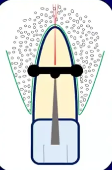
Answer
Observations:
- A gap between the fragments with bone visible between them.
- Rounding off of the peripheral edges.
- Pulp canal calcification.
Reasoning: Similar to the 12-year example, the gap with bone interposition, rounded edges, and calcification indicate a favorable healing response where the fragments did not unite with hard tissue but separated as the alveolus grew.
Takeaway: Long-term radiographic follow-up of root fractures in young patients may show a bony gap between fragments with rounded edges and pulp canal calcification.
Case: Root fracture, 4 months after trauma
Question
Scenario: A radiograph taken 4 months after a root fracture.
What’s shown: A radiograph of a fractured tooth root.
Consider: Identify the unfavorable tissue response and the radiographic signs of infection.
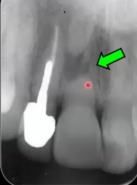
Answer
Observations:
- Radiolucency at the fracture line.
- The two fragments are forced apart slightly.
Reasoning: The radiolucency and separation of fragments indicate that the pulp in the coronal fragment has necrosed and become infected, leading to an inflammatory response and granulation tissue formation in the fracture line.
Takeaway: Pulp necrosis and infection of the coronal fragment can occur early after a root fracture, presenting as a radiolucency and separation of fragments due to granulation tissue.
Case: Root fracture, 25 years after trauma
Question
Scenario: A radiograph taken 25 years after a root fracture.
What’s shown: A radiograph of a fractured tooth root.
Consider: Identify the unfavorable tissue response and the radiographic signs of late infection.
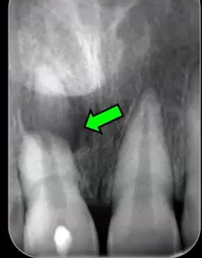
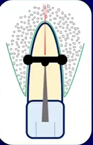
Answer
Observations:
- Radiolucency at the fracture line.
- The two fragments are forced apart.
Reasoning: The radiolucency and fragment separation occurring 25 years later indicate late pulp necrosis and infection, likely due to crown infractions or restoration breakdown, leading to granulation tissue formation in the fracture line.
Takeaway: Pulp necrosis and infection leading to granulation tissue at a root fracture line can occur many years after the initial trauma, often due to late crown infractions or restoration breakdown.
Case: Root fracture from squash racket, composite splint and long-term follow-up
Question
Scenario: A patient who was hit in the mouth with a squash racket, resulting in a root fracture.
What’s shown: A composite image of five sequential periapical radiographs showing a sub-crestal coronal third root fracture.
Consider: How to manage the fracture, monitor healing, and handle the pulp status over time.
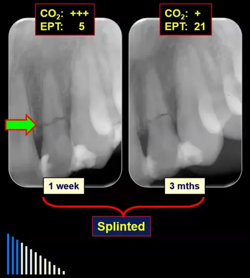
Answer
Observations:
- The initial dentist placed a composite splint attaching the loose fragment to the two adjacent teeth.
- At one week post-trauma, the tooth was sensitive to hot and cold, indicating reversible pulpitis.
- The splint was removed after three months, and the pulp was responding normally.
- Over a five-year follow-up, the tooth remained stable.
- Pulp canal calcification occurred in the coronal fragment, leading to a loss of response to CO2 but a normal response to electric pulp testing.
Reasoning: The management of a coronal third sub-crestal fracture involves repositioning, stabilizing, and monitoring. Since there were no signs of pulp necrosis, no further treatment was needed, and the pulp recovered and underwent calcification.
Outcome: The tooth remained stable over a five-year follow-up with normal pulp responses via electric pulp testing and pulp canal calcification.
Takeaway: For coronal third sub-crestal root fractures without pulp necrosis, conservative management with repositioning, stabilization, and monitoring is sufficient, often resulting in pulp canal calcification and long-term stability.
Case: Middle third root fracture with pulp necrosis, treated with calcium hydroxide and root canal treatment
Question
Scenario: A patient with a middle third root fracture where the pulp became necrotic and infected, with root canal treatment initially started by a referring dentist.
What’s shown: A series of periapical radiographs showing a middle third root fracture with a radiolucency between fragments, followed by calcium hydroxide placement and eventual root canal filling.
Consider: How to manage pulp necrosis and infection in a root fractured tooth with an open end at the fracture line.
Answer
Observations:
- A radiolucency representing granulation tissue was present between the two fragments.
- Root canal treatment was continued only to the fracture line.
- Calcium hydroxide was placed and changed every three months.
- At nine months, the radiolucency reduced in size and bone began to heal.
- At 15 months, a good hard tissue barrier had formed across the open end, allowing for the final root canal filling.
Reasoning: When pulp necrosis occurs early, the canal end at the fracture line is open, similar to an immature tooth. Long-term calcium hydroxide is used to encourage an apical barrier (hard tissue repair) to form before completing the root filling, without going beyond the fracture line.
Outcome: A hard tissue barrier formed across the fracture line, allowing successful root canal filling at 15 months, with reduction of the radiolucency and bone healing.
Takeaway: For early pulp necrosis in a root fractured tooth, root canal treatment should be limited to the coronal fragment, using long-term calcium hydroxide to induce a hard tissue barrier at the open fracture line before obturation.
Case: Apical third root fracture with pulp necrosis, root canal treatment and resolution of apical fragment
Question
Scenario: A patient with an apical third root fracture that developed pulp necrosis and infection.
What’s shown: A series of radiographs showing an apical third root fracture, root canal treatment to the fracture line, and subsequent follow-ups.
Consider: The management of pulp necrosis in an apical third root fracture and the potential reaction of the apical fragment.
Answer
Observations:
- The canal was wide at the fracture line when the injury occurred.
- Root canal treatment was performed to the fracture line, leaving the apical fragment untreated.
- The apical fragment resolved (resorbed) over time.
- A good hard tissue barrier formed across the end of the canal.
- The tooth became discoloured.
Reasoning: In apical third fractures with pulp necrosis, the coronal fragment is root filled to the fracture line while the apical fragment is left alone. It is common for a hard tissue barrier to form. In this unusual case, the apical fragment resorbed, which did not compromise the outcome.
Outcome: Root canal filling was completed after about 12 months. At a four-year review, the tooth was stable, the apical fragment had resolved, and a hard tissue barrier was present. The tooth was bleached to address discolouration.
Takeaway: In apical third root fractures with pulp necrosis, root canal treatment of the coronal fragment to the fracture line is the standard approach; the apical fragment may occasionally resorb without affecting the overall stability and success of the treatment.
Case: Maxillary central incisor with late pulp necrosis 25 years post-trauma
Question
Scenario: A patient who suffered trauma at age 8 (in 1969) resulting in fractured central incisors, presenting for evaluation 25 years later.
What’s shown: Periapical radiographs from 1994 (25 years post-trauma) and subsequent reviews up to 2016.
Consider: How to manage late pulp necrosis in a tooth with a history of root fracture and calcified canals.
Answer
Observations:
- Radiograph in 1994 shows calcification of canals in both coronal and apical fragments of both central incisors.
- Tooth 11 developed pulp necrosis, likely due to infractions reaching the dentine and allowing bacterial ingress.
- Root canal treatment in 1994 was routine despite a very narrow canal.
- Reviews in 2003, 2011, 2014, and 2016 show bone formation between fragments and stable tissues.
Reasoning: Late pulp necrosis can occur many years after trauma. Root canal treatment is usually straightforward if the canal can be located, as pulp canal calcification creates a narrow canal similar to an apical constriction. Long-term follow-up demonstrates stability and bone healing between the fragments.
Outcome: Root canal treatment completed in 1994. The tooth remained stable at the 22-year follow-up in 2016, with bone formed between the fragments.
Takeaway: Late pulp necrosis after a root fracture can be managed with routine root canal treatment if the calcified canal can be located, and long-term follow-up demonstrates stability.
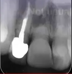
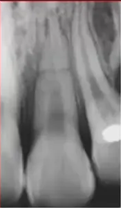
Case: Published case of root fracture with irreversible pulpitis at five years
Question
Scenario: A published case (from Sfek’s work/Andreasen’s book) of a tooth with a root fracture, evaluated at three and five years post-trauma.
What’s shown: Radiographs at three-year and five-year reviews following the initial trauma.
Consider: The management of a root fractured tooth that develops irreversible pulpitis due to caries.
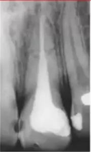
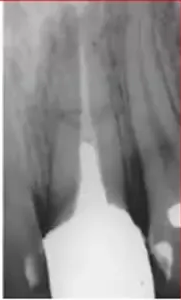
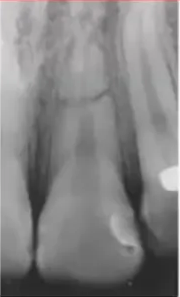
Answer
Observations:
- Three-year review shows normal healing, rounding off of edges, and some narrowing of the apical part of the canal.
- Five-year review reveals irreversible pulpitis resulting from mesial caries.
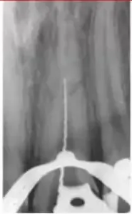
- Root canal treatment was performed through the fracture line.
- No extrusion of root canal filling material occurred through the fracture line.
Reasoning: The lack of material extrusion indicates that the tooth had healed internally with hard tissue (dentine laid down on internal walls) prior to the pulp becoming inflamed. Treating through the fracture line is possible when internal healing has occurred, though it carries risks of pushing debris into the fracture line if healing hasn’t happened.
Outcome: Root canal treatment completed through the fracture line to the apex without extrusion of material.
Takeaway: If a root fractured tooth heals internally with hard tissue, root canal treatment can be performed through the fracture line without extrusion of material, though this approach is unusual and risks a foreign body reaction if debris is pushed into the fracture line.
Case: Published case of root fracture with early necrosis and surgical removal of apical fragment
Question
Scenario: A published case (from Sfek/Spec’s work) of a root fracture where pulp necrosis developed early, leading to a complication.
What’s shown: Radiographs at baseline, four months, five months, and a clinical photograph showing granulation tissue.
Consider: The consequences of performing root canal treatment without establishing a hard tissue barrier in a fractured tooth.
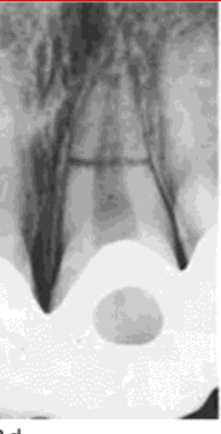
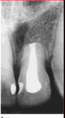
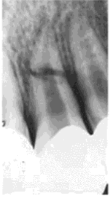
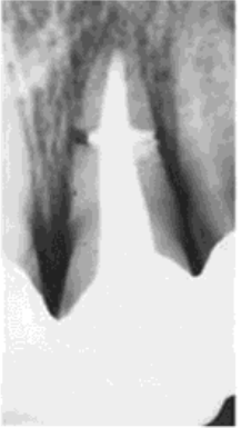
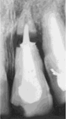
Answer
Observations:
- Pulp necrosis and infection were evident at the four-month review.
- Root canal treatment was performed at five months without using long-term calcium hydroxide to encourage an apical barrier.
- This resulted in significant extrusion of root canal material into the fracture line.
- A clinical photo shows enlarging granulation tissue and a larger radiolucency.
Reasoning: Without a hard tissue barrier, root canal filling material extrudes into the fracture line, causing a foreign body reaction and inflammation. This delays healing, worsens the situation, and causes the root canal filling to effectively dislodge from the apical fragment.
Outcome: Surgery was required to remove the apical fragment and the excess extruded material, leaving the tooth with a very short root.
Takeaway: When managing pulp necrosis in a root fractured tooth, treating to the fracture line with long-term calcium hydroxide is preferred to avoid extrusion of materials into the fracture line, which can necessitate surgical removal of the apical fragment.
Case: Central incisor with a history of trauma and recent root fracture
Question
Scenario: A male patient presents with a recent root fracture of a central incisor. The tooth had a history of trauma many years prior, resulting in a short root and pulp canal calcification.
What’s shown: Clinical photos and radiographs showing the fractured tooth, the initial root canal treatment, the orthodontic extrusion setup with a temporary post and composite retainer, and the progress at six weeks and three months.
Consider: What factors influence the decision for orthodontic extrusion in this patient, and how is the extrusion and retention managed?
Answer
Observations:
- The tooth has a history of trauma resulting in a very short root and pulp canal calcification, with clinical discoloration.
- There is gingival recession, bone loss, and natural spacing between the teeth.
- The tooth is not suitable for a bridge or partial denture due to the spacing, and the patient is not a candidate for an implant.
- Radiographs at six weeks show the tooth pulled down out of its socket, with a composite crown acting as a retainer.
- At three months, the retention device is removed and the tissue has come down with the tooth.
Reasoning:
- The presence of recession and bone loss, along with natural spacing, makes orthodontic extrusion favorable as the emergence profile is less critical and the crown width can be reduced.
- The coronal fragment is removed, and root canal treatment is performed on the remaining root.
- A temporary post with a hook is placed, and orthodontic brackets are used on adjacent teeth for anchorage.
- Chain elastic modules are used to pull the tooth down over six weeks.
- A composite crown over the wires holds the tooth in its extruded position for three months to allow the periodontal ligament to reorient and bone to repair.
Outcome:
- The tooth was successfully extruded and retained for three months.
- Periodontal recontouring surgery was necessary after the tissue came down with the tooth.
Takeaway:
- Orthodontic extrusion is a viable management option for supracrestal root fractures, especially when factors like recession, bone loss, and spacing make the emergence profile favorable and other prosthetic options unsuitable.
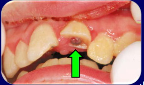
Case: Tooth with a deep palatal crown root fracture (Heithersay case)
Question
Scenario: A published case report by Professor Heithersay involving a tooth with a complicated crown root fracture.
What’s shown: A clinical photograph showing the initial fracture and the long-term outcome.
Consider: How was this deep crown root fracture managed, and what was the long-term outcome?
Answer
Observations:
- The fracture was quite deep on the palatal aspect.
- The tooth was orthodontically extruded and retained in place.
- It was subsequently restored with a post core crown.
Reasoning:
- A deep palatal fracture in a crown root fracture can be managed using the same principles as a supracrestal root fracture, utilizing orthodontic extrusion to bring the fracture margin to a restorable position.
Outcome:
- At a 20-year review, the tooth looked quite normal, with good soft tissue and aesthetics.
Takeaway:
- Orthodontic extrusion followed by a post core crown can provide excellent long-term aesthetic and functional outcomes for deep crown root fractures.
Footnotes
-
Original PDF page 1: L13 Dento-Alveolar Trauma 6 - Managing Root Fractures, p.1 ↩
-
Original PDF page 2: L13 Dento-Alveolar Trauma 6 - Managing Root Fractures, p.2 ↩
-
Original PDF page 3: L13 Dento-Alveolar Trauma 6 - Managing Root Fractures, p.3 ↩
-
Original PDF page 4: L13 Dento-Alveolar Trauma 6 - Managing Root Fractures, p.4 ↩
-
Original PDF page 7: L13 Dento-Alveolar Trauma 6 - Managing Root Fractures, p.7 ↩
-
Original PDF page 8: L13 Dento-Alveolar Trauma 6 - Managing Root Fractures, p.8 ↩
-
Original PDF page 9: L13 Dento-Alveolar Trauma 6 - Managing Root Fractures, p.9 ↩
-
Original PDF page 10: L13 Dento-Alveolar Trauma 6 - Managing Root Fractures, p.10 ↩
-
Original PDF page 11: L13 Dento-Alveolar Trauma 6 - Managing Root Fractures, p.11 ↩
-
Original PDF page 12: L13 Dento-Alveolar Trauma 6 - Managing Root Fractures, p.12 ↩
-
Original PDF page 13: L13 Dento-Alveolar Trauma 6 - Managing Root Fractures, p.13 ↩
-
Original PDF page 14: L13 Dento-Alveolar Trauma 6 - Managing Root Fractures, p.14 ↩
-
Original PDF page 15: L13 Dento-Alveolar Trauma 6 - Managing Root Fractures, p.15 ↩
-
Original PDF page 16: L13 Dento-Alveolar Trauma 6 - Managing Root Fractures, p.16 ↩
-
Original PDF page 17: L13 Dento-Alveolar Trauma 6 - Managing Root Fractures, p.17 ↩
-
Original PDF page 18: L13 Dento-Alveolar Trauma 6 - Managing Root Fractures, p.18 ↩
-
Original PDF page 19: L13 Dento-Alveolar Trauma 6 - Managing Root Fractures, p.19 ↩
-
Original PDF page 20: L13 Dento-Alveolar Trauma 6 - Managing Root Fractures, p.20 ↩
-
Original PDF page 22: L13 Dento-Alveolar Trauma 6 - Managing Root Fractures, p.22 ↩
-
Original PDF page 21: L13 Dento-Alveolar Trauma 6 - Managing Root Fractures, p.21 ↩
-
Original PDF page 23: L13 Dento-Alveolar Trauma 6 - Managing Root Fractures, p.23 ↩
-
Original PDF page 24: L13 Dento-Alveolar Trauma 6 - Managing Root Fractures, p.24 ↩
-
Original PDF page 25: L13 Dento-Alveolar Trauma 6 - Managing Root Fractures, p.25 ↩
-
Original PDF page 26: L13 Dento-Alveolar Trauma 6 - Managing Root Fractures, p.26 ↩
-
Original PDF page 27: L13 Dento-Alveolar Trauma 6 - Managing Root Fractures, p.27 ↩
-
Original PDF page 28: L13 Dento-Alveolar Trauma 6 - Managing Root Fractures, p.28 ↩
-
Original PDF page 29: L13 Dento-Alveolar Trauma 6 - Managing Root Fractures, p.29 ↩
-
Original PDF page 31: L13 Dento-Alveolar Trauma 6 - Managing Root Fractures, p.31 ↩
-
Original PDF page 36: L13 Dento-Alveolar Trauma 6 - Managing Root Fractures, p.36 ↩
-
Original PDF page 40: L13 Dento-Alveolar Trauma 6 - Managing Root Fractures, p.40 ↩
-
Original PDF page 41: L13 Dento-Alveolar Trauma 6 - Managing Root Fractures, p.41 ↩
-
Original PDF page 42: L13 Dento-Alveolar Trauma 6 - Managing Root Fractures, p.42 ↩
-
Original PDF page 43: L13 Dento-Alveolar Trauma 6 - Managing Root Fractures, p.43 ↩
-
Original PDF page 44: L13 Dento-Alveolar Trauma 6 - Managing Root Fractures, p.44 ↩
-
Original PDF page 45: L13 Dento-Alveolar Trauma 6 - Managing Root Fractures, p.45 ↩
-
Original PDF page 46: L13 Dento-Alveolar Trauma 6 - Managing Root Fractures, p.46 ↩
-
Original PDF page 47: L13 Dento-Alveolar Trauma 6 - Managing Root Fractures, p.47 ↩