L17 Management of Incompletely Developed Tooth 2
Management of Immature Teeth1
Dr Mostafa Elkholy, BDS, MSc (Endo), PhD (Endo)
Senior Lecturer and Discipline Lead in Endodontics
Dental School, UWA
Management Overview23
The management approach is determined first by pulp status:
- Vital Pulp
- Small Exposure → Partial Pulpotomy
- Large Exposure → Pulpotomy
- Pulpless and infected RCS
- Regeneration
- Apexification
Related trauma navigation: Root Fractures and Developing Teeth.
Lecturer — Immature Teeth and Regeneration
Immature teeth have short roots, thin dentinal walls, and large incompletely formed apices that cannot be sealed properly; behavior management must also be considered because these patients are usually young.
- Regeneration may fail over time, its long-term predictability remains uncertain, and the tissue formed may not be true pulp or have normal pulp-like vascularity.
- Histological studies have found mixtures of cementum-like, dentin-like, bone-like, and other cells, suggesting that induced bleeding may produce a healing response rather than normal pulp.
- Stem-cell treatment remains primarily a research concept requiring a scaffold, growth factors, pulp-like tissue-forming cells, and tissue capable of laying down dentin and reforming the tooth.
- Apexogenesis is used when vital pulp is maintained so root formation can continue, whereas apexification is used for non-vital teeth with incompletely formed roots to create a calcific barrier at the apex.
Dr Mostafa Elkholy
Historical Management Of Necrotic Immature Roots
Early Treatment Approaches4
-
Custom fitting of the filling material Friend LA. The root treatment of teeth with open apices. Proc R Soc Med 1966;19:1035–6.
-
Paste fills Friend LA. Treatment of immature teeth and non-vital pulps. J Br Endod Soc 1967;1:28–33
-
Apical surgery Ingle JI. Endodontics. Philadelphia: Lea and Febiger; 1965:504
-
Molding gutta-percha with chloroform or heat
Limitations Of Early Approaches5
- Custom fitting of the filling material
- Not advisable as the apical portion of the root is frequently wider than the coronal portion, making proper condensation of the GP impossible.
- Apical surgery
- Difficulty of obtaining the necessary apical seal in the young pulpless tooth with its thin, fragile, irregular walls at the root apex
Lecturer — Early Apical Control
The wide apex made control of the filling material unpredictable.
- There was no reliable apical stop.
- Condensation of gutta-percha could result in extrusion beyond the apex.
- Apical surgery was difficult to manage predictably in young patients because of the irregular, thin apical area.
Leo Grudin6
- When should endodontics start at a University level?
- What should an endodontic course include?
- Should there be a standardization of teaching methods?

J. Henry Kaiser
- Implantation of luxated teeth
- Treatment of wide open canals with partially degenerated pulps
Lecturer — Kaiser’s Proposal
The treatment was first proposed by J. Henry Kaiser at an American Association of Endodontists meeting in 1964 for teeth with wide apices and partially degenerated pulp. This approach became associated with the development of apexification.
F. J. Marshall
- Patient history and its relevance.
- Antibiotics, medications and contra-indications.
C. G. Maurice
- To culture or not to culture in endodontic treatment
- Disinfection and sterilization of endodontic instruments and materials
- Root canal filling: materials, methods, technique
- The standardization of root canal instrument size and its impact on endodontics
- Causes of treatment failures
Richard M. Moodnik
- Histopathology of pulp; pulp reactions to operative procedures
- Endo-periodontal problems
- Recent findings of research project comparing N-2 and standard endodontic procedures
Continued Apical Formation Therapy78
See - Frank 1966 Continued Apical Formation
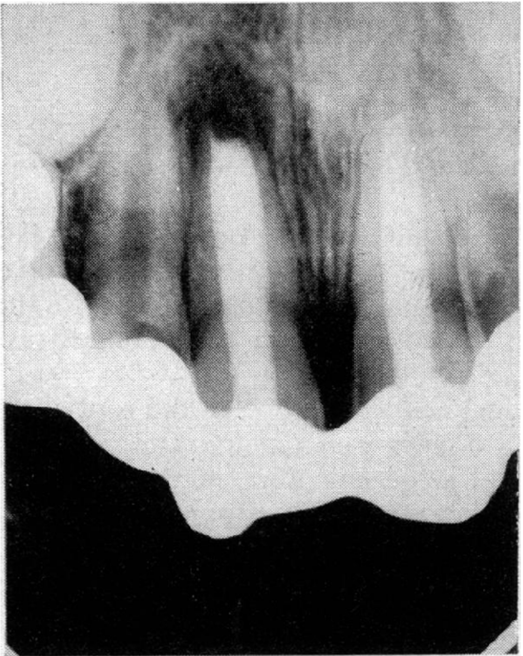
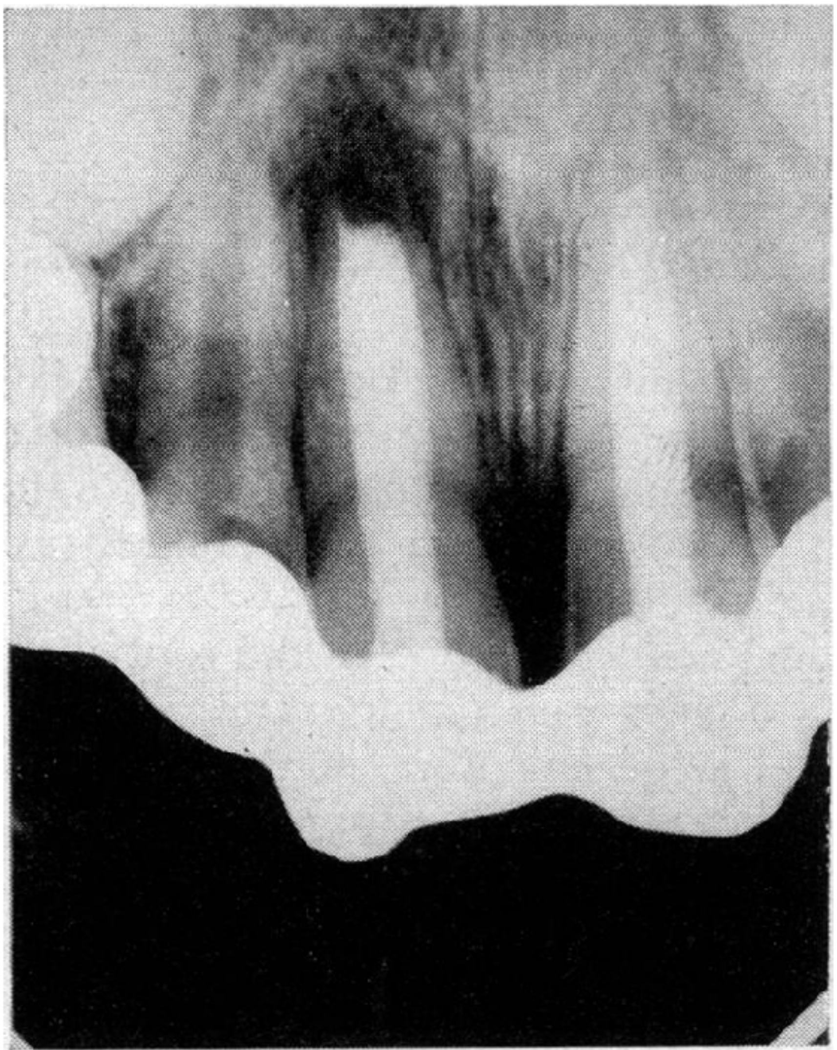
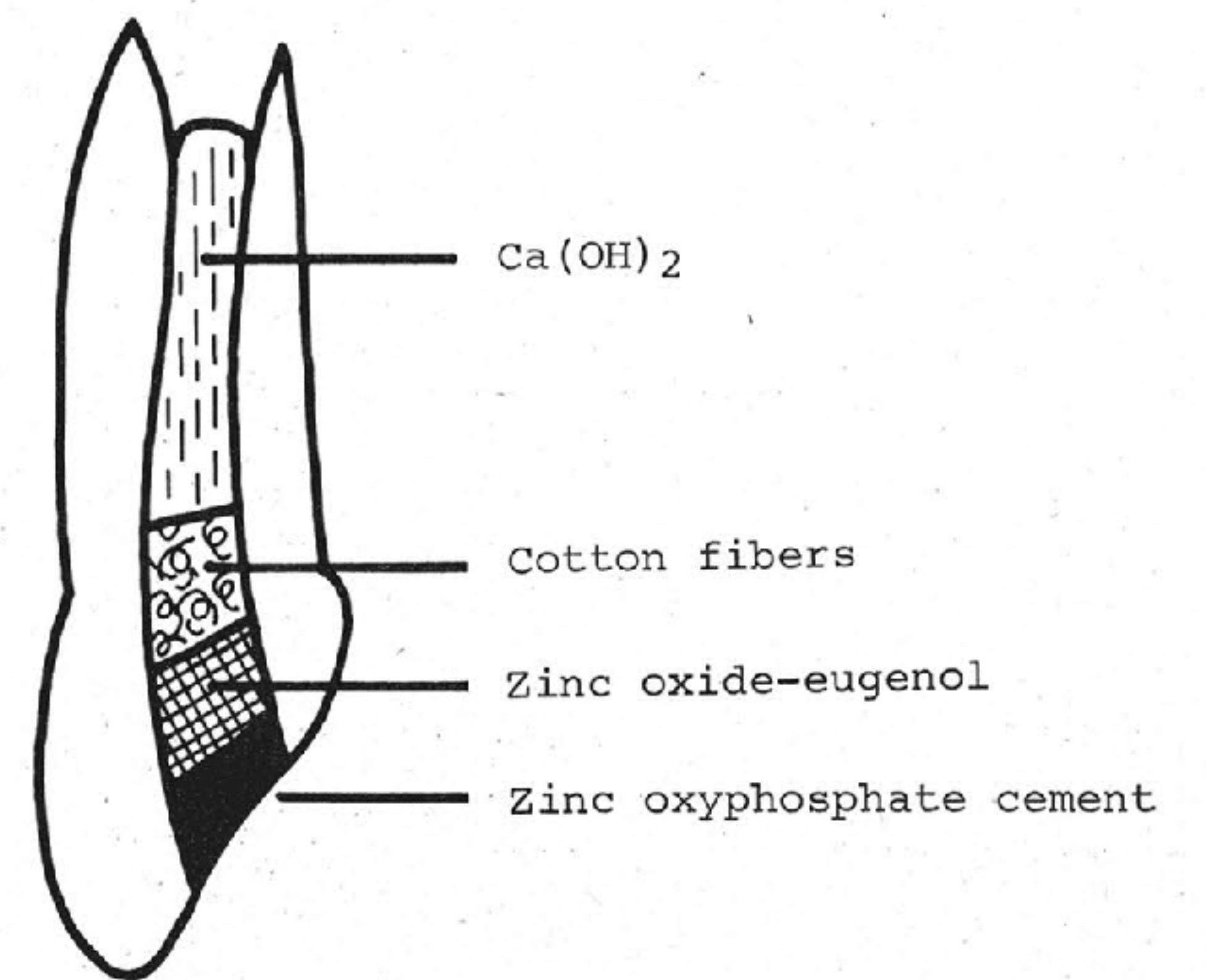
Frank’s historical sequence placed Ca(OH)2 in the divergent pulpless canal and open apex under a provisional seal, then used the resulting calcific barrier as a stop for later root filling; see Frank 1966 - Continued Apical Formation for the full material list, excerpt, and original PDF pages.
- Calcium hydroxide is used initially because infection control is a primary treatment goal in pulpless and infected root canals.
Lecturer — Apical Barrier
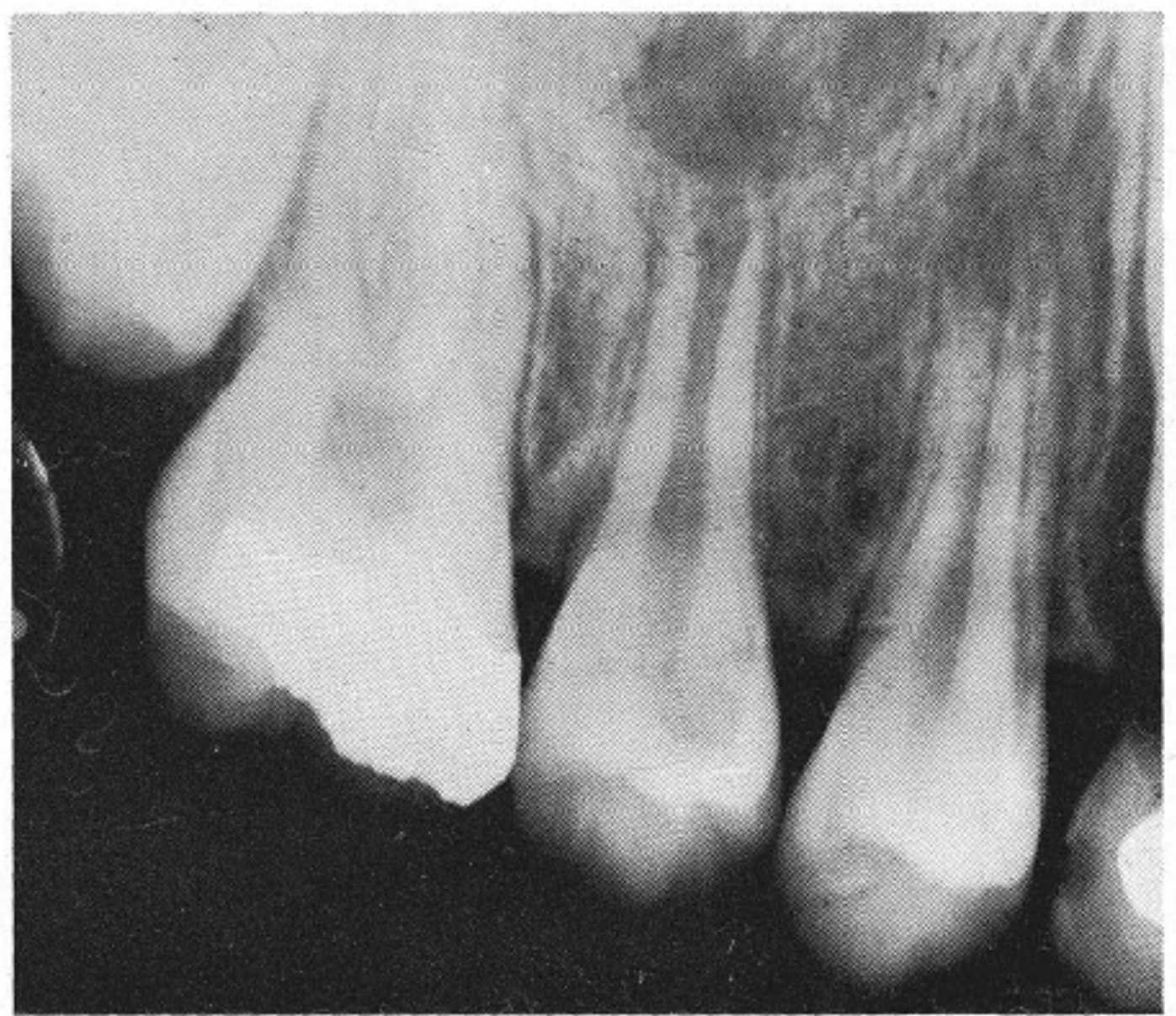
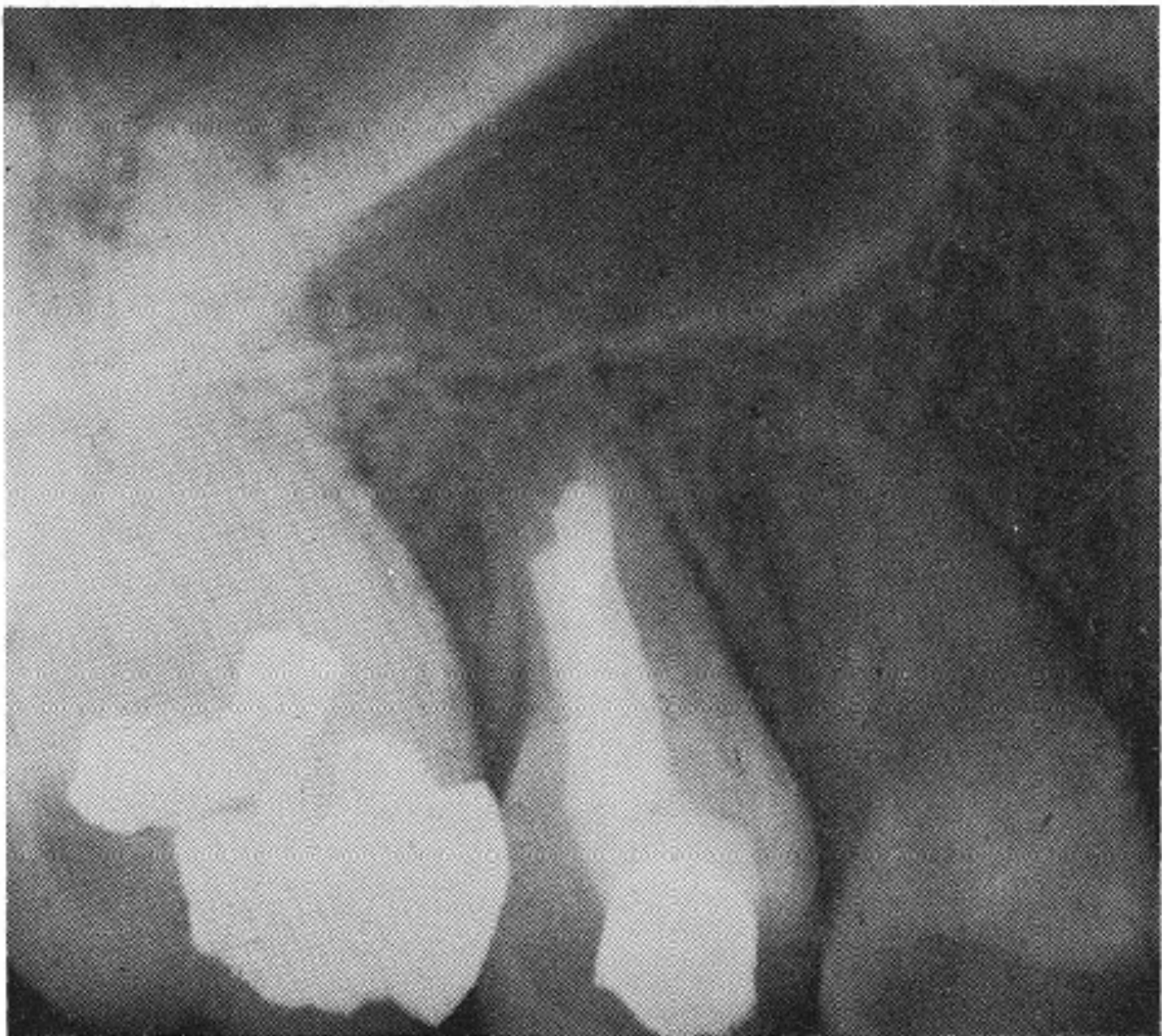
The calcific barrier is not a continuation of the root. It may resemble bone, cementum, or another type of calcified tissue, but its principal purpose is to provide an apical stop so that the root canal filling material can be compacted.
See - Calcium Hydroxide Replacement Interval
See Treatment Protocol Note for how often the dressing is replaced and why.
Lecturer — Fracture Risk
The lecturer noted that the methods used in some fracture studies had limitations. Some methodologies may not have represented clinical treatment accurately or simulated real clinical situations, raising questions about the external validity of their findings. Although fracture risk remains described in the literature as a proposed shortcoming, the lecturer did not personally believe that long-term calcium hydroxide medication in a properly treated tooth necessarily decreases fracture resistance.
 |  |
 |
Calcium Hydroxide Apexification
Antimicrobial And Chemical Effects9
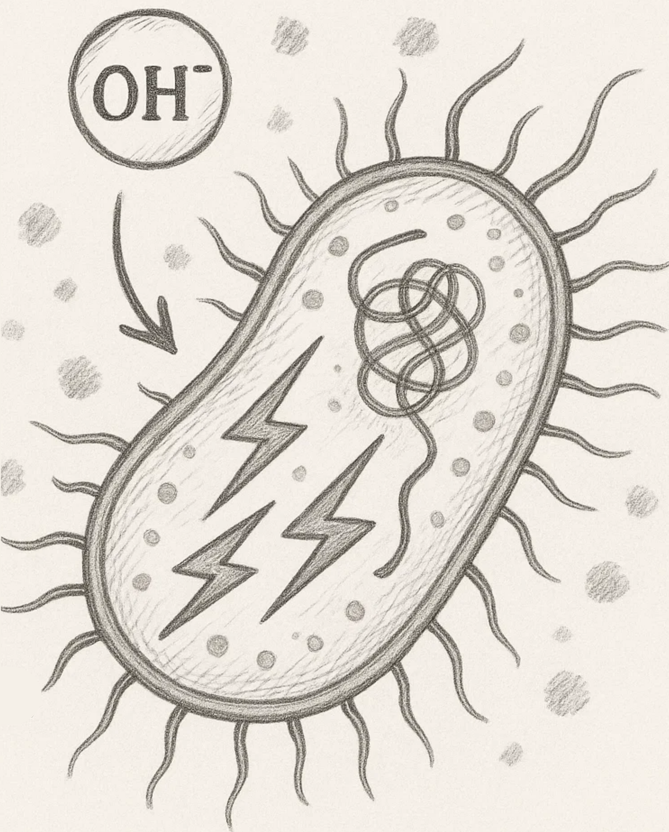
OH⁻
-
Ca(OH)₂ is antimicrobial because of its release of hydroxyl ions, which can cause damage to the bacterial cellular components. Safavi KE, Nichols FC (1993). Journal of Endodontics 19, 76–8.
-
Ca(OH)₂ chemically alters LPS, which affects its various biological properties. Safavi KE, Nichols FC (1994). Journal of Endodontics 20, 127–9.
Osteo-Inductive Properties10
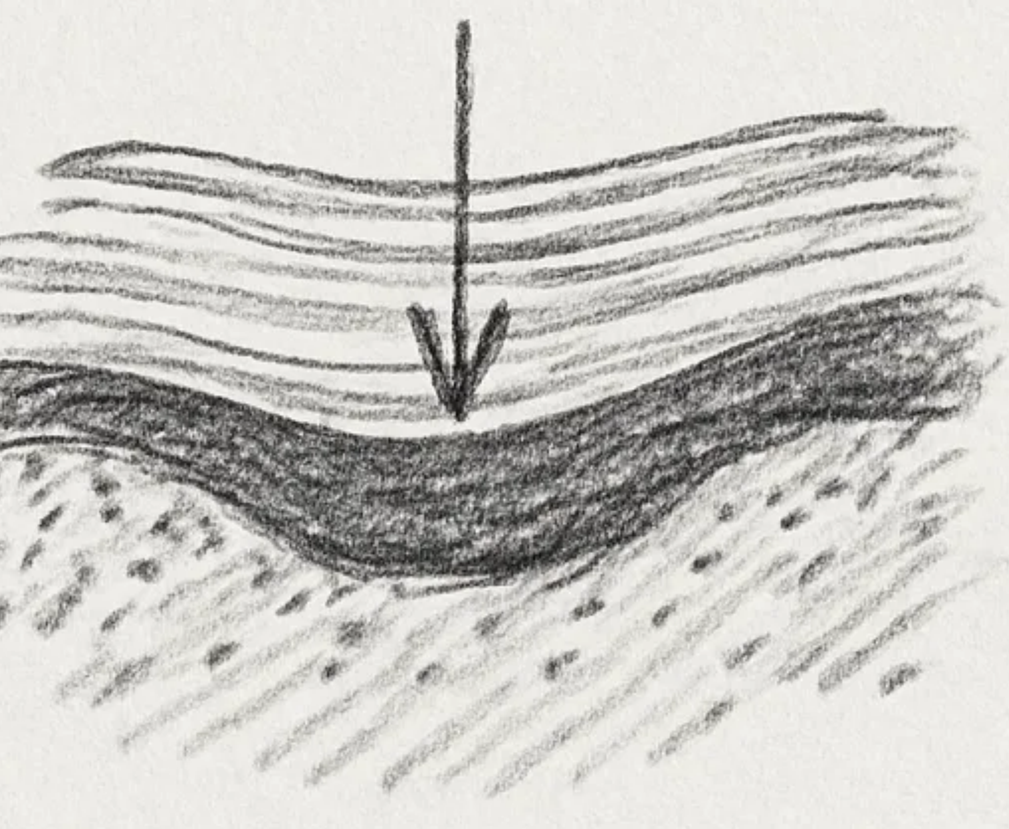
Ca(OH)₂ has been suggested to have osteo-inductive properties, considering that the high pH may be a contributing factor for the induction of hard tissue formation.
Javelet J, Torabinejad M, Bakland LK (1985). Journal of Endodontics 11, 375–8.
 |  |
Apexification Timeframe And Protocol
Apexification Timeframe11
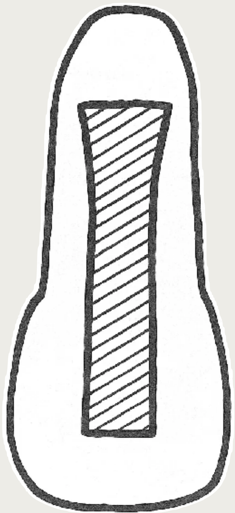
- Apical barrier formation with Ca(OH)₂ can be considerable — often up to 20 months.
Factors That Affect Time Needed
-
Patient age
-
Presence of symptoms
-
Presence of periradicular radiolucencies
-
Patient cooperation
-
Access to dental services
-
Availability of specialist dental services
-
The clinician’s skills and knowledge
Treatment Protocol Note
- Ca(OH)₂ replacement interval: Rafter (2005) gives roughly every 3 months; the lecturer prefers monthly (“every month is better”), because dentinal fluid buffers the paste’s alkalinity — e.g. for a planned 6-month medication, change the dressing monthly.
Shortcomings Of Calcium Hydroxide Apexification1213
- Long overall treatment time
- Multiple visits, creating high demands on patients/carers and increased clinical costs
- Higher risk of tooth fracture when Ca(OH)₂ is used as a long-term root-canal dressing
Andreasen JO, Farik B, Munksgaard EC. Endod Dent Traumatol 2002;18, 134–7. Cvek M. Endod Dent Traumatol 1992;8:45–55.
See - Cvek 1992 apexification study
The stage-specific fracture table attributed to Cvek is archived from L16 PDF page 7 in Cvek 1992 - Luxated Nonvital Incisors Apexification. L17’s broader long-term calcium hydroxide fracture-risk claim above retains its joint Cvek and Andreasen attribution.
Lecturer — Moisture and Condensation
Hydraulic calcium silicate materials were described as useful for sealing open apices and as performing better than gutta-percha and resin sealer in a moist environment.
- The material should be properly compacted to reduce voids and create a dense mass.
- With very wide apices, immediate barrier placement may cause inadequate condensation, extrusion, or a poorly controlled result.
- A previously formed calcific barrier can provide a stop that improves control when the final bioceramic plug and filling are placed.

Bioceramic Materials
Biological Characteristics14151617
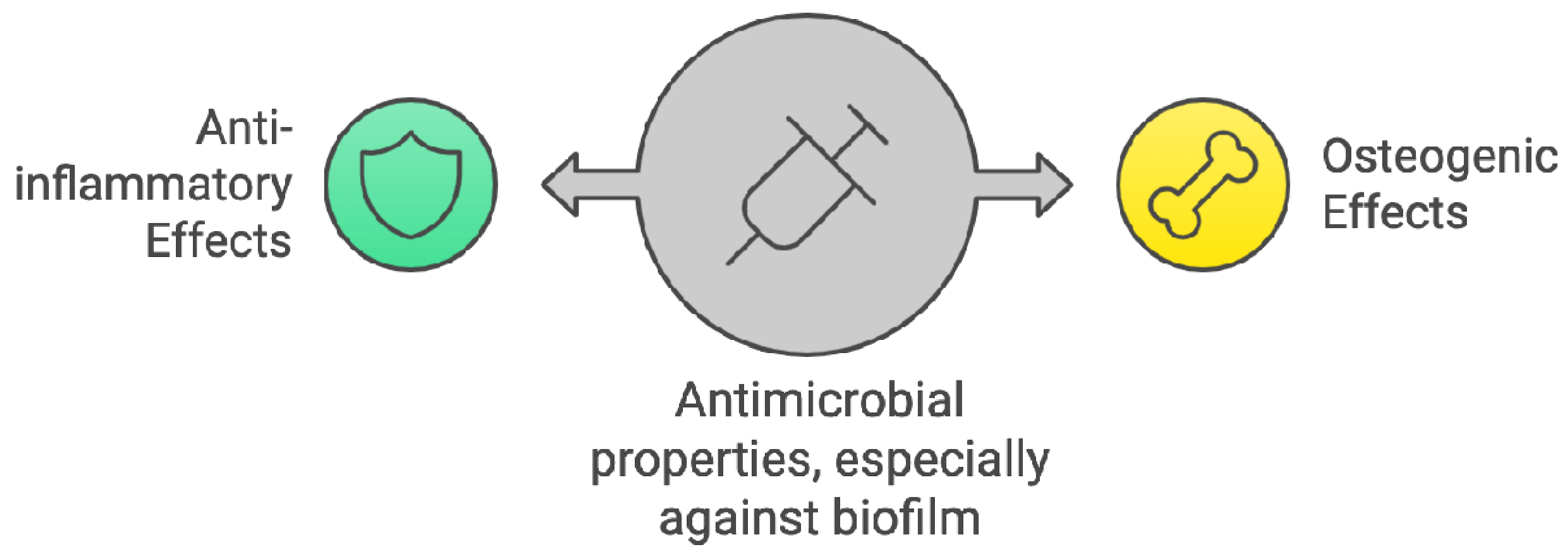
- Anti-inflammatory effects
- Osteogenic effects
- Antimicrobial properties, especially against biofilm
Int Endod J. 2022;55(Suppl. 3):710–777.
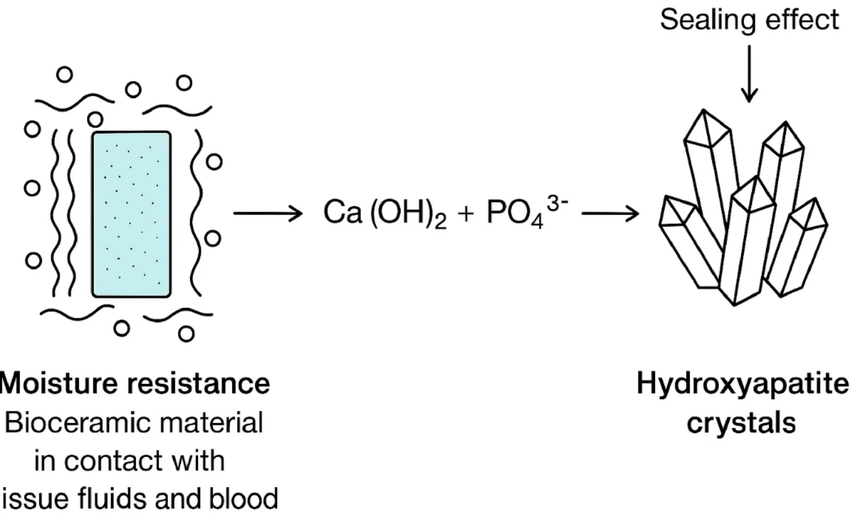
- Moisture resistance — bioceramic material in contact with tissue fluids and blood
- Ca(OH)₂ + PO₄³⁻ → sealing effect via hydroxyapatite crystals
Int Endod J. 2022;55(Suppl. 3):710–777.
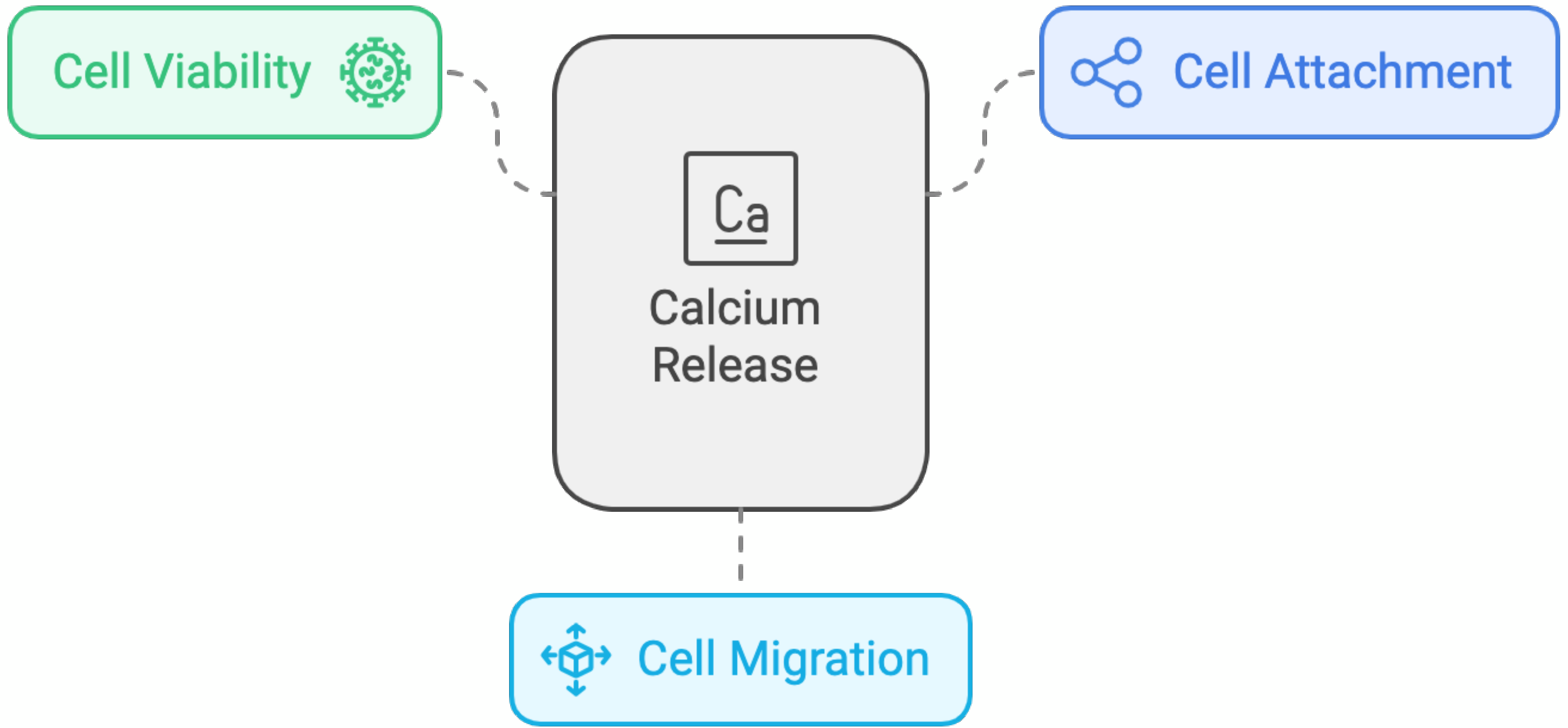
- Cell viability
- Calcium (Ca) release
- Cell attachment
- Cell migration
Int Endod J. 2022;55(Suppl. 3):710–777.
Lecturer — Biological Effects
The lecturer linked the biological effects of these materials to their high pH and setting reaction.
- As the material sets, it releases hydroxide groups and calcium into the surrounding environment.
- Calcium release may support bone formation when an apical lesion is present and periodontal ligament attachment in the area.
- Interaction with tissue phosphate may produce hydroxyapatite crystals within dentinal tubules, improving the apical seal.
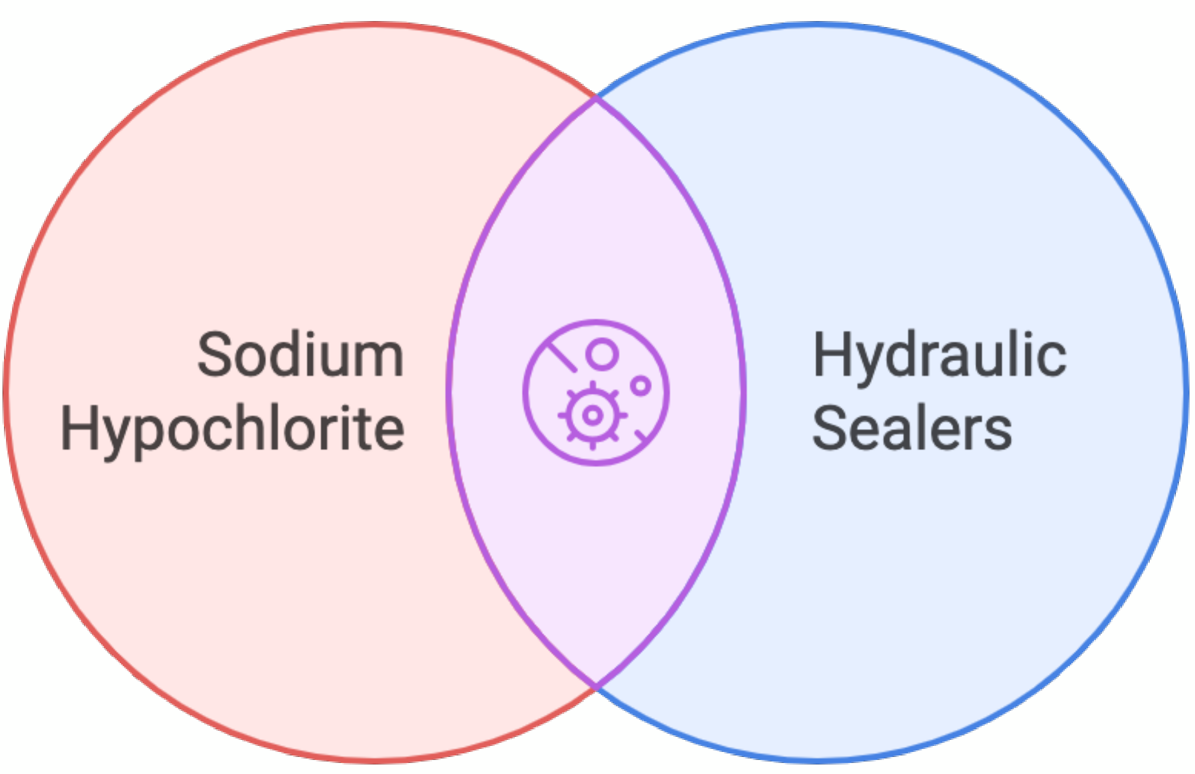
- Enhanced antimicrobial efficacy: sodium hypochlorite combined with hydraulic sealers
Int Endod J. 2022;55(Suppl. 3):710–777.
Clinical Performance And Material Interactions18
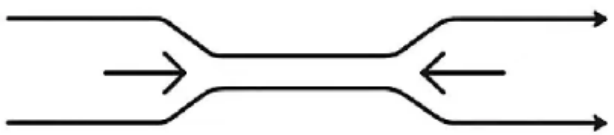
Irrigating Solutions — Impact on Hydraulic Sealers19
- Sodium hypochlorite — enhanced antimicrobial activity
- Calcium chelators (EDTA) — hindered mineral interaction; chemical alterations
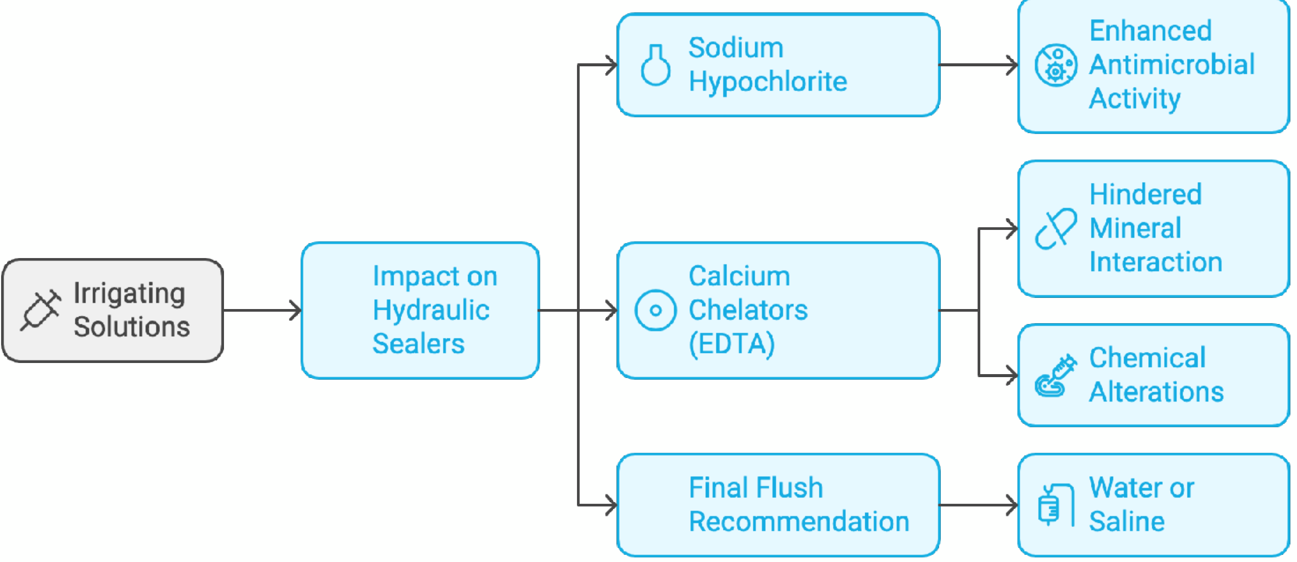
Final Flush Recommendation
Water or saline.
Int Endod J. 2022;55(Suppl. 3):710–777
- Stable and insoluble
- Superior long-term sealing
- Biocompatibility
- Material expansion during setting
Int Endod J. 2022;55(Suppl. 3):710–777
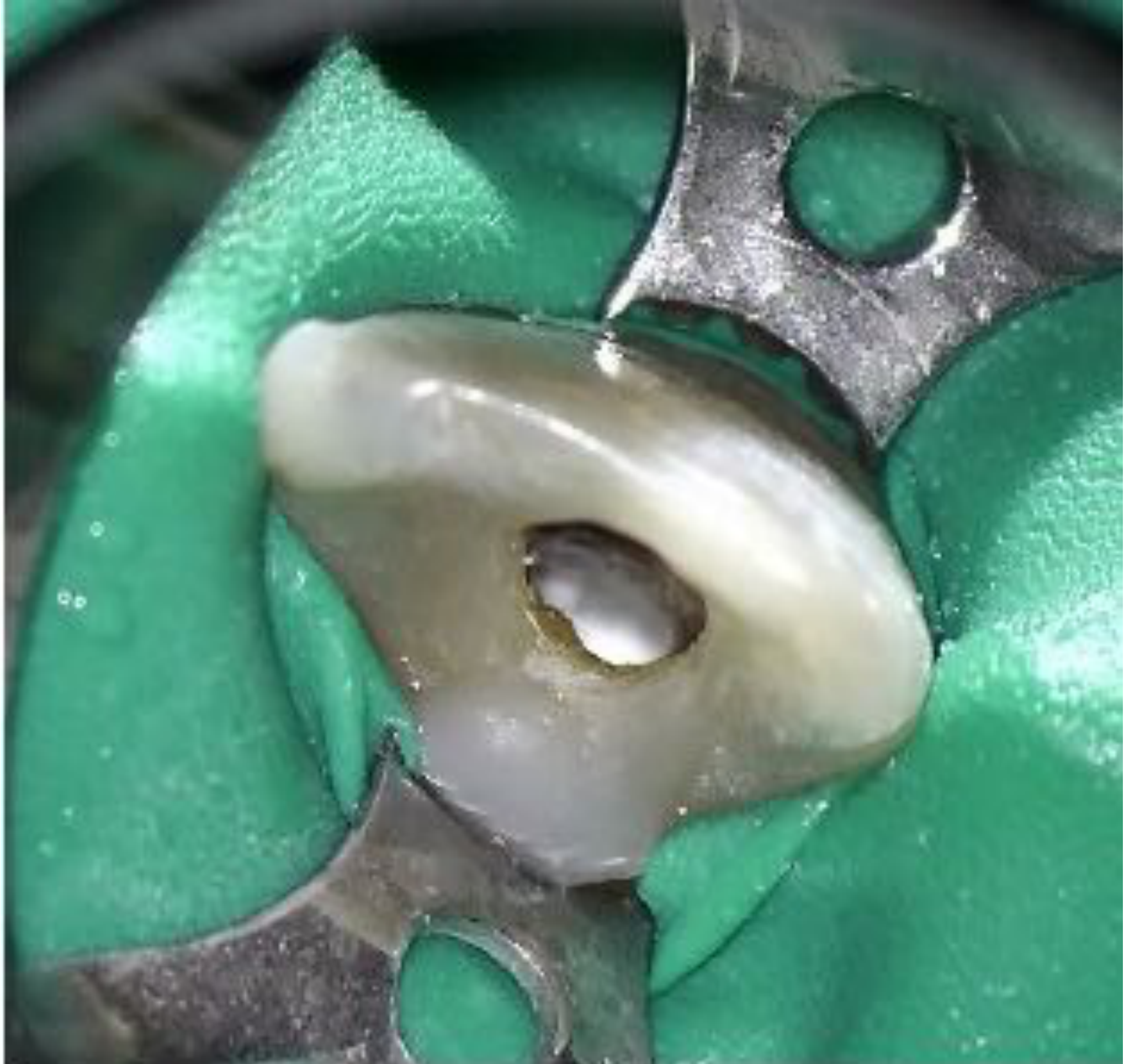
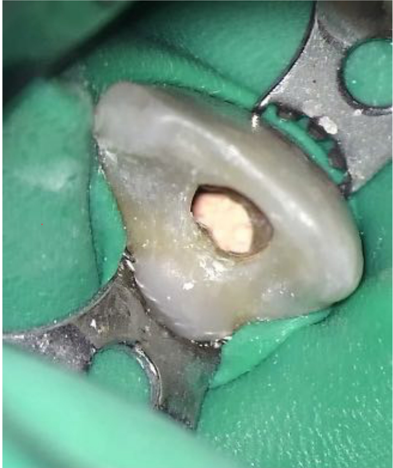
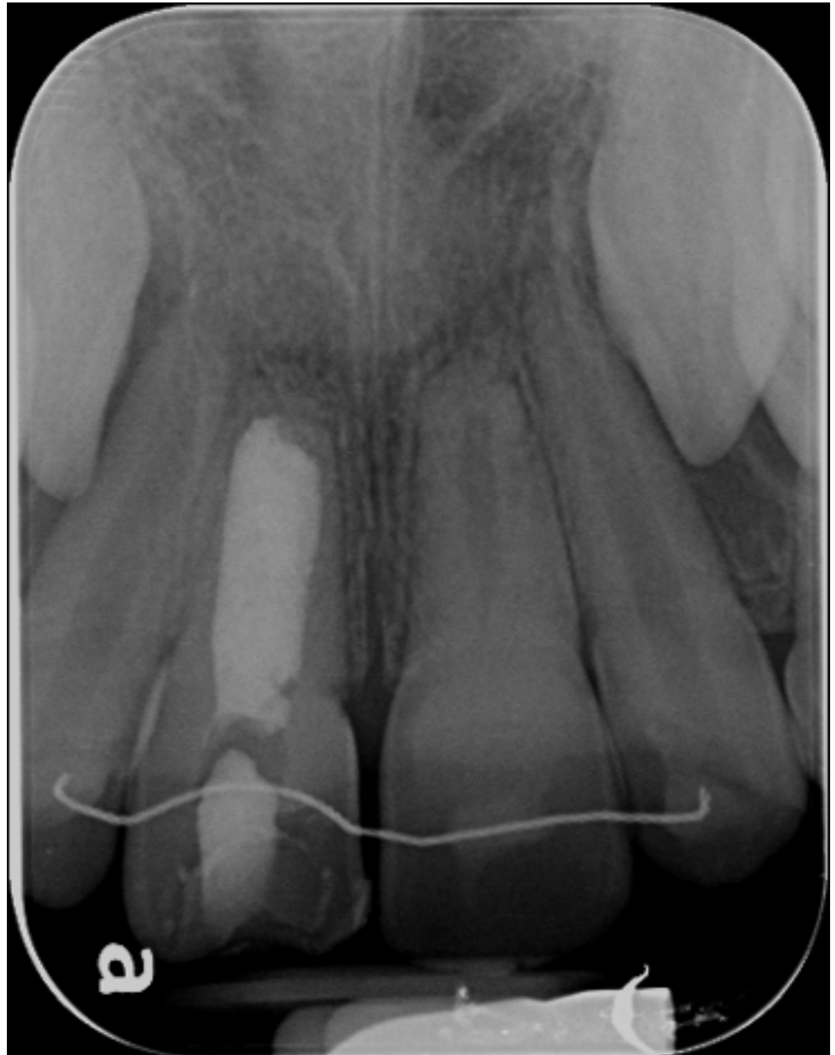
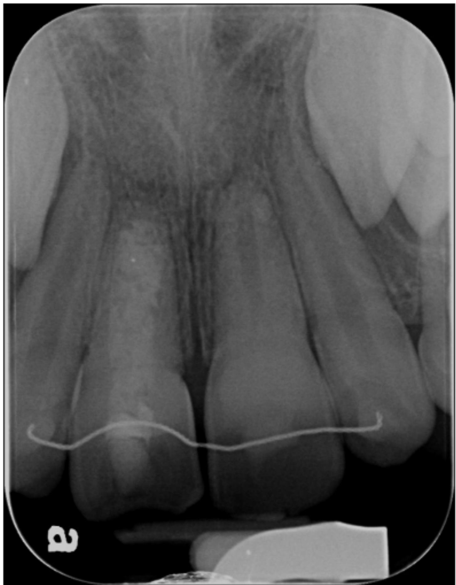
Apical Plug Case Report2021
 |  |
 |
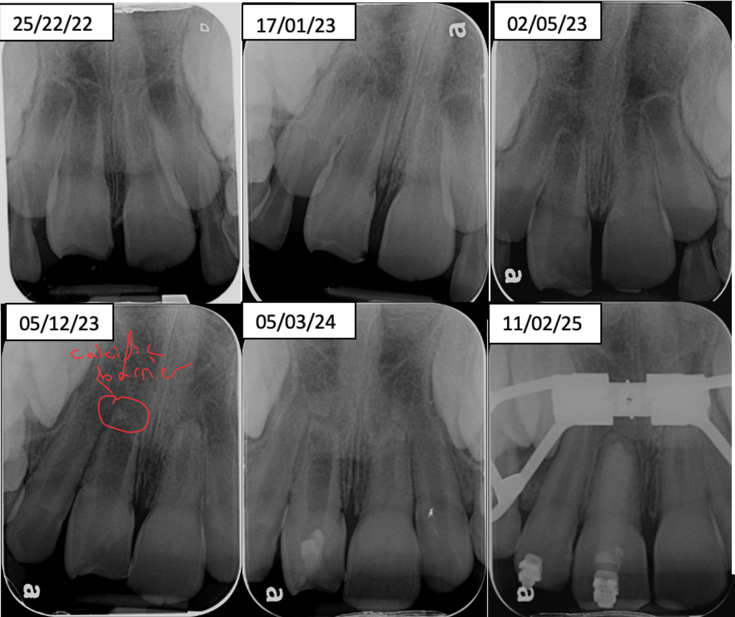
Case Timeline22
- Concussed tooth — apical plug
- 25/22/22
- 17/01/23
- 02/05/23
- 05/12/23 — calcified barrier
- 05/03/24
- 11/02/25
Ryan Butler, Endodontics Registrar
See - Open-Apex Trauma Case
See Case: Traumatized anterior teeth with open apices and different pulp responses for the full findings and sequence of this case.
Ryan Butler, Endodontics Registrar
See - Perforating Internal Resorption with a Wide Apex
This separate case concerns perforating internal resorption with a wide apex, not an incompletely formed root. The lecturer reserves “apexification” for long-term calcium hydroxide and barrier formation, calling the localized seal an “apical plug”; see Case: Perforating internal inflammatory resorption with a wide apex for the detailed findings and procedure.
 |  |
Treatment Flowcharts
Vital Pulp Treatment Pathway23
-
Healthy Tooth → Restored Tooth → Vital pulp treatment
-
If a tooth has been traumatized, the priority is to preserve the tooth whenever possible.
-
Vital pulp treatment may include partial pulpotomy or complete pulpotomy.
-
The choice depends on the size of the pulp exposure and the time at which the patient presents for treatment.
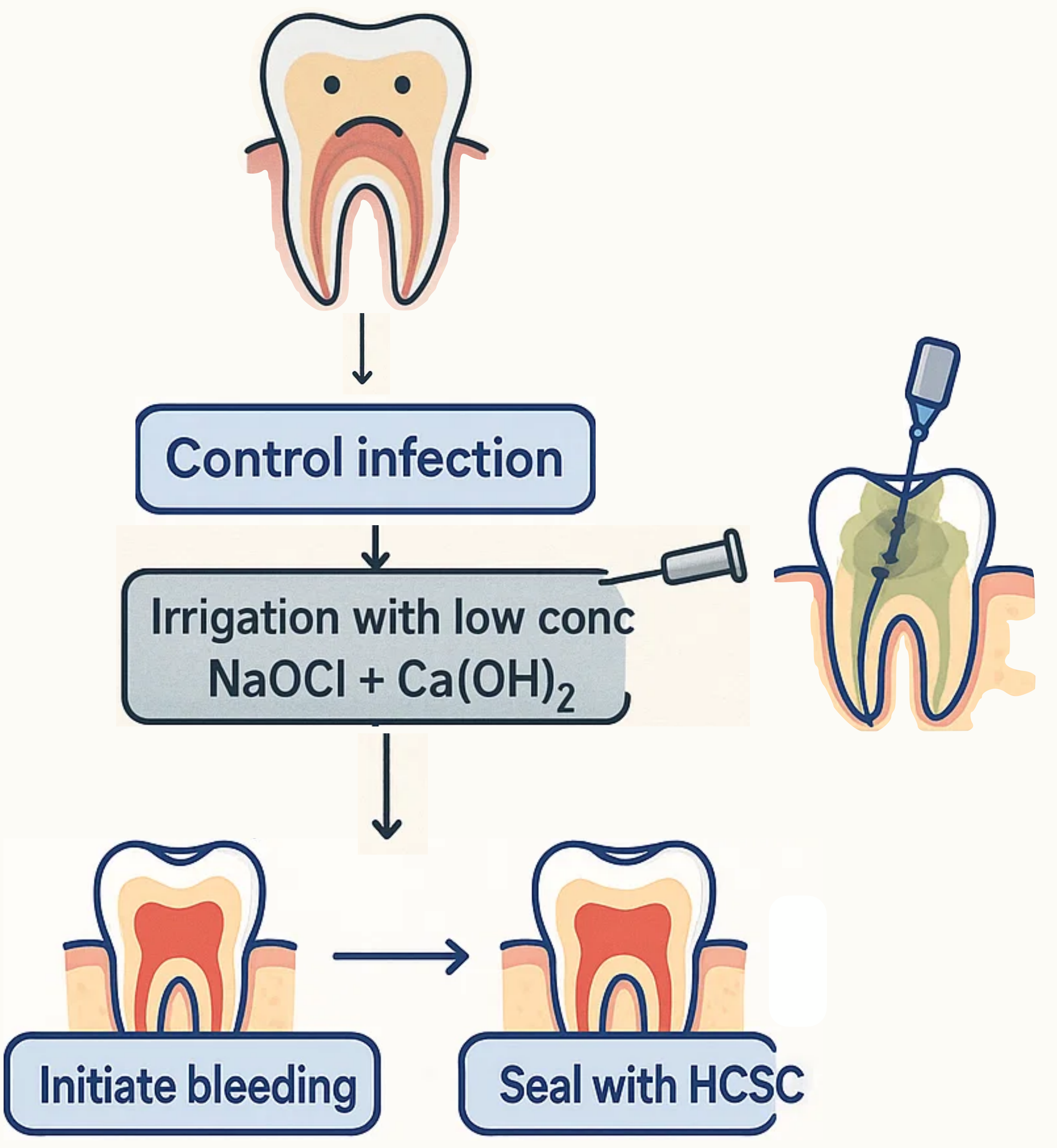
Infected Tooth Pathway
- Infected Tooth → Control infection
- Irrigation with low conc NaOCl + Ca(OH)₂
- Initiate bleeding
- Seal with HCSC
Lecturer — Apexification Options
For a pulpless and infected root canal system, infection must first be controlled before regeneration is considered.
- Apexification is another treatment option and was described as more predictable when calcium hydroxide is used until a calcific barrier forms.
- A bioceramic or MTA material can then be condensed over the barrier.
- Regeneration was described as less predictable.
Vital Pulp Treatment24
-
Healthy Pulp → Vital pulp treatment
-
The aim is to preserve pulp vitality and allow continued root development.
Regenerative Endodontic Treatment
- Infected Pulp → Control infection
- Irrigation with low conc NaOCl + Ca(OH)₂
- Initiate bleeding
- Seal with HCSC
Lecturer — Regeneration Outcomes
Regeneration may be attempted even though its outcome is not predictable.
- If regeneration fails, treatment can be changed to apexification.
Vital Pulp Treatment Branch25
-
Tooth diagram → Tooth diagram → Vital pulp treatment
-
For a traumatized tooth with vital pulp, preserve the pulp through partial or complete pulpotomy, selected according to the size of the exposure and the timing of treatment.
-
Maintaining pulp vitality allows continued root formation.
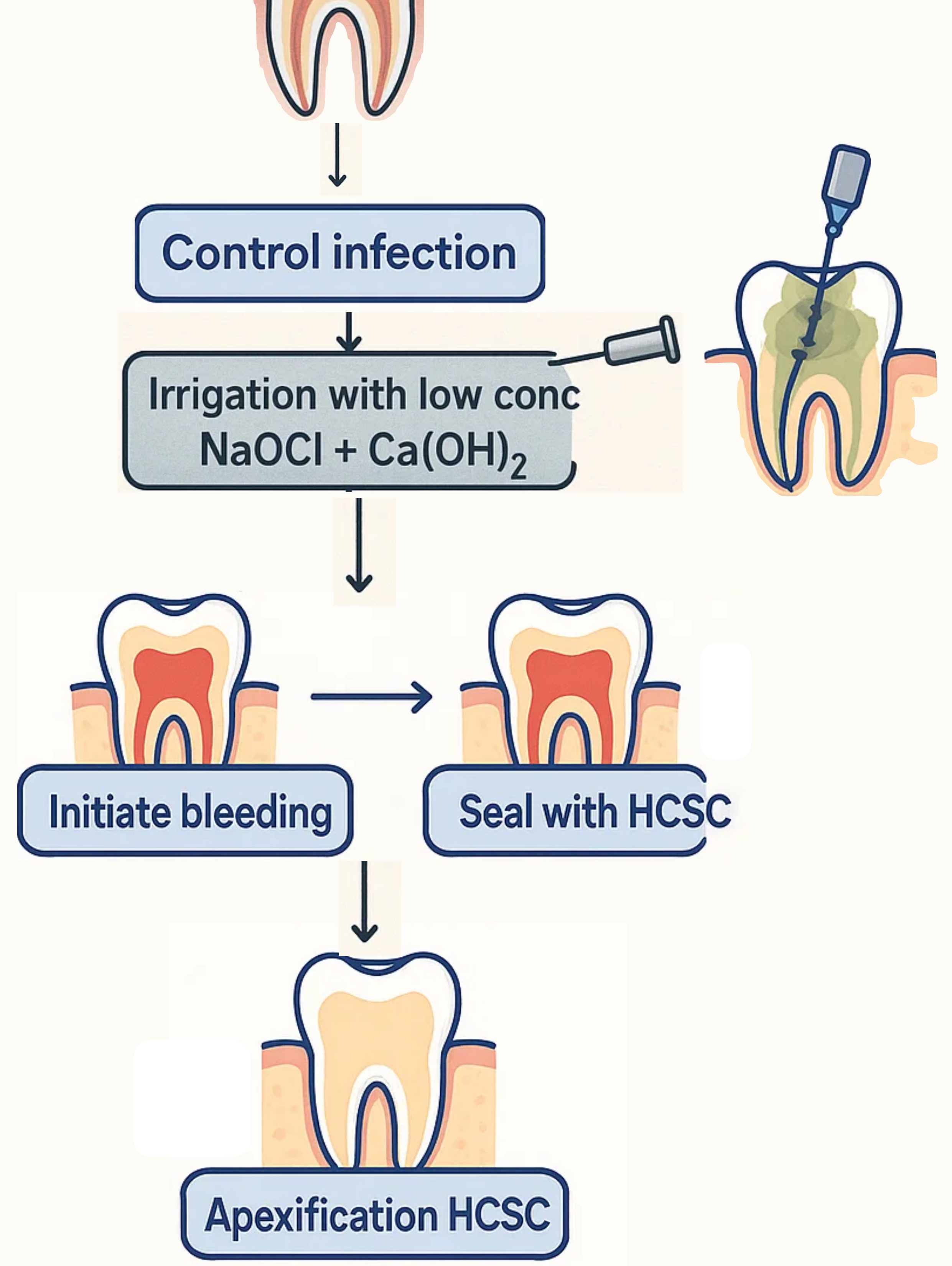
Regenerative Endodontic Branch
- Tooth diagram → Control infection
- Irrigation with low conc NaOCl + Ca(OH)₂
- Initiate bleeding, then either:
- Seal with HCSC
- Apexification HCSC
Lecturer — Treatment Sequencing
For a non-vital tooth, clinicians may consider regenerative treatment after controlling the infection, but the sequence affects the remaining options.
- If apexification is performed first, returning to regeneration is not possible.
- If regeneration is attempted first and fails, apexification remains available.
- Both approaches were described as potentially acceptable given the continuing debate over the preferred sequence.
- The treatment decision should be discussed with the patient’s parents or carers, including the potential for additional dentin thickness, calcific-like tissue formation, risks and benefits, uncertainty of regeneration, and the greater predictability of apexification.
- The family may prefer extraction or a more predictable option, and clinicians should accept diversity in treatment planning rather than adopting a fixed rule.
Questions26
Questions?
Lecturer — Apical Barrier
The apical barrier is not necessarily a uniform dentinal constriction covered by cementum.
- Histological studies show that it may contain bone-like tissue, cementum-like tissue, or other calcified tissue.
- Radiographically, one part of the apex may appear more completely formed than another.
- Its purpose is to provide a barrier against which the final filling material can be compacted.
- Even when infection is controlled, the calcific barrier alone is not a complete final seal; the apical area must still be sealed with hydraulic calcium silicate material.
The lecturer also stated that this was the final lecture in the students’ university journey. Further examination information would be announced on the learning management system, including the dental traumatology section’s case-based and theory multiple-choice questions, Dr. Fu’s approximately five or six short-essay questions, and the contribution of each component as 5% of the total unit assessment.
Clinical Cases
Case: Traumatized anterior teeth with open apices and different pulp responses
Question
Scenario: A patient with a history of trauma and concussion to the anterior teeth (11 and 21) is evaluated.
What’s shown: A series of sequential periapical radiographs showing the initial presentation and follow-up visits over time.
Consider: Identify the different pulp responses to the trauma and determine the appropriate management for the tooth that lost vitality.
Answer
Observations:
- Initial radiographs show open apices at teeth 11 and 21.
- Follow-up radiographs reveal two different pulp responses: one tooth exhibits pulp canal obliteration (calcification), while the other has lost vitality and retained its open apex.
- After long-term calcium hydroxide medication on the non-vital tooth, a calcific barrier forms around the apex and increases in density over four months.
Reasoning: The calcific barrier induced by long-term calcium hydroxide is not an ideal, continuous root apex closure, but it provides a necessary stop to properly condense and compact restorative materials without the risk of extrusion. A bioceramic (calcium silicate-based) material is chosen for the apical plug because of its biological properties and superior performance in a moist environment compared to gutta-percha and resin sealers. Attempting to place an apical barrier straight away without the calcific barrier would lack a strong apical stop, risking material extrusion and making the procedure messy.
Outcome: A putty bioceramic material was packed to form at least a 3mm apical plug over the calcific barrier. The bioceramic plug provides the apical seal; gutta-percha was then injected to backfill the canal, and the access was sealed coronally with a restoration.
Takeaway: Combining long-term calcium hydroxide apexification to form a calcific barrier with a subsequent bioceramic apical plug provides a strong stop for condensation, prevents material extrusion, and ensures a proper apical seal in teeth with open apices and necrotic pulps.
Case: Large apex with some apical closure, treated with an MTA plug
Question
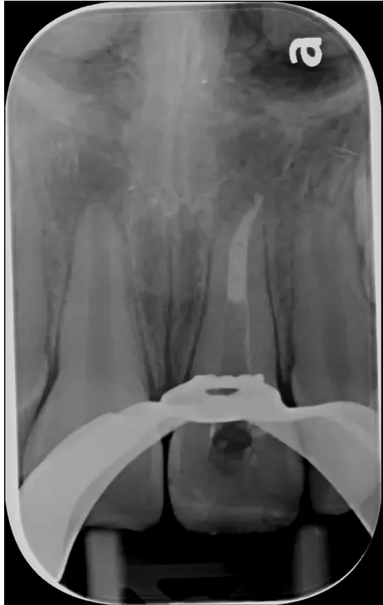
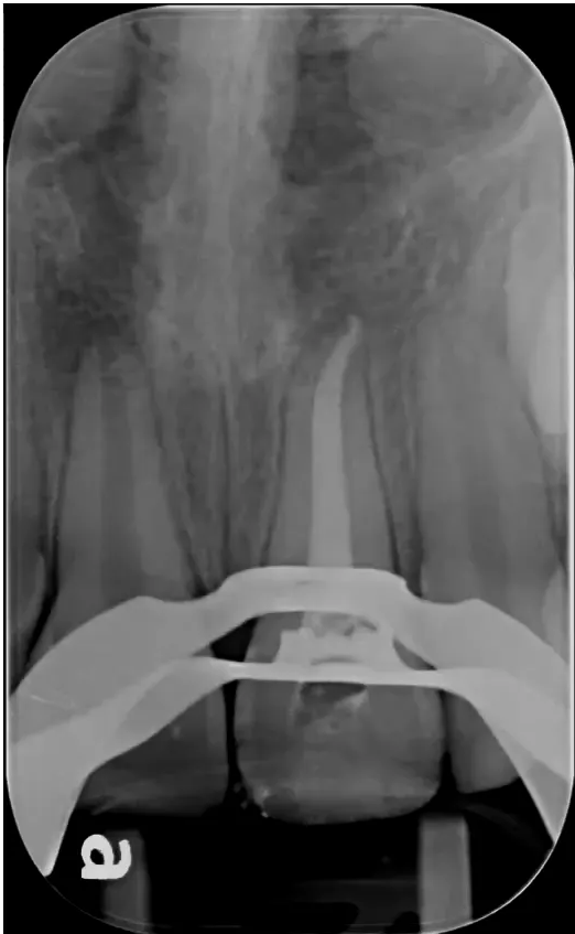
Scenario: A tooth with a very large apex (larger than size 70) but with some existing apical closure is presented for management.
What’s shown: A radiograph showing the apex with some apical closure but significant width.
Consider: How to manage the apical portion to ensure a proper seal given the potential for fluid exchange.
Answer
Observations:
- The apex is larger than size 70 but still has some apex formation and closure.
- There is potential for significant fluid exchange within the periodontium.
Reasoning: Because of the fluid exchange and the wide apex, gutta-percha (GP) alone is not ideal. An MTA or bioceramic apical plug is preferred due to its better performance in a moist environment and biological effects, providing a strong barrier to condense material against.
Outcome: A 3 to 5 mm MTA plug was placed, and GP was injected on top. The material was condensed properly without extrusion.
Takeaway: For large apices with some closure, an MTA/bioceramic apical plug provides a superior seal and biological effect compared to GP alone.
Case: Wide apex requiring an apical plug, complicated by extrusion
Question
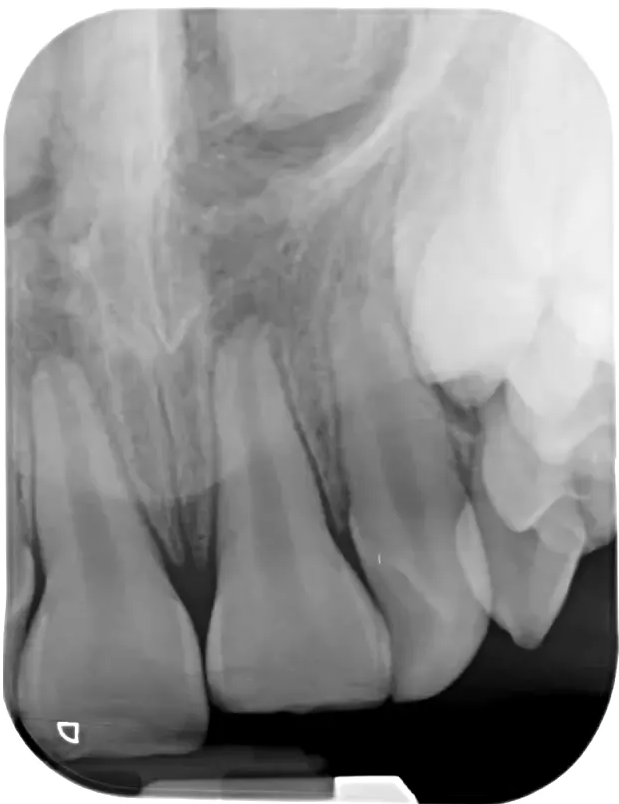
Scenario: A second tooth with a wide apex requiring an apical plug is presented to contrast with the previous case.
What’s shown: A radiograph of the second case showing a wide apex.
Consider: The challenges of condensing an apical plug in a wide apex.
Answer
Observations:
- The tooth has a wide apex.
Reasoning: Condensing the apical plug material in this specific anatomy is more difficult than in the previous case.
Outcome: The material was harder to condense, resulting in extrusion beyond the apex.
Takeaway: Condensing apical plugs in wide apices can be technically challenging and carries a risk of material extrusion.
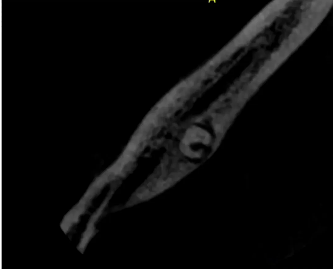
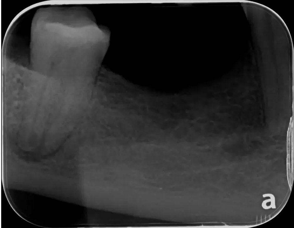
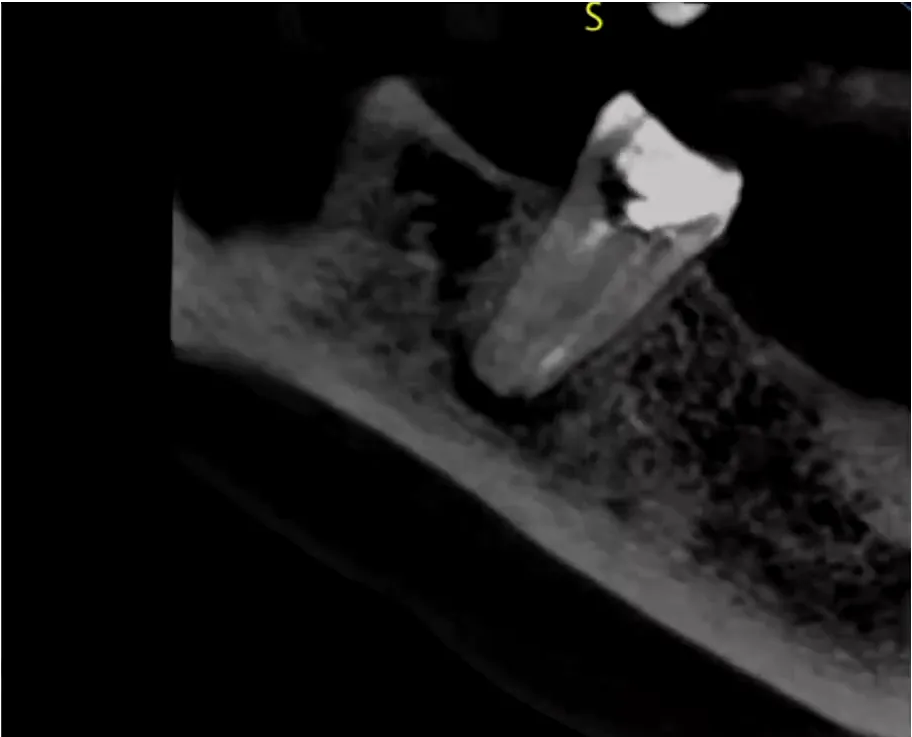
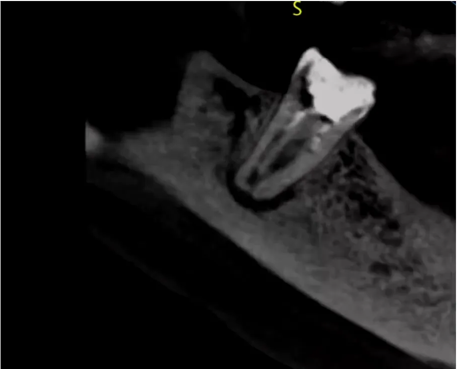
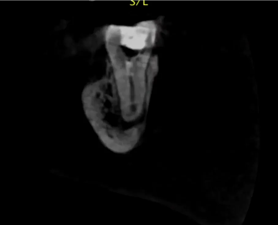
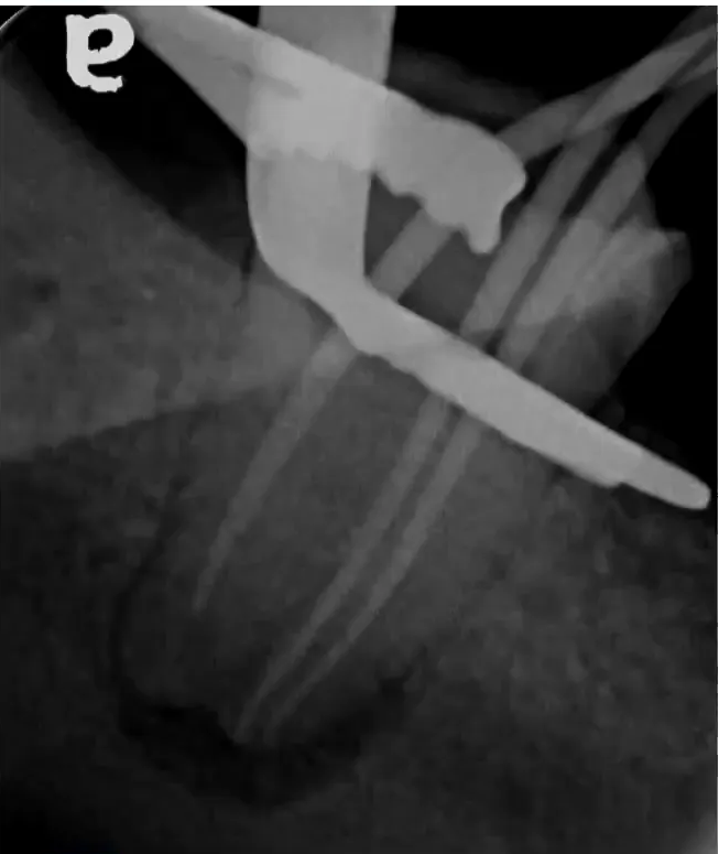
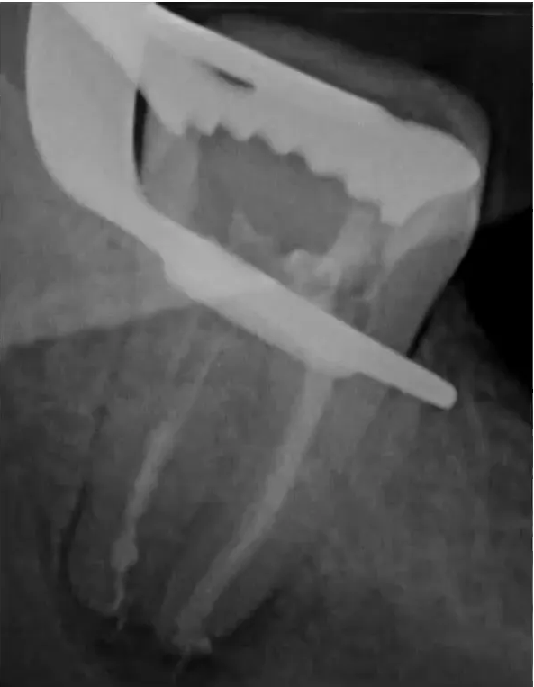
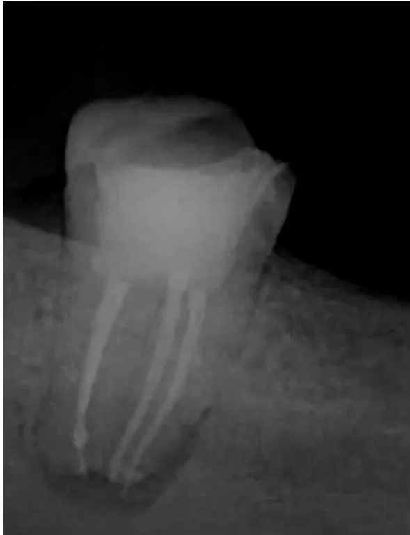
Case: Perforating internal inflammatory resorption with a wide apex
Question
Scenario: A tooth with perforating internal inflammatory resorption at the apical portion and a very wide apex is presented. It is not an incompletely formed root.
What’s shown: CBCT and radiographs showing the resorptive defect, the wide apex, and the master gutta-percha (GP) cone in the canal.
Consider: How to manage the perforating resorptive defect and determine the working length in the presence of a wide apex and bleeding.
Answer
Observations:
- CBCT shows perforating internal resorption open toward the furcal area at the apical portion of the root.
- The apex is very wide.
- Inserting a paper point deeper into the canal resulted in bleeding, indicating the boundary between the root canal and the periodontium.
Reasoning: Placing GP straight away is uncomfortable because it does not provide a substantial seal in this inflammatory area and wide apex, even with a thin layer of bioceramic. The bleeding line marks the end of the root canal. A more biological, injectable material is needed to seal the resorptive defect.
Outcome: The master GP was stopped at the bleeding line. Injectable MTA/bioceramic material was injected to seal the resorptive defect, followed by injectable GP to complete the fill.
Takeaway: In cases of perforating internal resorption with a wide apex, an injectable bioceramic/MTA is necessary to adequately seal the defect, as GP alone cannot provide a sufficient seal in the inflammatory environment.
Footnotes
-
Original PDF page 1: L17 Management of Incompletely Developed Tooth 2, p.1 ↩
-
Original PDF page 2: L17 Management of Incompletely Developed Tooth 2, p.2 ↩
-
Original PDF page 3: L17 Management of Incompletely Developed Tooth 2, p.3 ↩
-
Original PDF page 4: L17 Management of Incompletely Developed Tooth 2, p.4 ↩
-
Original PDF page 5: L17 Management of Incompletely Developed Tooth 2, p.5 ↩
-
Original PDF page 6: L17 Management of Incompletely Developed Tooth 2, p.6 ↩
-
Original PDF page 7: L17 Management of Incompletely Developed Tooth 2, p.7 ↩
-
Original PDF page 8: L17 Management of Incompletely Developed Tooth 2, p.8 ↩
-
Original PDF page 9: L17 Management of Incompletely Developed Tooth 2, p.9 ↩
-
Original PDF page 10: L17 Management of Incompletely Developed Tooth 2, p.10 ↩
-
Original PDF page 11: L17 Management of Incompletely Developed Tooth 2, p.11 ↩
-
Original PDF page 12: L17 Management of Incompletely Developed Tooth 2, p.12 ↩
-
Original PDF page 13: L17 Management of Incompletely Developed Tooth 2, p.13 ↩
-
Original PDF page 14: L17 Management of Incompletely Developed Tooth 2, p.14 ↩
-
Original PDF page 15: L17 Management of Incompletely Developed Tooth 2, p.15 ↩
-
Original PDF page 16: L17 Management of Incompletely Developed Tooth 2, p.16 ↩
-
Original PDF page 17: L17 Management of Incompletely Developed Tooth 2, p.17 ↩
-
Original PDF page 19: L17 Management of Incompletely Developed Tooth 2, p.19 ↩
-
Original PDF page 18: L17 Management of Incompletely Developed Tooth 2, p.18 ↩
-
Original PDF page 21: L17 Management of Incompletely Developed Tooth 2, p.21 ↩
-
Original PDF page 22: L17 Management of Incompletely Developed Tooth 2, p.22 ↩
-
Original PDF page 20: L17 Management of Incompletely Developed Tooth 2, p.20 ↩
-
Original PDF page 23: L17 Management of Incompletely Developed Tooth 2, p.23 ↩
-
Original PDF page 24: L17 Management of Incompletely Developed Tooth 2, p.24 ↩
-
Original PDF page 25: L17 Management of Incompletely Developed Tooth 2, p.25 ↩
-
Original PDF page 26: L17 Management of Incompletely Developed Tooth 2, p.26 ↩