L6 Vital Pulp Treatment 1
Vital Pulp Treatment12
Dr Mostafa Elkholy
Dr Mostafa Elkholy, BDS, MSc, PhD
Lecturer — Vital Pulp Treatment
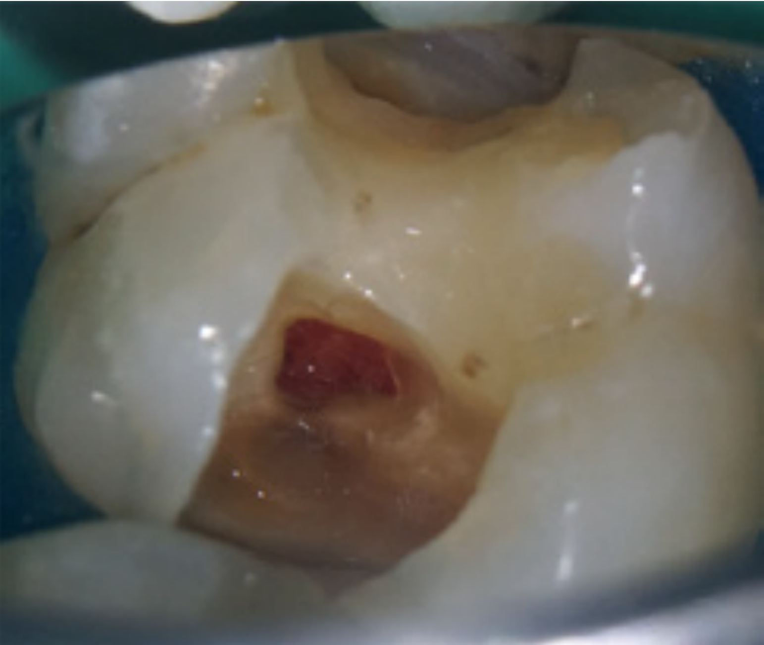
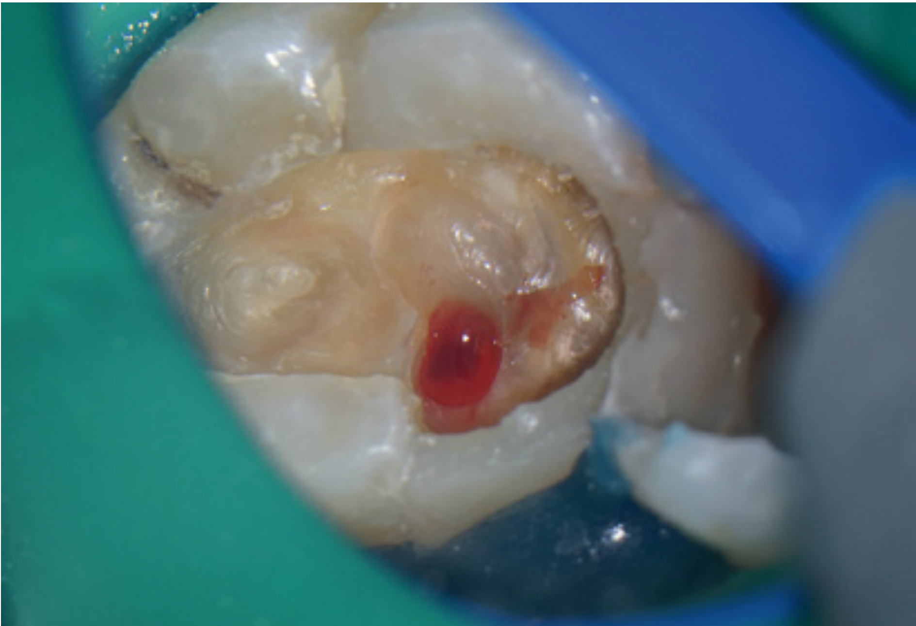
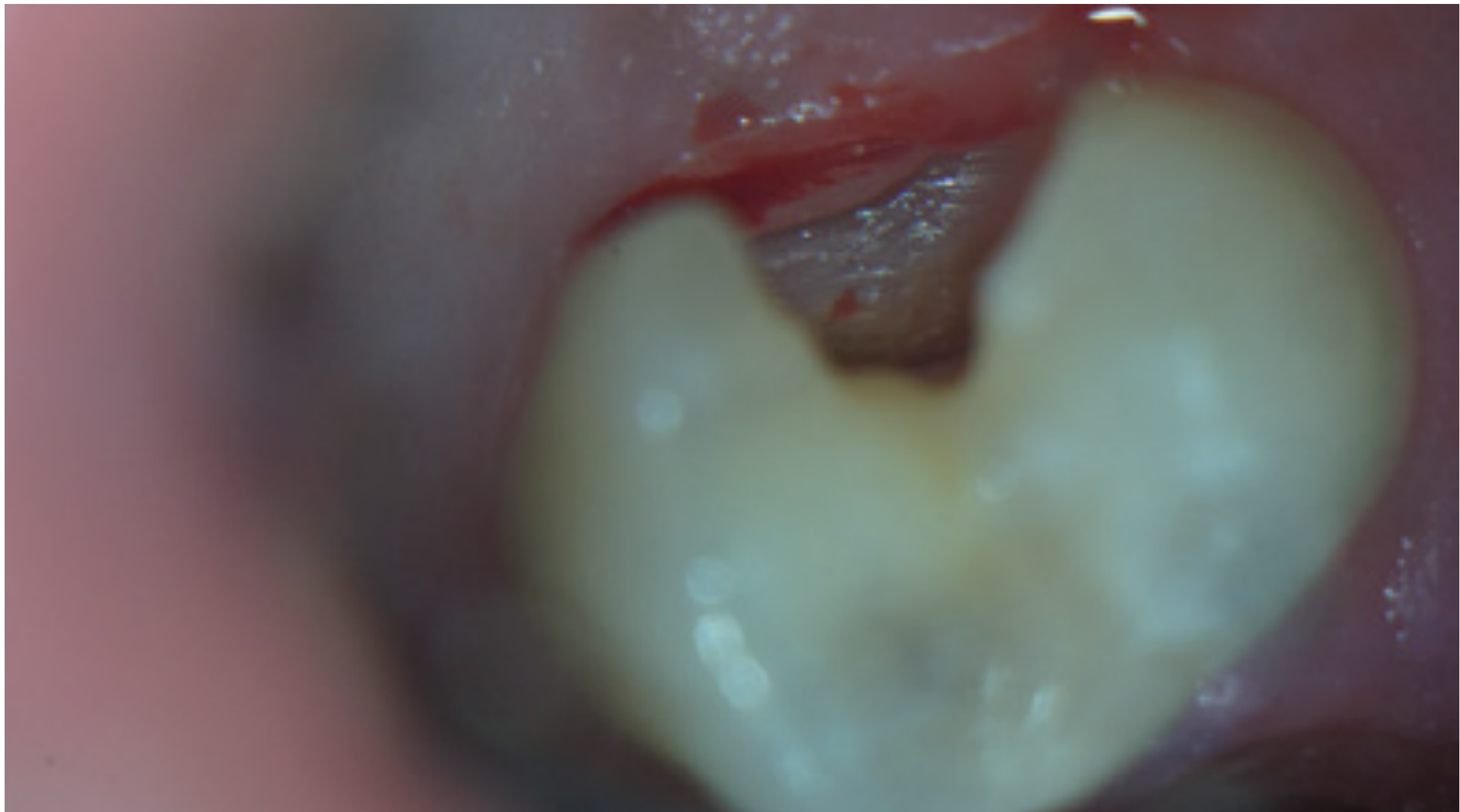
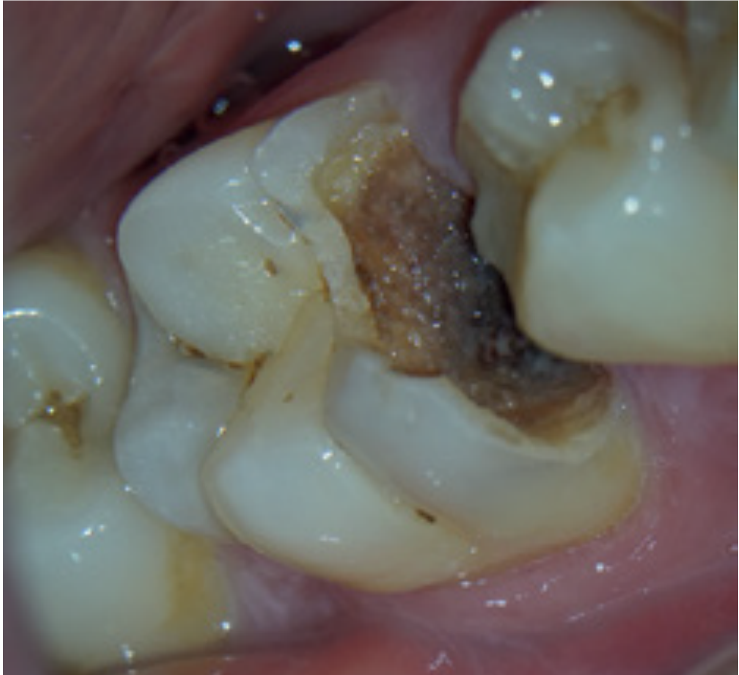
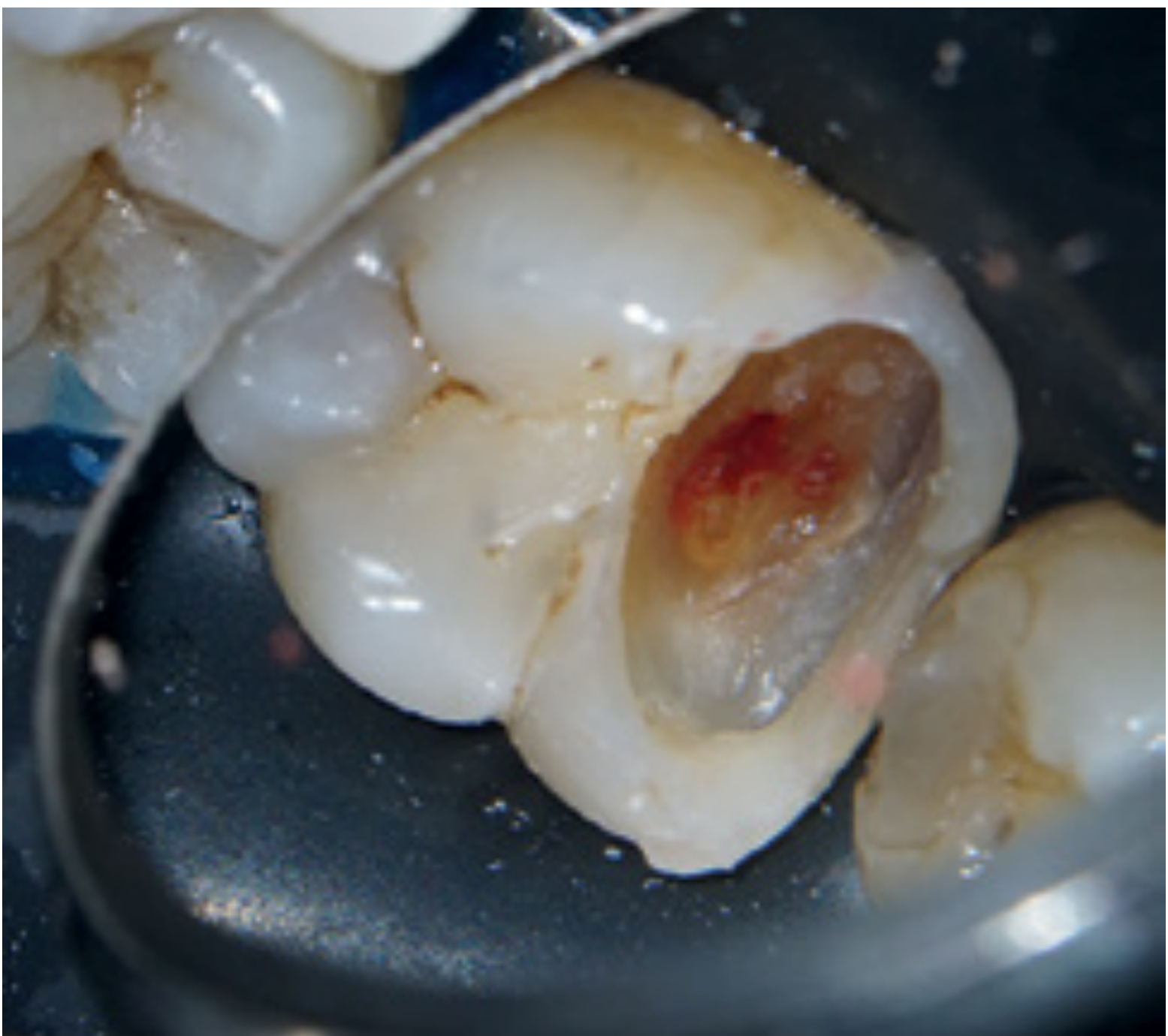
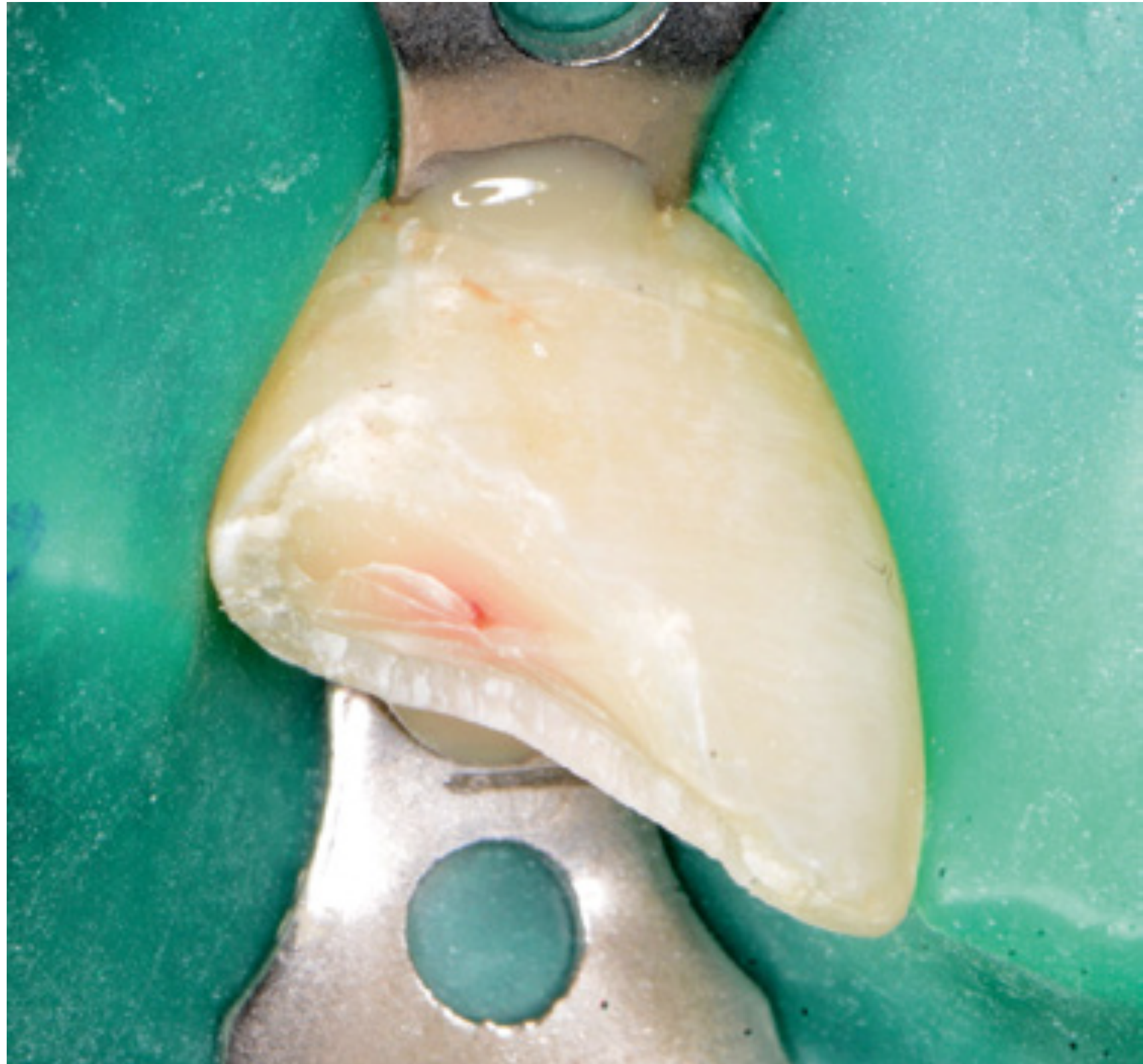
Vital pulp treatment is a clinically important and increasingly demanded area of endodontic practice. It is also called vital pulp therapy or conservative pulp treatment.
- The practical questions include whether to remove all caries or leave the deepest carious dentine, whether pulp exposure should be avoided, and whether root canal treatment is needed immediately after exposure.
- Decisions also involve the materials and procedures that may preserve a healthy pulp, balanced against the patient’s medical, financial, and practical circumstances.
- Vital pulp treatment should not replace root canal treatment when root canal treatment is indicated; teeth requiring root canal treatment should receive it, while suitable teeth should be managed with vital pulp treatment.
 |  |  |
 |  |  |
History of Pulp Capping3
- In the 18th century, a piece of gold was placed over the exposed vital pulp in an attempt to promote healing. J Dent Res 1949;39:97-107.
- In 1922, it was stated that ‘the exposed pulp is a doomed organ’. Heft 1922;16:3-83.
- Studies in the 1950s reported a successful outcome of between 60 to 70% for direct pulp capping compared with over 80% for conventional RCT. Acta Odontol Scand 1955;13:1-130. Odontol Tidsk 1958;66:296-364. Acta Odontol Scand 1956;14:1-175.
- Later studies demonstrated a successful outcome of 80 to 90%. J Am Dent Assoc 1978;97:607-612. Int Dent J 1981;31:251-260 J Am Dent Assoc 2008;139:305-315. J Clin Pediatr Dent 2006;31:72-76.
Lecturer — Pulp Capping Practice
The lecturer described how the historical view of pulp exposure shaped clinical practice.
- Pulp exposure was commonly followed by root canal treatment or extraction for more than a century.
- Clinicians differ in their support for pulp preservation, while some endodontists have traditionally recommended root canal treatment whenever the pulp is exposed.
- There is a gap between academic teaching and private clinical practice; decisions should also consider the patient’s medical condition, financial burdens, ability to attend appointments, likely treatment burden, and the clinician’s responsibility for the patient’s overall physical and mental well-being.
Lecturer — Dentine Resilience
Moist dentine contributes to the resilience that helps the tooth withstand mastication and other forces.
- After pulp removal, dentine inside the root canal becomes dry and stiffer.
- Although enamel is the hardest structure in the human body, dentine provides the tooth’s resilience.
Lecturer — Progressive Structure Loss
Tooth structure may be lost progressively over the life cycle of a tooth.
- A restoration may be placed and later removed because of inflammation, infection, or recurrent caries.
- Remaining caries may then be removed and an access cavity prepared for root canal treatment.
- Placement of a post, core, and crown can cause further unavoidable loss of tooth structure and reduce structural integrity.
Lecturer — Curved Canal Risks
Preserving the pulp may avoid the need to shape a complex or curved root canal system.
- Root canal treatment in a curved canal may result in instrument fracture or perforation.
- These procedures can create additional treatment complications, so preserving the pulp may be advantageous when the tooth is suitable for vital pulp treatment.
Sequela of Loss of Pulp Vitality
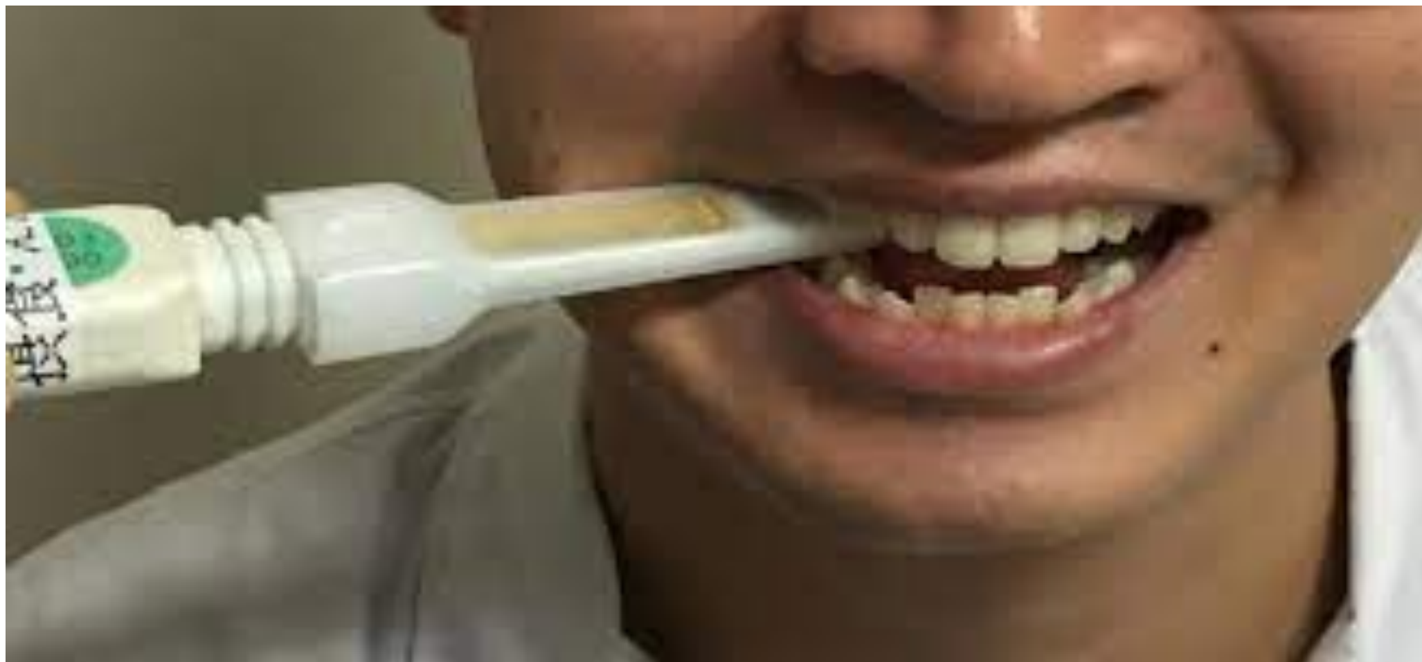
The tooth becomes more vulnerable to irreparable fracture because of the loss of proprioceptive mechanisms.
Lecturer — Fracture Evidence
One study examined whether loss of pulp vitality changes biting behavior.
- Bite forces were compared on an endodontically treated tooth and its contralateral tooth using a bite fork.
- Some patients may bite more heavily on the tooth after pulp removal because mechanoreceptors or proprioceptive receptors have been lost.
- The lecturer considered the evidence to exist but questioned its strength because the available literature was limited.
Fracture Vulnerability After Loss of Vitality4
J Endod 2017;43: 871.
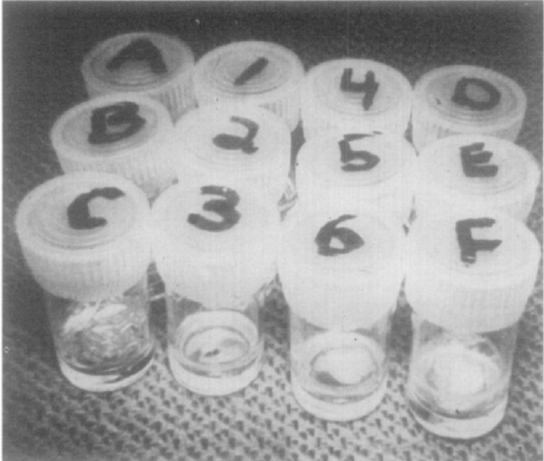
Moisture Depletion and Reduced Tooth Stiffness5
Moisture depletion from dentine and reduction of tooth stiffness after root canal treatment due to moisture depletion from dentine.
Oral Surg Oral Med Oral Pathol 1972;34: 661.
Cumulative Loss of Tooth Structure6
The cumulative loss of tooth structure from endodontic and restorative treatment weakens teeth.
J Endod 2021 Sep;47(9):1472-1480.
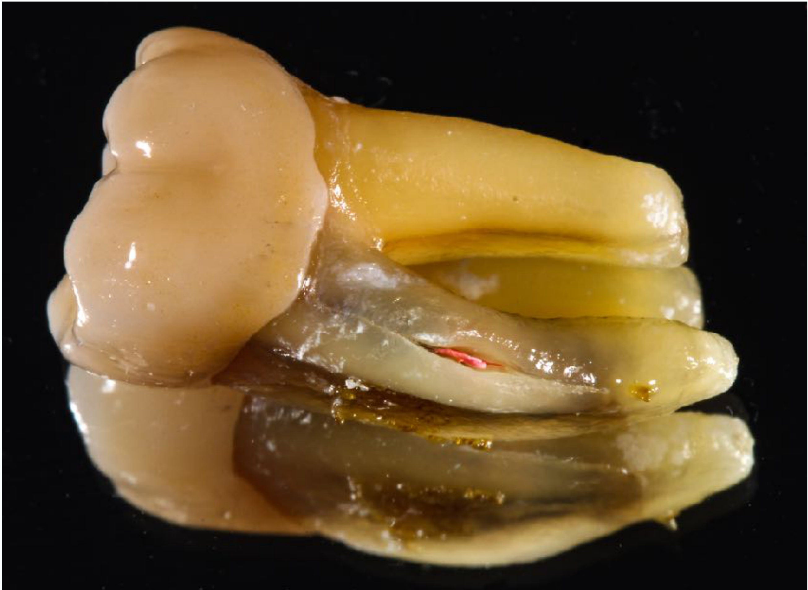
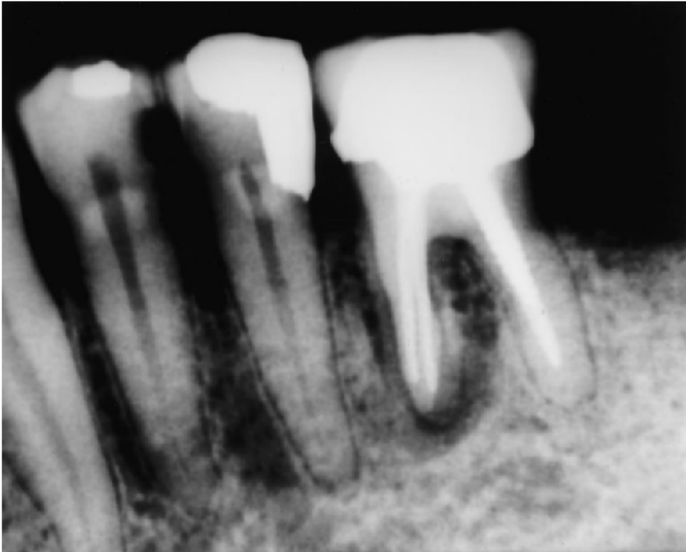
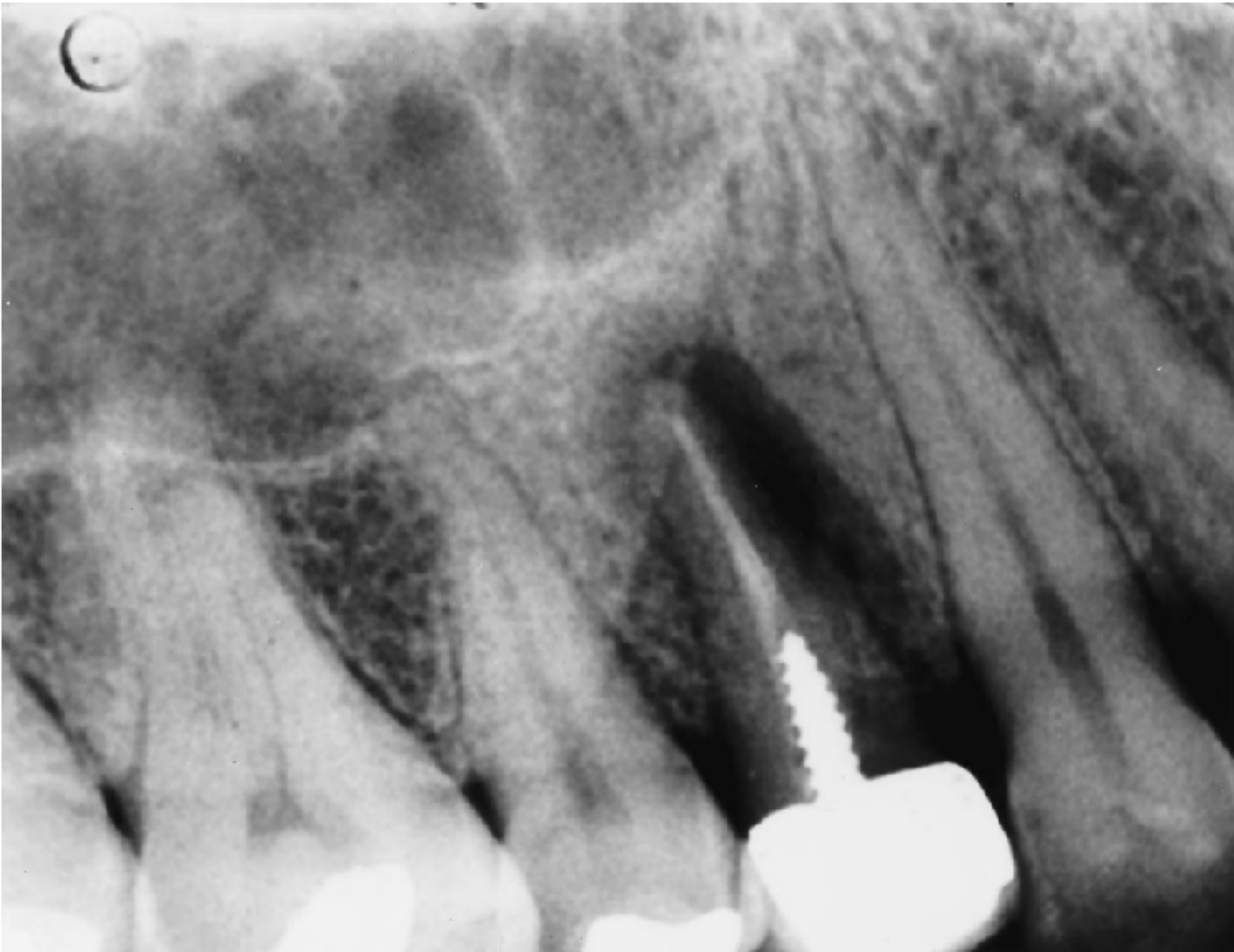
Changes in Root Canal Geometry7
Changes in root canal geometry are inevitable in the course of canal preparation and may lead to a higher incidence of fractures.
J Endod 2001;27: 46.
 |  |
Additional Potential Problems8
-
Tooth discoloration. (Dent Traumatol 2013;29: 2.)
-
Susceptibility to recurrent caries because of poor marginal integrity of restorations or modification of their biologic environment. (J Public Health Dent 1997;57: 31.)
-
Root canal treatments may be more complex than initially thought, while procedures for maintaining vitality are conservative, easy to implement, and often cost-effective. (J Endod 2014;40: 1764.)
-
A possible eventual outcome is loss of the tooth.
Rationale of Vital Pulp Treatment9
- Maintaining a healthy pulp is preferable so it can do its developmental, defensive, and proprioceptive functions.
- Preserving the dental pulp, especially in vital teeth with exposed pulp or immature teeth with incomplete root formation, supports continued root development.
- There has been a call for a more conservative approach in endodontics, emphasizing the importance of preserving rather than removing pulp tissue.
- Preserving the pulp is encouraged in modern endodontics due to its conservative nature, biological acceptance, and lower technical demands compared to complete pulpectomy, and it is more cost effective.
Lecturer — Treatment Selection
The lecturer emphasized that preserving the pulp may be justified even when the predicted success is very low, because there may otherwise be little to lose.
- A mature tooth with a completely formed root differs from an immature tooth, where preserving the pulp allows root formation to continue.
- Vital pulp treatment should not be chosen merely to avoid a technically difficult root canal procedure; if root canal treatment is indicated but cannot be performed, the patient should be referred to an appropriate clinician.
Biological Basis of Vital Pulp Treatment
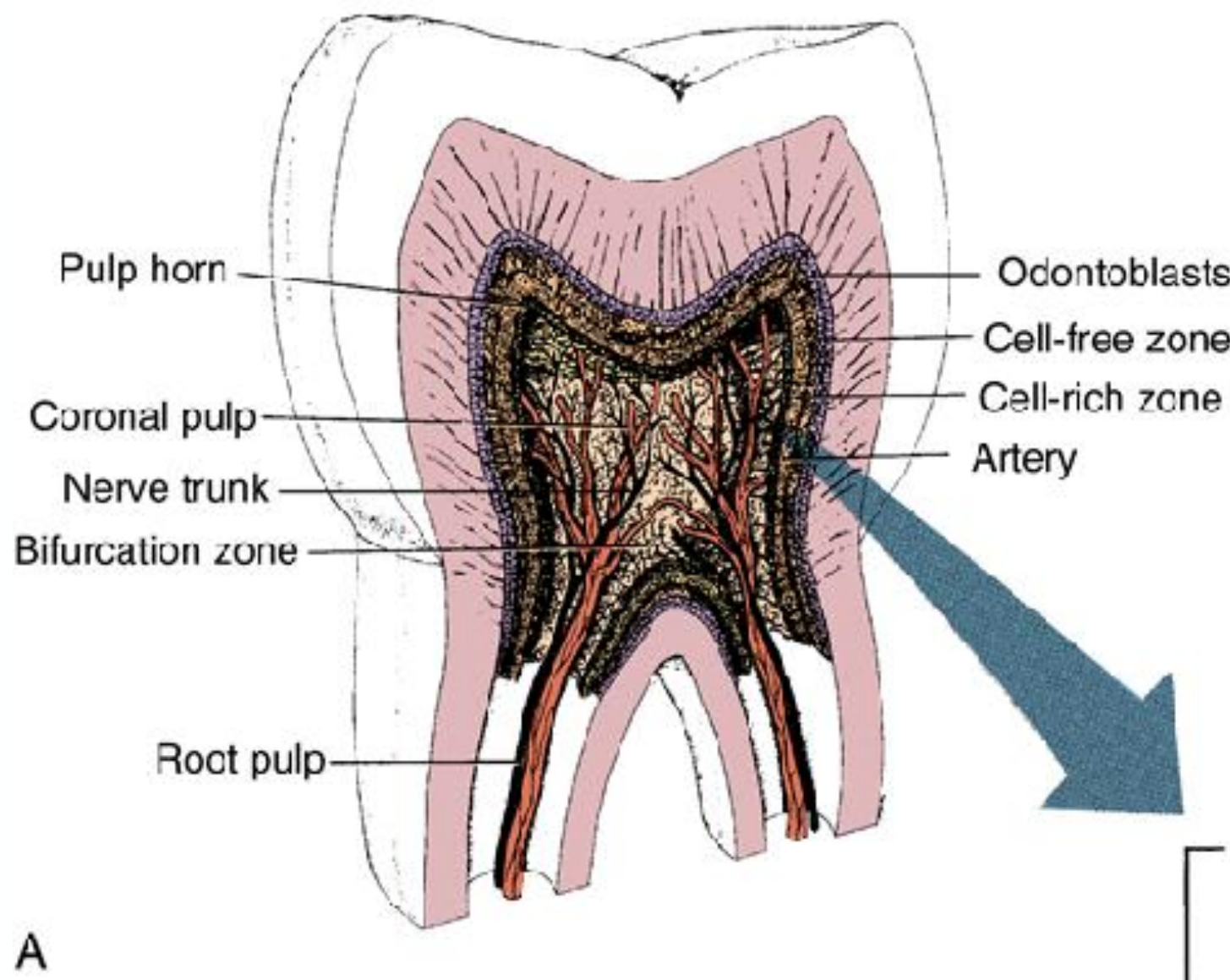
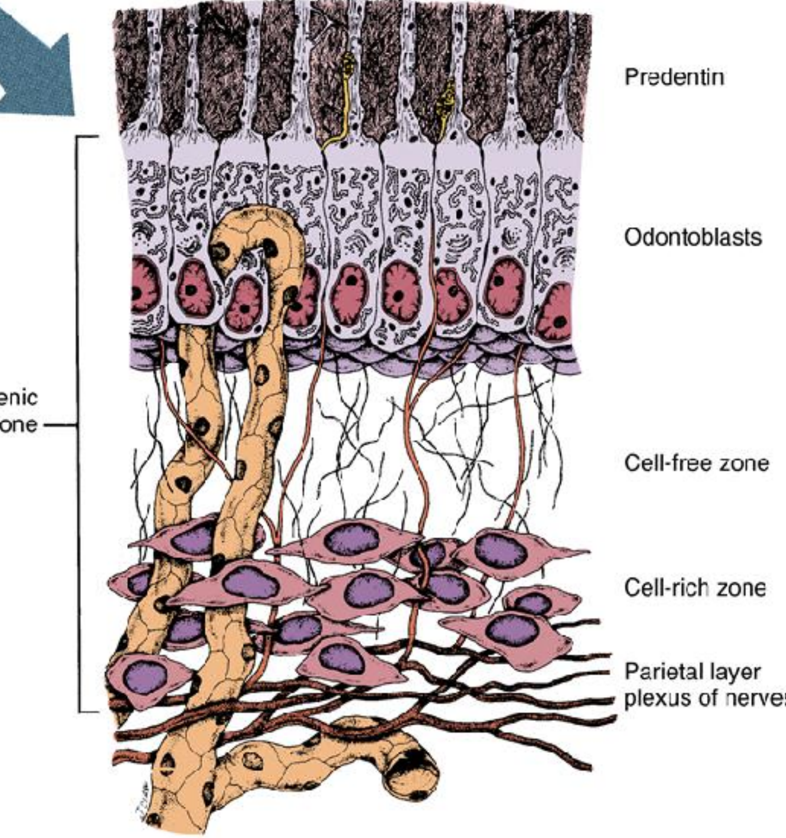
The dental pulp comprises the following structural and functional components:
Dental Pulp Structure and Function10
-
Odontoblasts
-
Fibroblasts
-
Immune cells
-
Extracellular matrix (ECM)
-
Nerves
-
Vasculature
-
Odontoblasts were described as the main constituent of pulp tissue.
 |  |
Healing Capability of Dental Pulp
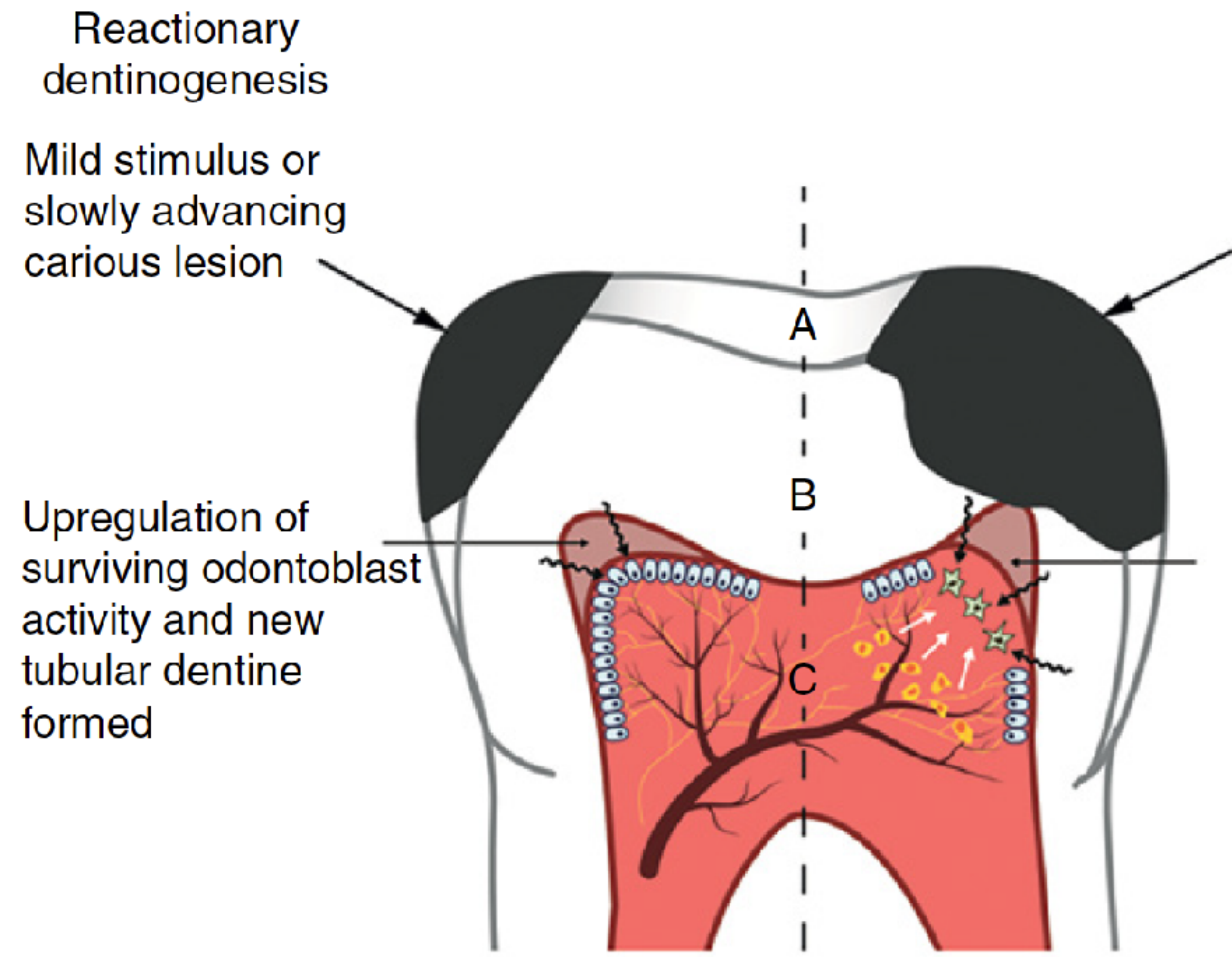
Reactionary Dentinogenesis11
- Trigger: mild stimulus or slowly advancing carious lesion
- Mechanism: upregulation of surviving odontoblast activity
- Result: new tubular dentine formed
Lecturer — Reactionary Dentinogenesis
Reactionary dentinogenesis occurs when the pulp responds to an external stimulus by laying down normal tubular dentine.
- The odontoblasts remain able to organize themselves and form dentine with the normal tubular pattern.
Reparative Dentinogenesis
- Trigger: severe or aggressive stimulus or rapidly advancing carious lesion
- Mechanism: death of primary odontoblast and replacement by odontoblast-like cell (recruited from progenitor/stem cells)
- Result: low-quality atubular mineralized tissue formed
Lecturer — Reparative Dentinogenesis
Reparative or tertiary dentine may be more resistant to caries than well-organized reactionary dentine.
- Normal dentinal tubules form relatively direct pathways from the external dentine surface toward the pulp.
- In reparative dentine, the tubules are less organized and more twisted, making it more difficult for caries to progress directly toward the pulp.
Figure legend (A–C): odontoblast; odontoblast-like cell; progenitor/stem cell; blood vessel; neuron; dentine matrix component.
Pulpal Diagnosis12
The conundrum: What do we have?
Lecturer — Pulpal Diagnosis
Diagnosis is based on clinical findings rather than direct knowledge of the pulp’s histological condition.
- Relevant information includes pulp sensibility testing, pain history, cold testing, electric pulp testing, percussion, palpation, radiographic examination, assessment of the periapical tissues, and visual examination for possible pulp exposure.
- Sensibility tests do not directly establish the histological status of the pulp, and a negative response to cold or electric testing does not automatically prove necrosis.
- Testing should be repeated and compared with adjacent and contralateral teeth. If the tooth repeatedly fails to respond while control teeth respond normally, this supports a diagnosis of a necrotic pulp or at least a pulp that is not clinically normal.
- For electric pulp testing, test tooth structure rather than a large metallic restoration. For cold testing, the location on the crown is less important because the stimulus is very cold; the gingiva should not be touched, and the test should be applied consistently and repeated when necessary.
Pulpal Sensibility Tests
- Pain history
- Character of pain
- Signs & Symptoms
Asymptomatic Molar With Faulty Restoration
A patient had an asymptomatic, mobile molar with a large faulty restoration, no caries, and no radiographic finding; the tooth was being considered for crowning.
- The tooth did not respond to cold or electric pulp testing and was not tender to percussion.
- Other teeth in the quadrant responded normally, but the contralateral tooth had not initially been tested.
- Repeat the cold and electric tests, test adjacent and contralateral teeth for comparison, and confirm that the lack of response is reproducible.
- If the tooth continues not to respond while control teeth respond, regard the pulp as not clinically normal and consider endodontic treatment. Testing should be performed before removing the amalgam where possible.
Vital Pulp Treatment Modalities Aiming Not to Expose the Pulp13

Caries Removal Strategies
Non-Selective Caries Removal14
Complete removal of soft and firm carious dentine from the periphery and central aspects of the cavity until hard dentine is reached.
Selective caries removal
A collective name for several treatments: selective caries removal in one-stage and stepwise excavation.
Adapted from the European Society of Endodontology
Selective Caries Removal
Selective caries removal is a collective name for several treatments: selective caries removal in one-stage and stepwise excavation.
Selective Caries Removal in One-Stage
Application of a biomaterial onto a dentine barrier in an indirect one-stage selective carious-tissue removal technique. Removal of soft or firm dentine. Immediate placement of a permanent restoration.
Stepwise Excavation
Application of a biomaterial in an indirect two-stage selective carious-tissue removal technique. Temporary restoration placement between visits and re-entry after 6–12 mo. The first stage involves selective carious removal to soft dentine to the extent that it facilitates proper placement of a temporary restoration, and the second stage involves removal to firm dentine – the final placement of a permanent restoration.
Adapted from the European Society of Endodontology
Indirect Pulp Capping15
Application of a biomaterial onto a thin dentine barrier in a one-stage carious-tissue removal technique generally to hard dentine.
Leaves neither soft nor firm carious dentine behind.
Considered more aggressive than selective carious-tissue removal in one-stage and stepwise excavation. However, if the excavation is successful, pulp exposure is avoided. The problem is that the frequency of unintended exposures will be more frequent than for selective caries removal in one-stage or stepwise excavation.
Adapted from the European Society of Endodontology
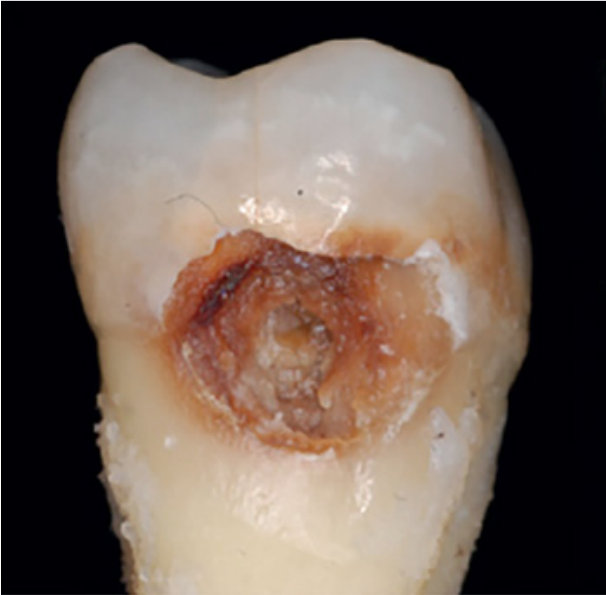
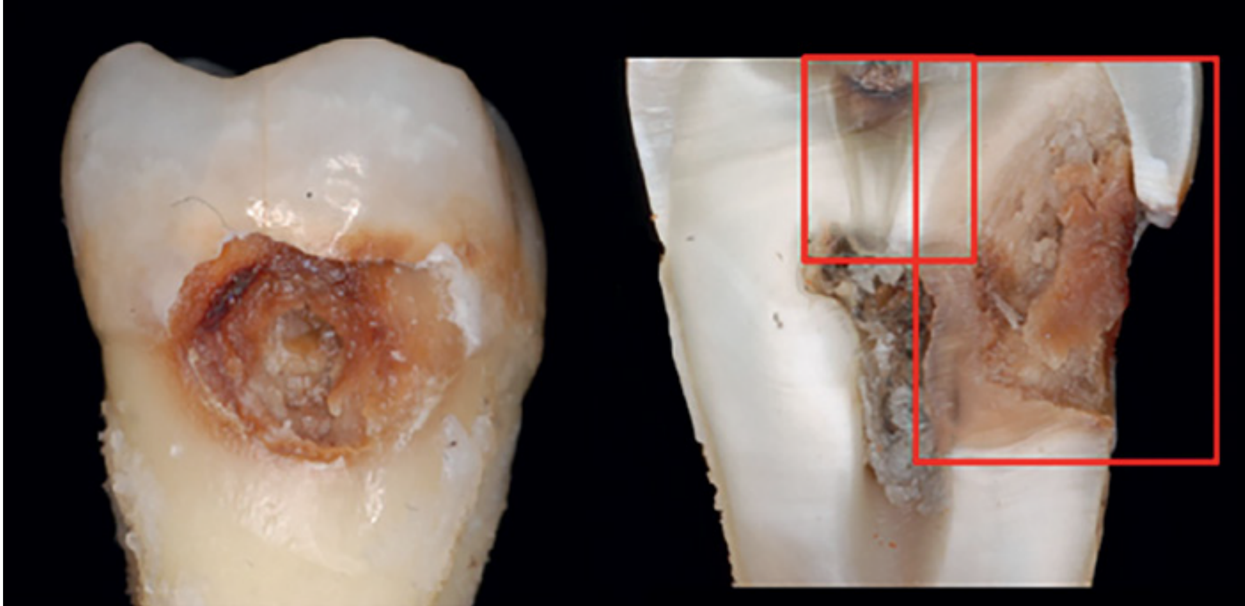
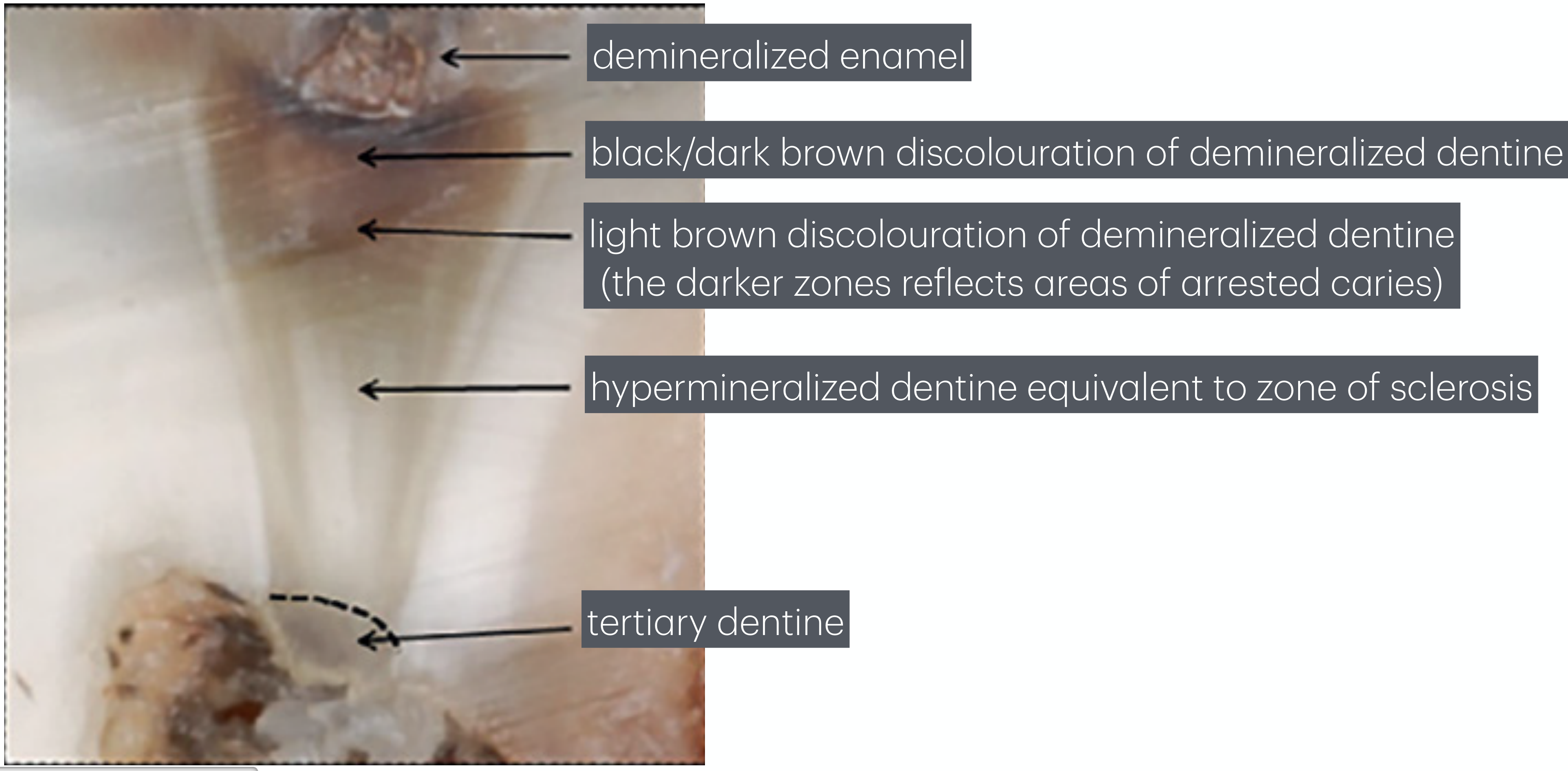
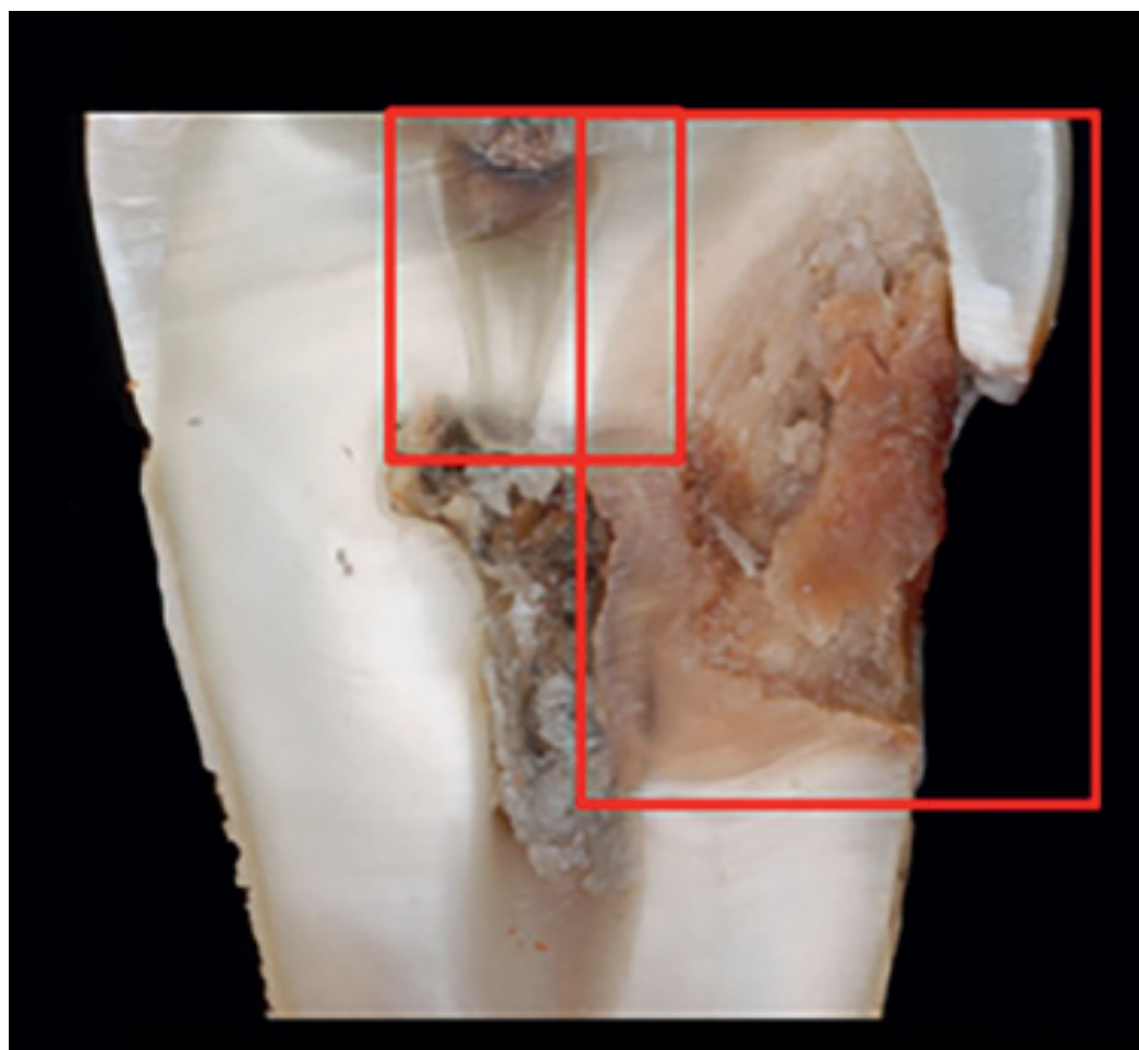
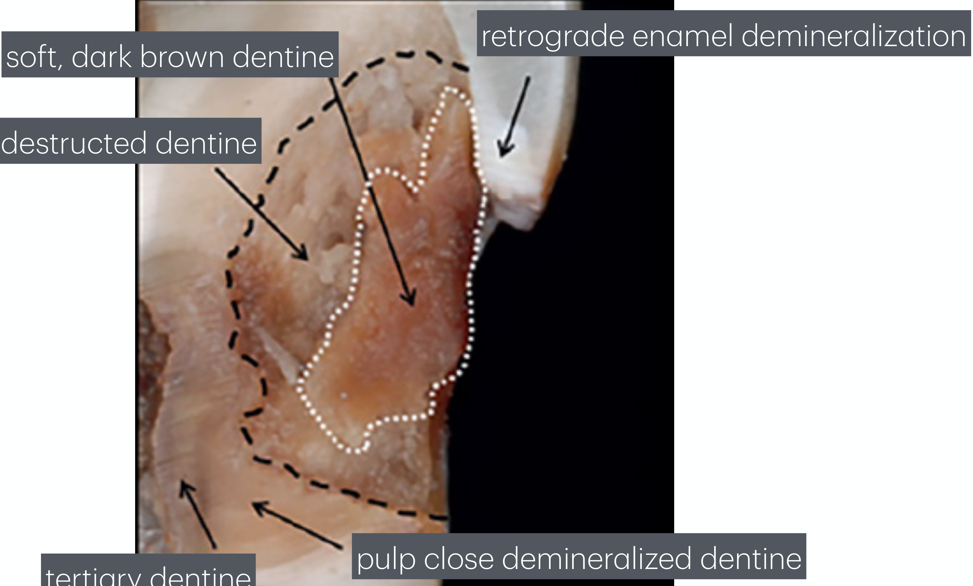
Caries Excavation16171819
Source: John Wiley & Sons, Inc.
- demineralized enamel
- black/dark brown discolouration of demineralized dentine
- light brown discolouration of demineralized dentine (the darker zones reflects areas of arrested caries)
- hypermineralized dentine equivalent to zone of sclerosis (described in the audio as near the pulp)
- tertiary dentine
Source: John Wiley & Sons, Inc.
- soft, dark brown dentine
- destructed dentine
- retrograde enamel demineralization
- tertiary dentine
- pulp close demineralized dentine
Source: John Wiley & Sons, Inc.
Management of Deep Carious Lesions
Radiographic Features20
This stage is defined as a lesion penetrating 2/3–3/4 of the dentine, with a radiopaque zone separating the advancing front of the lesion and the pulp.
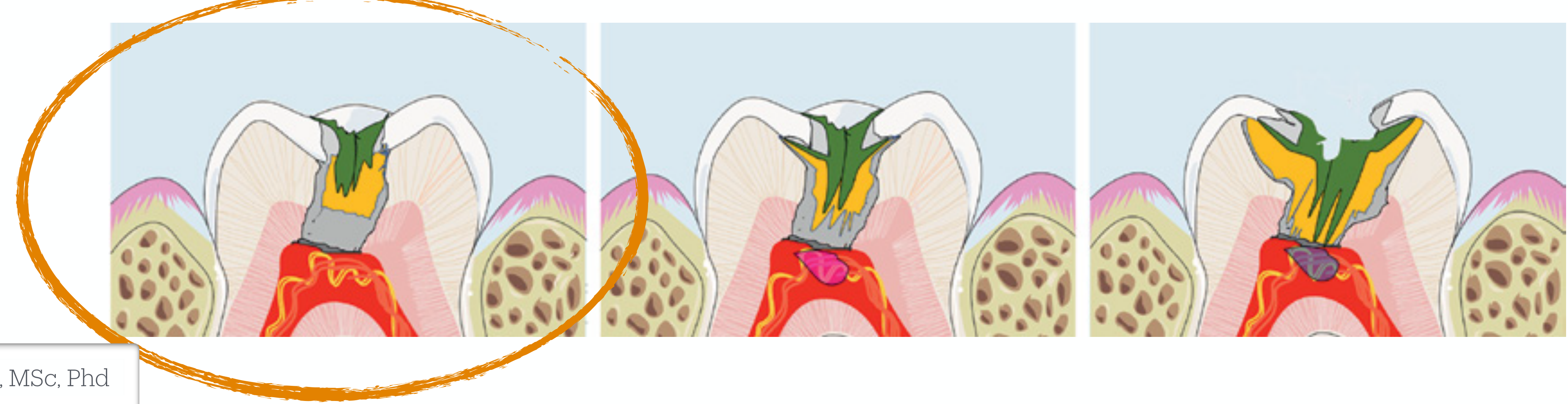
Histologic Features
Microorganisms are located in the carious dentine. Evidence of chronic pulp inflammation, but not associated with invasion of microorganisms into the pulp cavity.
Recommended Treatment
Selective carious removal in one step to either soft or firm dentine. This particular lesion class is most often treated with a selective caries removal strategy.
Deep Caries
Radiographic Features
This stage, described as deep caries, is defined as a lesion penetrating 3/4 or more of the dentine, but still with a radiopaque zone separating the advancing front of the lesion and the pulp.
Histologic Features
Microorganisms are located in the carious dentine. Evidence of chronic pulp inflammation with a low probability of the presence of invading microorganisms in the pulp cavity.
Recommended Treatment
Stepwise excavation. Evidence for selective carious removal in one step to either soft or firm dentine is available but mainly based on children and adolescent patients.
Extremely Deep Caries
Radiographic Features
This stage, described as extremely deep caries, is defined as a lesion penetrating the entire thickness of the dentine without a radiopaque zone separating the advancing front of the lesion and the pulp.
Histologic Features
Microorganisms are located in the carious dentine, tertiary dentine and within the pulp cavity. The pulpal status includes severe inflammation associated with partial or full necrosis in the pulp cavity.
Recommended Treatment
Pulpectomy or root canal treatment. Pulpotomy including partial pulpotomy may also be considered, but comparative studies with longer follow-up are lacking.
Stepwise Excavation Case Illustrations21222324252627
Lim ZE, Duncan HF, Moorthy A, McReynolds D. Br Dent J. 2023;234(4):233–40.
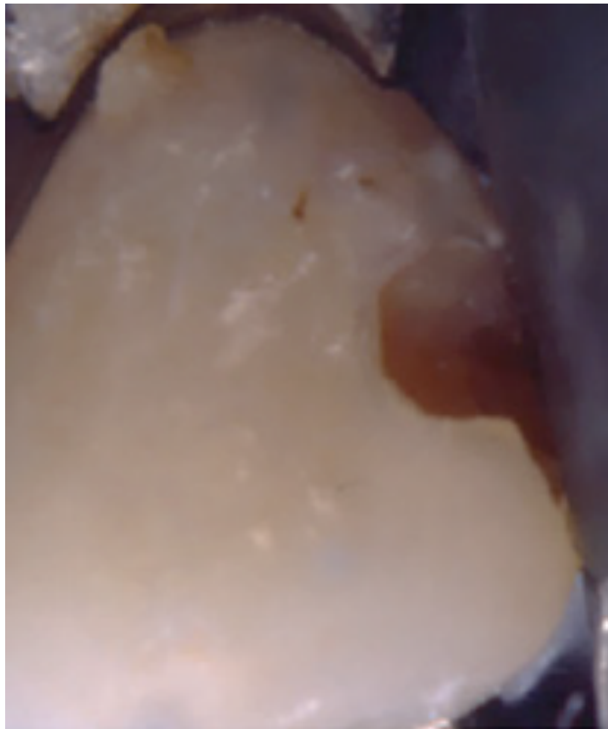
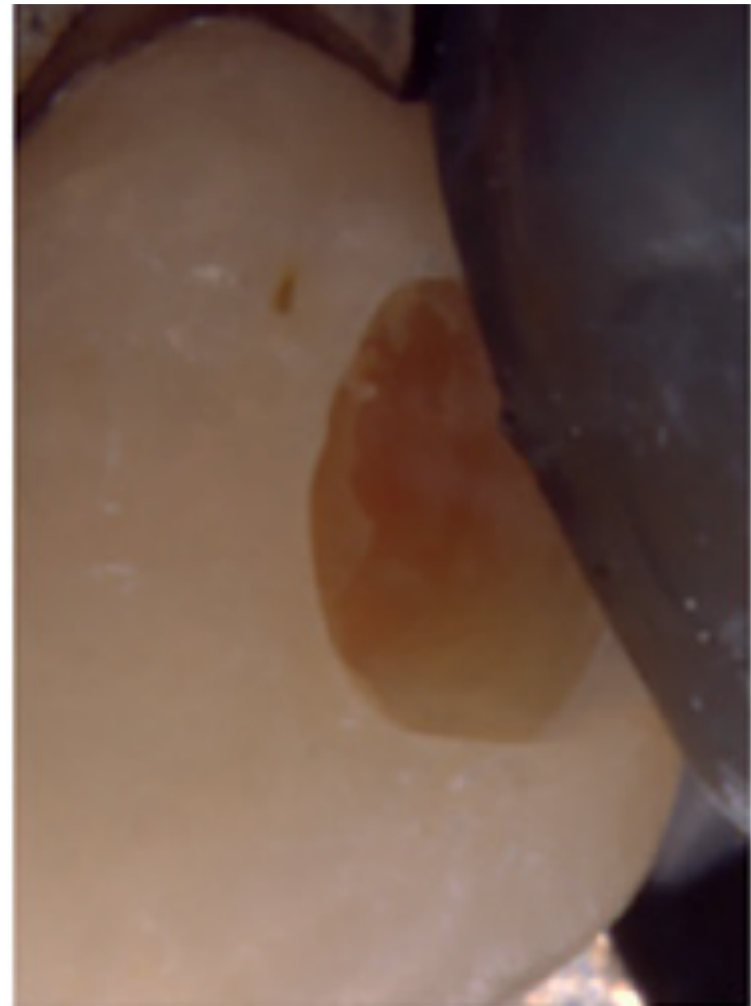
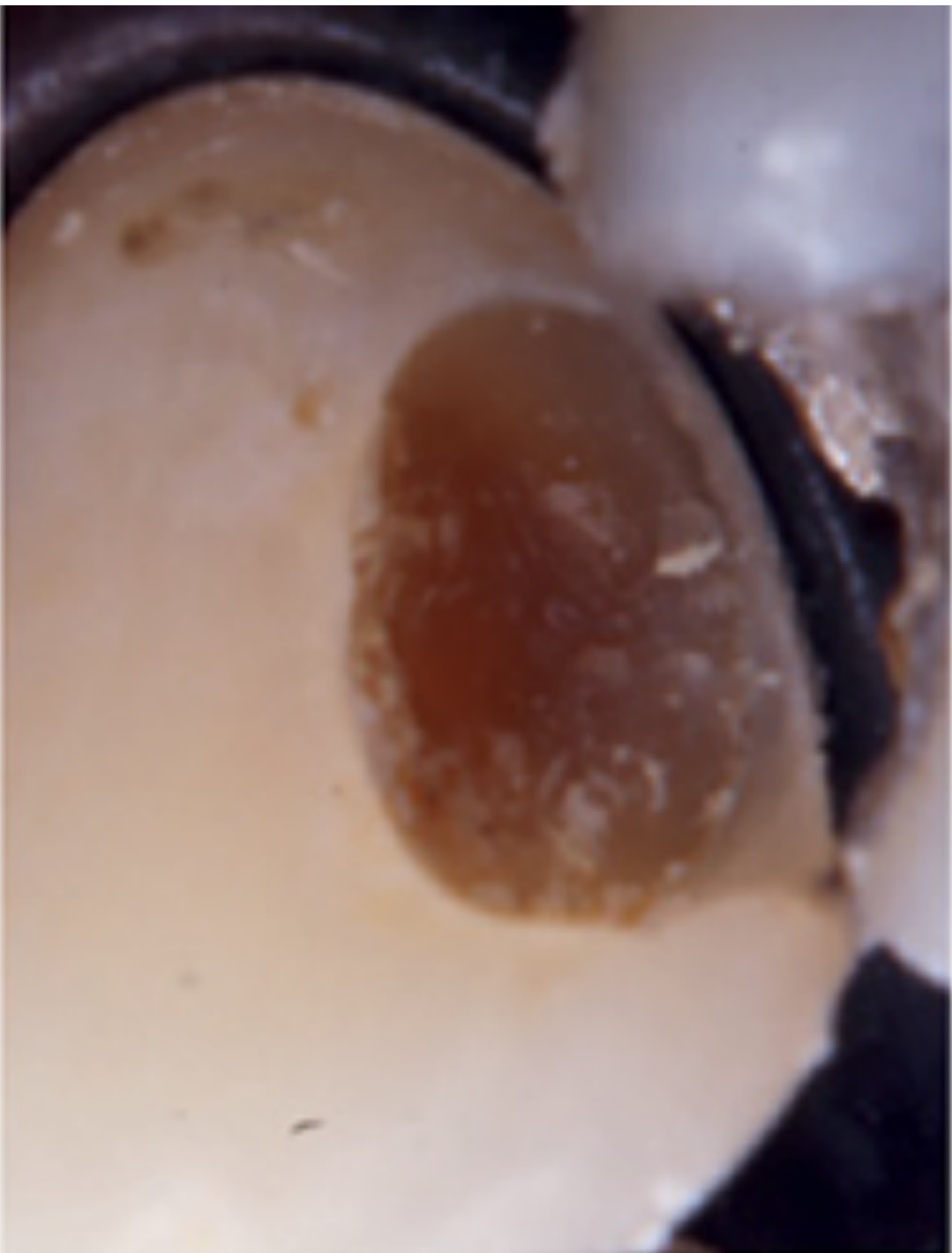
After the first stage selective carious removal to soft dentine to an extent that facilitates proper placement of a temporary restoration.
Lim ZE, Duncan HF, Moorthy A, McReynolds D. Br Dent J. 2023;234(4):233–40.
Following a treatment interval and removal of temporary restoration the retained dentine is darker reflecting lesion arrest.
The periphery of the cavity should be excavated to hard dentine in order to facilitate the proper placement of a temporary restoration.
Lim ZE, Duncan HF, Moorthy A, McReynolds D. Br Dent J. 2023;234(4):233–40.
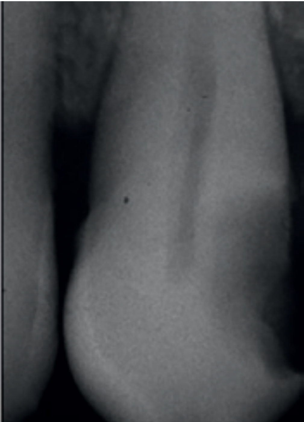
The carious lesion before treatment is categorized as a radiographically well-defined deep carious lesion penetrating to the pulpal quarter but with a radiopaque zone separating the carious lesion to the pulp.
Lim ZE, Duncan HF, Moorthy A, McReynolds D. Br Dent J. 2023;234(4):233–40.
Parts of the overlaying enamel have been removed.
Note the outermost disintegrated carious dentine with a dark brown, dry and soft appearance.
Lim ZE, Duncan HF, Moorthy A, McReynolds D. Br Dent J. 2023;234(4):233–40.
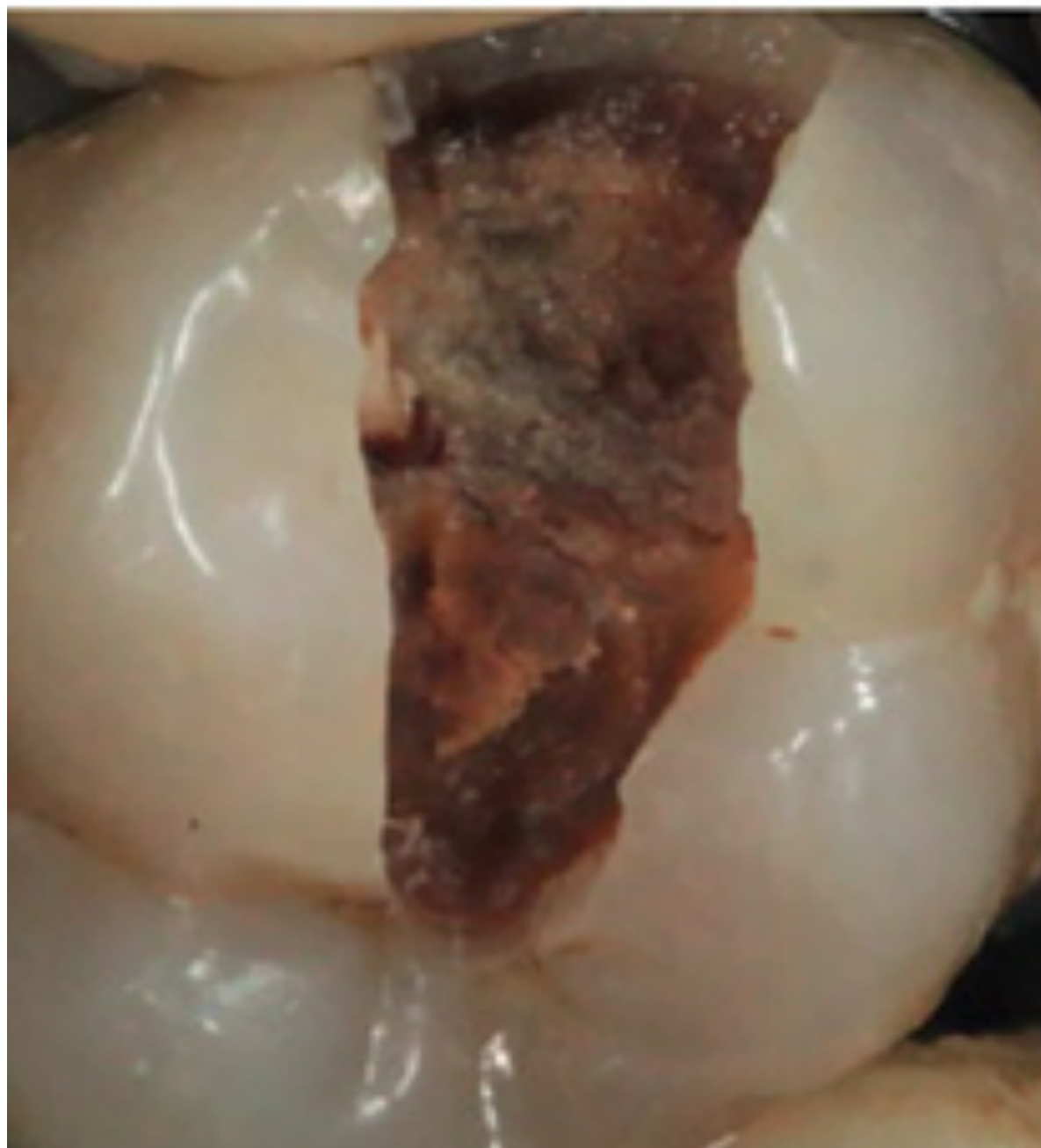
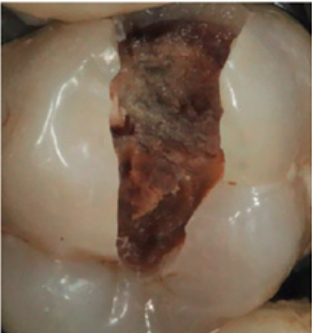
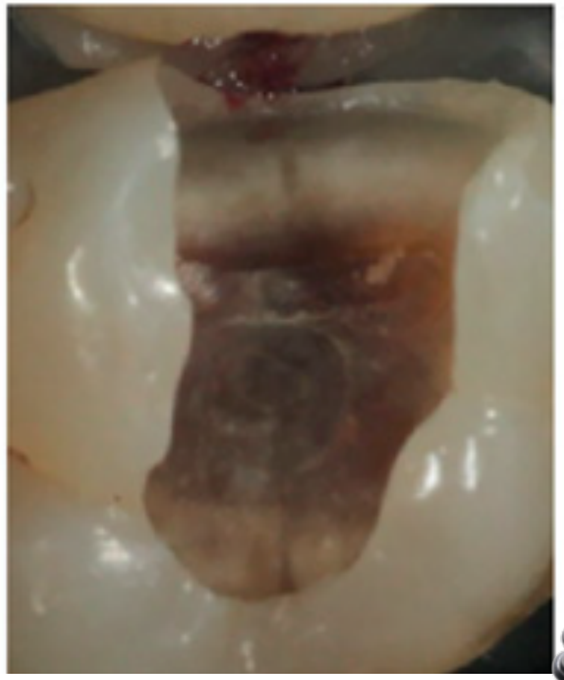
Completion of selective removal using burs and hand excavator to firm dentine.
The periphery of the cavity should be excavated to hard dentine in order to facilitate the proper placement of a temporary restoration.
Lim ZE, Duncan HF, Moorthy A, McReynolds D. Br Dent J. 2023;234(4):233–40.

Selective Caries Removal Case Illustrations28
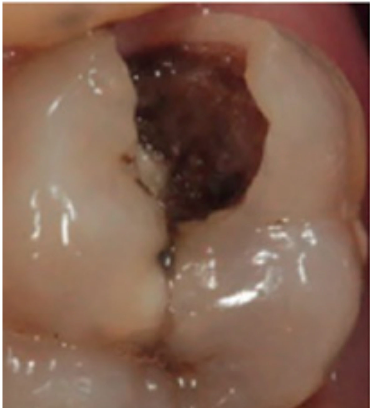
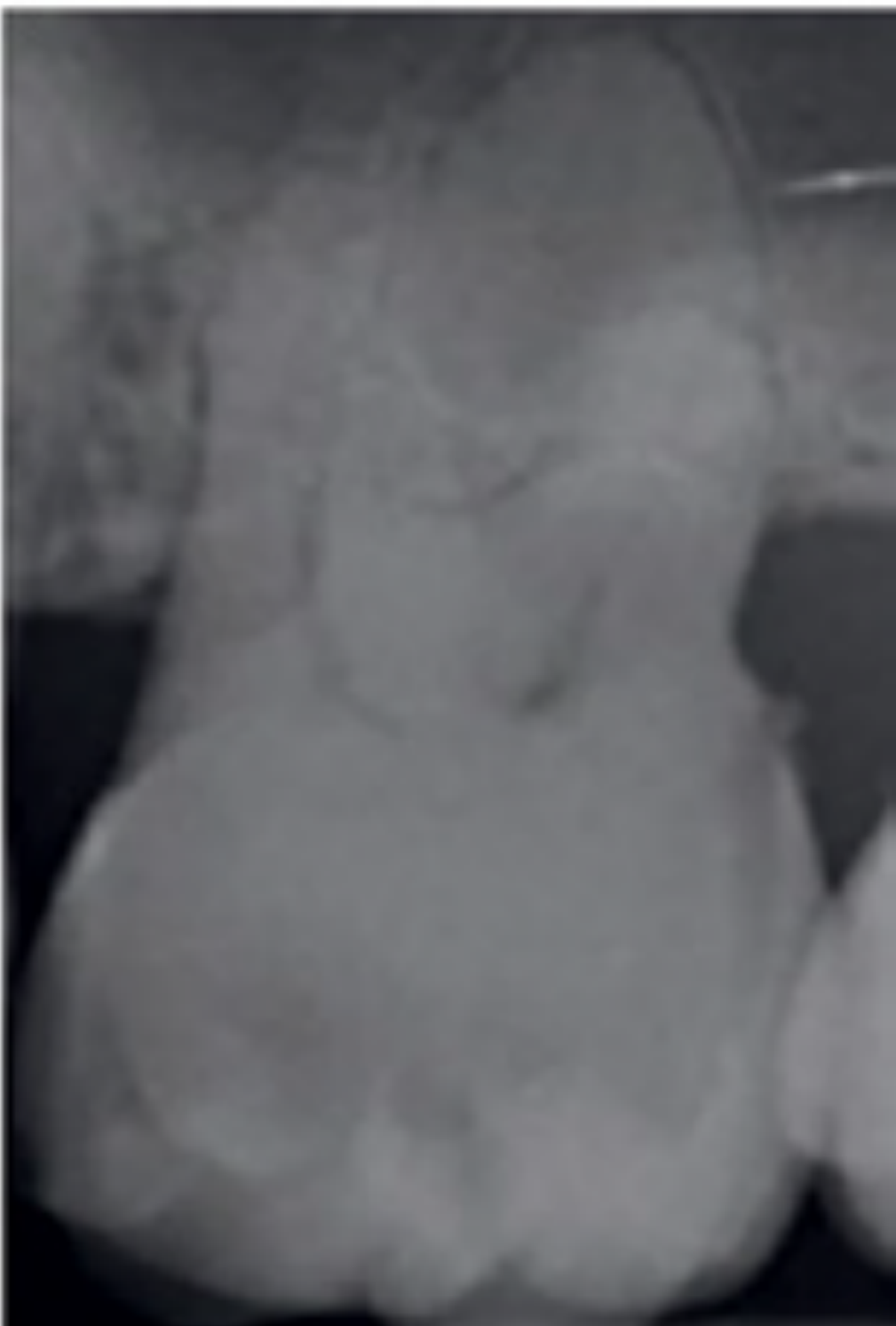
The carious lesion before treatment is categorized as a radiographically well-defined carious lesion penetrating to the middle third of the dentine.
Lim ZE, Duncan HF, Moorthy A, McReynolds D. Br Dent J. 2023;234(4):233–40.
 |  |
Evidence and Clinical Outcomes
European Society of Endodontology position statement: Management of deep caries and the exposed pulp. International Endodontic Journal, doi:10.1111/iej.13080.
European Society of Endodontology (ESE) developed by: H. F. Duncan¹, K. M. Galler², P. L. Tomson³, S. Simon⁴, I. El-Karim⁵, R. Kundzina⁶, G. Krastl⁷, T. Dammuschke⁸, H. Fransson⁹, M. Markvart¹⁰, M. Zehnder¹¹ & L. Bjørndal¹⁰
¹Division of Restorative Dentistry, Dublin Dental University Hospital, Trinity College Dublin, Dublin, Ireland; ²Department of Conservative Dentistry and Periodontology, University Hospital Regensburg, Regensburg, Germany; ³School of Dentistry, Institute of Clinical Sciences, University of Birmingham, Birmingham, UK; ⁴Paris Diderot University, Paris 7, Paris, France; ⁵School of Medicine Dentistry and Biomedical Sciences, Queen’s University Belfast, Belfast, UK; ⁶Faculty of Health Sciences, Institute of Clinical Odontology, UiT the Arctic University of Norway, Tromsø, Norway; ⁷Department of Conservative Dentistry and Periodontology, University Hospital of Würzburg, Würzburg; ⁸Department of Periodontology and Operative Dentistry, Westphalian Wilhelms-University, Münster, Germany; ⁹Faculty of Odontology, Department of Endodontics, Malmö University, Malmö, Sweden; ¹⁰Cariology and Endodontics, Faculty of Health and Medical Sciences, Department of Odontology, University of Copenhagen, Copenhagen, Denmark; and ¹¹Department of Preventive Dentistry, Periodontology and Cariology, University of Zurich, Zurich, Switzerland
AAE Position Statement on Vital Pulp Therapy29
Distribution Information: AAE members may reprint this position statement for distribution to patients or referring dentists. ©2021
About This Document: The following statement was prepared by the AAE Special Committee on Vital Pulp Therapy.
Introduction: The American Association of Endodontists is dedicated to excellence in the art and science of endodontics and to the highest standards of patient care. The basis for endodontic treatment utilizes the best available evidence from scientific and clinical studies in concert with the accumulated clinical knowledge and judgment of the practitioner.
ESE Position Statement on Deep Caries30
How Should We Manage Deep Caries to Avoid Pulp Exposure?
One-stage selective carious-tissue removal to soft or firm dentine (Maltz et al. 2012) and two-stage stepwise excavation (Bjørndal et al. 2017) are indicated in deep carious lesions when the tooth is asymptomatic or has signs and symptoms indicative of reversible pulpitis. Non-selective (complete) caries removal is no longer regarded the treatment of choice in deep carious lesions, with recent consensus considering it to represent overtreatment (Schwendicke et al. 2016).
These selective removal VPT approaches have demonstrated a 3-year success, defined as the absence of clinical symptoms and maintenance of pulp health, of 90% for one-stage selective removal and 70% for stepwise excavation (Maltz et al. 2012); however, within this study, many of the stepwise cases did not return for the second visit, making comparison
Caries Management (Contrasting View)
Complete caries removal is essential to eliminate infected tissues and visualize pulp tissue conditions under magnification when pulpal exposures occur.¹⁹,²⁰ Residual caries compromises necessary observations of pulpal inflammation levels and areas of potential necrosis. Accordingly, predictable management of vital pulp tissue should not be performed without complete removal of both demineralized enamel and infected dentin.
Hard or firm dentin and dentin below white spot enamel lesions is infected by bacteria in both active and arrested lesions. Specifically, histobacteriological studies have consistently shown the presence of chronic inflammatory cell infiltrates and subclinical pulp inflammation where carious tissues are retained, thus potentially compromising pulp vitality.²¹,²² Additionally, adhesion of bonding resins to sound dentin
Randomized Trial Outcomes
At 3 years, success (no pulpitis, necrosis, exposure, osteitis, extraction or fracture) was higher after one-stage selective (partial) caries removal than after stepwise excavation: 91% vs 69%, adjusted p = .004. Maltz et al. 2012
Trial Flow (n=299)
See - Trial flow
Numbers Analyzed and Outcomes
See - Analyzed groups and outcomes
Source check needed: The adjacent citation J Dent 2020 Sep:100:103430 remains unresolved; proximity does not establish that it belongs to the 2012 trial.
Treatment Considerations and Follow-Up
Expected Outcomes and Risks
Condition: Carious Lesion Extending Towards the Pulp
Stepwise excavation or selective caries removal
- Expected outcome: vital pulp without any sign of pulp inflammation or apical periodontitis
- Benefits: technically easy and quick to perform
- Potential risks and disadvantages:
- Persistent pulp inflammation
- Infection leading to pulp necrosis and apical periodontitis
- Symptoms
- Root canal treatment with inferior prognosis than pulpectomy
Pulpectomy
- Expected outcome: healthy periapical conditions with no signs of apical periodontitis
- Benefits: removal of potential irreversibly damaged pulp
- Potential risks and disadvantages:
- Infection leading to apical periodontitis
- Symptoms
- Technically difficult and time consuming
- Fractures
- Extraction
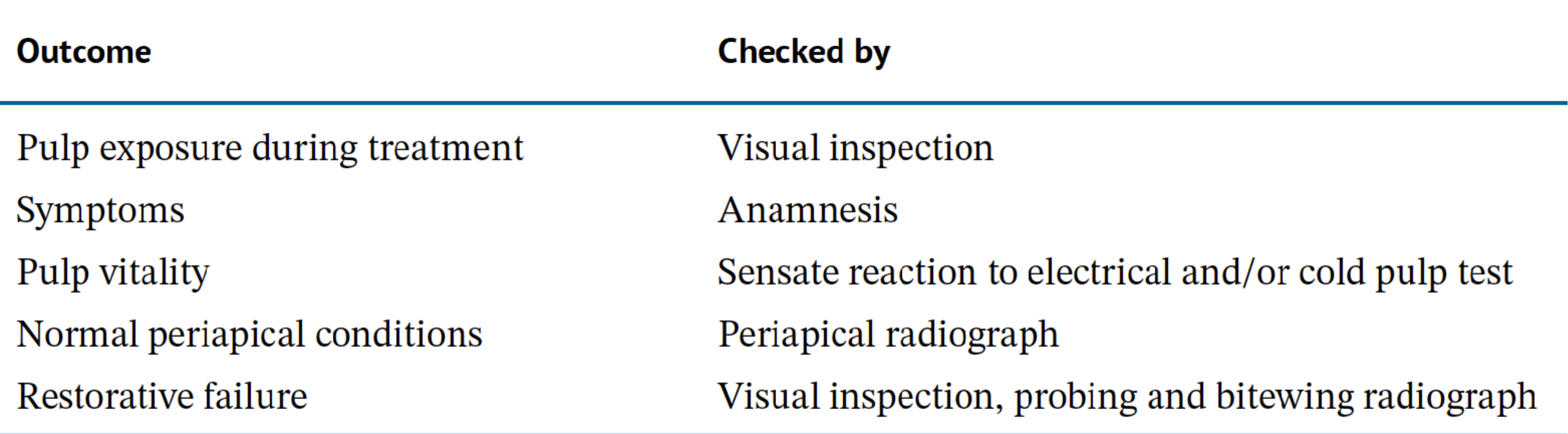
Outcome Monitoring31
| Outcome | Checked by |
|---|---|
| Pulp exposure during treatment | Visual inspection |
| Symptoms | Anamnesis |
| Pulp vitality | Sensate reaction to electrical and/or cold pulp test |
| Normal periapical conditions | Periapical radiograph |
| Restorative failure | Visual inspection; probing when restorative failure is suspected; bitewing radiograph |
Summary of Modalities3233
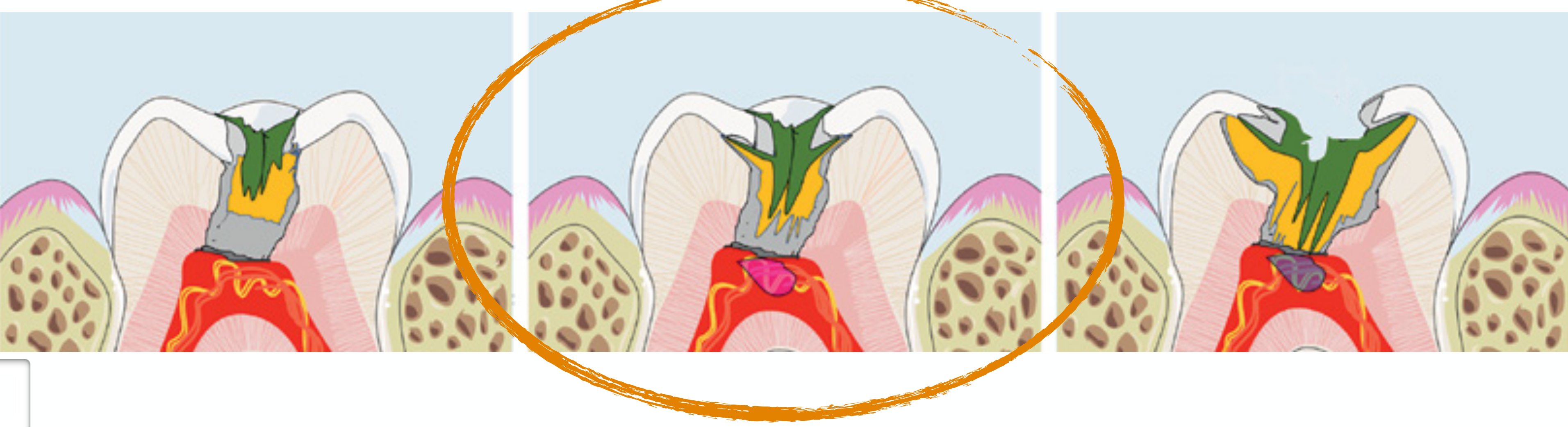
- Non-selective caries removal
- Selective caries removal
- Selective caries removal in one-stage
- Stepwise excavation

Key Challenges in Managing Deep Carious Lesions34
- Managing deep carious lesions is challenging because infected dentine cannot be objectively measured.
- It is not possible to monitor pulpal inflammation at the chairside based on patient symptoms or clinical findings.
- The term “deep carious lesion” lacks a clear definition.
- Traditional terms like “complete” and “partial” removal of carious tissue are imprecise and ambiguous.
- It is unclear whether “partial caries removal” refers to the removal of microorganisms or demineralized dentine.
Audio Appendix
Additional Audio Content
The following sections from the lecture audio did not correspond to any heading in the main document.
Vital Pulp Treatment Modalities Aiming Not to Expose the Pulp
The lecture focused on treatment modalities intended to avoid pulp exposure by leaving a final layer of dentine over the pulp.
Caries Removal Strategies
Non-Selective Caries Removal
- Non-selective caries removal involves removing all soft and firm carious dentine from both:
- The periphery of the cavity.
- The central area of the cavity.
- If complete caries removal exposes the pulp, treatment proceeds according to the exposure, such as pulpotomy, pulpectomy, or root canal treatment. See L8 pulpotomy procedures for the exposed-pulp pathway.
- If the pulp is not exposed, the clinician proceeds with placement of the selected material and restoration.
- The lecturer preferred the terminology complete caries removal rather than non-selective caries removal.
Selective Caries Removal
Selective caries removal involves leaving the deepest layer of soft carious dentine over the pulp.
It may be performed in:
- One stage.
- Two stages, known as stepwise excavation.
In one-stage selective caries removal:
- Soft carious dentine is left over the pulp.
- A bioactive material, such as a bioceramic material or MTA, is placed.
- A final restoration is placed.
In stepwise excavation:
- The deepest soft carious dentine is left over the pulp.
- A biomaterial and provisional restoration are placed.
- The patient returns at a later stage.
- The provisional restoration is removed.
- The remaining caries is excavated.
- The biomaterial and final restoration are placed.
The proposed rationale for stepwise excavation is that the provisional restoration allows the pulp to heal and move away from the carious lesion.
The lecturer questioned this rationale and expressed concern that re-entry may expose the pulp, particularly when a thin bridge of soft carious dentine remains.
Indirect Pulp Capping
- Indirect pulp capping was described as a collective term for procedures in which a biomaterial is placed over the area closest to the pulp without exposing the pulp.
- Some or all of the soft carious dentine may be left.
- The exact terminology varies according to whether the procedure is performed in one stage or in a stepwise manner.
Caries Excavation
The International Caries Consensus Collaboration’s lesion-area list is in the main section:
See - Caries Excavation
The lecturer questioned how reliably these areas can be identified clinically:
- He stated that he could not reliably distinguish all of these areas in clinical practice.
- The appearance of each tooth and the degree of discoloration vary.
- Terms such as “leathery dentine” and “soft dentine” depend on:
- Operator skill.
- Operator experience.
- The force used during excavation.
- It is unclear how these classifications translate into improved treatment for an individual patient.
The lecturer also questioned the distinction between infected and affected dentine:
- Historically, clinicians were taught to remove infected dentine while leaving affected dentine.
- It is difficult to determine clinically which dentine is infected and which is affected.
- Bacteria cannot be directly seen in the patient’s mouth.
- A caries-detecting dye detects porous or demineralized dentine rather than necessarily detecting infected dentine.
- Some studies suggest that removing all demineralized dentine increases the likelihood that infected dentine has also been removed, but this does not provide certainty.
- Discolored dentine may be bacteria-free.
- Conversely, dentine that appears acceptable may still have bacterial penetration.
Management of Deep Carious Lesions
Radiographic Features35
Radiographic assessment may show:
- A carious lesion extending through approximately two-thirds to three-quarters of the dentine.
- A radiopaque zone separating the carious lesion from the pulp.
- A dentine bridge between the lesion and the pulp.
- A very deep lesion approaching the pulp.
- A lesion with no apparent radiopaque bridge and possible pulp involvement.
Radiographic depth does not provide a complete clinical assessment because:
- A certain amount of demineralization is required before caries becomes visible radiographically.
- Demineralized dentine may extend deeper than the area visible on the radiograph.
- The apparent depth depends on:
- The size of the pulp chamber.
- The quality of the remaining dentine.
- The mineralization of the remaining dentine.
- The status of the pulp beneath the lesion.
Histologic Features
The described clinical scenarios included:
- Microorganisms located within carious dentine.
- Chronic inflammatory cells within the pulp.
- In deeper lesions, evidence of chronic inflammation approaching or involving the pulp.
- In the deepest lesions, apparent invasion of the pulp by caries and the possibility of pulp exposure during excavation.
The lecturer identified several unresolved questions:
- Whether bacteria left beneath a restoration will die or continue to progress through the dentinal tubules.
- Whether chronic inflammation already present in the pulp will progress.
- Whether bacteria left in dentine can act as a reservoir for exotoxins that irritate the pulp.
- Whether the pulp beneath the lesion is clinically healthy, inflamed, or already damaged.
- Whether caries will recur beneath a restoration placed over soft carious dentine.
Recommended Treatment
For a deep carious lesion in an asymptomatic tooth with normal sensibility responses, the proposed options include:
- Selective caries removal in one stage.
- Stepwise excavation.
- Complete caries removal followed by treatment according to whether the pulp is exposed.
- Pulpectomy or root canal treatment.
The lecturer’s clinical preference was:
- Remove all caries.
- If the pulp is exposed, proceed with root canal treatment.
- Do not leave caries based solely on an assumption that it will remain inactive.
- Use the patient’s circumstances and the clinical findings in treatment planning.
Deep Caries
Deep caries was discussed as a situation in which:
- The lesion extends close to the pulp.
- A thin layer of dentine may remain.
- There may be a risk of pulp exposure during excavation.
- The clinician cannot objectively determine the depth of bacterial penetration.
- Pulp inflammation cannot be directly monitored chairside.
The lecturer emphasized that the pulp is a dynamic organ:
- It may move through reversible and irreversible inflammatory stages.
- It may move between chronic and acute phases.
- The clinical presentation represents the status observed at the time of diagnosis and does not reveal the entire histological history of the pulp.
Extremely Deep Caries
In an extremely deep lesion:
- The caries may extend through most of the dentine.
- Chronic inflammation may be present in the pulp.
- Excavation may expose the pulp.
- Root canal treatment or pulpectomy was suggested when the pulp is involved or exposed.
The lecturer questioned whether the proposed radiographic classifications can be applied reliably because the clinical boundaries between categories are unclear.
Stepwise Excavation Case Illustrations
Case Study: Proximal Carious Lesion in a Maxillary Canine
See - Full Clinical Case
Summary
Slides: deep approximal lesion to the pulpal quarter with a radiopaque zone still separating it from the pulp. Stage 1: selective removal to soft dentine centrally, periphery to hard dentine, temporary restoration. Stage 2 (after the treatment interval): retained dentine is darker, reflecting lesion arrest; definitive restoration. Lecturer: questioned whether stepwise excavation suits a mature canine and warned that re-entry risks pulp exposure.
Selective Caries Removal Case Illustrations
Case Study: Deep Caries in a Maxillary Molar
See - Full Clinical Case
Summary
Slides: open occlusal lesion to the middle third of dentine; one-stage selective removal with burs and hand excavator to firm dentine, periphery to hard dentine. Lecturer: described the retained dark central dentine as likely discoloration rather than active caries (not settled by the slides).
Evidence and Clinical Outcomes
AAE Position Statement on Vital Pulp Therapy
- The American Association of Endodontics was described as supporting complete caries removal for preservation of the pulp.
- The American approach was contrasted with the European position favoring selective caries removal in suitable deep carious lesions.
- The lecturer also referred to research showing bacteria remaining within dentinal tubules even after caries removal.
- Remaining bacteria may create a risk of:
- Caries recurrence.
- Continued bacterial activity.
- Exotoxin release.
- Pulpal irritation.
ESE Position Statement on Deep Caries
- The European Society of Endodontology was described as supporting:
- One-stage selective caries removal.
- Two-stage selective caries removal, or stepwise excavation.
- This approach was described as applicable to asymptomatic deep carious lesions.
- The European position was contrasted with the American recommendation for complete caries removal.
- The lecturer described the terms “selective” and “non-selective” as partly involving word choice:
- Selective caries removal may mean partial caries removal.
- Non-selective caries removal may mean complete caries removal.
- He questioned whether the terminology could make one treatment appear more acceptable than another.
Randomized Trial Outcomes
- A clinical study involving approximately 300 patients compared selective caries removal with stepwise caries removal.
- Lecturer (audio) summarised the trial as showing no significant difference; this does not match the published adjusted result.
- Follow-up was approximately three years.
- The lecturer cautioned against accepting the conclusion without examining the study details:
- Each group initially contained approximately 140 patients.
- There was a high attrition rate over three years.
- Approximately one-third of the patients were lost to follow-up.
- It is therefore unknown what happened to the patients who did not attend review.
- The results of studies with high attrition rates should be interpreted carefully.
Treatment Considerations and Follow-Up
Expected Outcomes and Risks
For selective caries removal or vital pulp treatment, the expected outcome is:
- A vital pulp.
- No signs of inflammation.
Risks include:
- Persistent pulpal inflammation.
- Persistent infection.
- Pulpal necrosis.
- Periapical inflammation.
- Symptoms developing after treatment.
- The eventual need for root canal treatment.
For root canal treatment, the expected outcome is:
- A healthy periapical condition.
- No signs of apical inflammation.
Risks and disadvantages include:
- Further loss of tooth structure.
- Tooth fracture associated with loss of vitality and structural weakening.
- Technical difficulty.
- Greater treatment time.
- Possible residual infection if treatment is not performed adequately.
Treatment should not be selected solely because it is technically easier for the clinician. If the clinician cannot perform a technically difficult root canal treatment, referral should be considered.
Outcome Monitoring
See - Outcome Monitoring
The audio also lists palpation and percussion.
All studies described by the lecturer involving selective or non-selective caries removal disinfected the tooth before treatment:
- The tooth was isolated.
- A disinfectant such as sodium hypochlorite or another disinfecting solution was used.
- The procedure was described as potentially aseptic rather than completely aseptic.
When a deep lesion is suspected, the lecturer recommended:
- Rubber dam isolation.
- Aseptic preparation.
- Having sodium hypochlorite available.
- Being prepared to control bleeding and disinfect the cavity if pulp exposure occurs.
Summary of Modalities
See - Summary of Modalities
The lecturer’s clinical position was:
- Complete caries removal should be performed.
- If the pulp is exposed, root canal treatment should generally be performed.
- The lecturer did not feel comfortable leaving caries because the extent of bacterial penetration and the condition of the underlying pulp cannot be determined.
- Root canal treatment should not be avoided solely because it is difficult.
- In immature teeth, preserving the pulp remains particularly important, although complete caries removal is still preferred.
- If pulp exposure occurs in an immature tooth, vital pulp procedures such as pulp capping, partial pulpotomy, or pulpotomy may be considered.
Materials discussed included:
- Calcium hydroxide.
- Calcium silicate materials.
- MTA.
- Bioceramic putty.
- Injectable MTA.
- Hydraulic cement materials.
The lecturer stated that:
- Calcium hydroxide is not obsolete and may still be used.
- More recent evidence favors calcium silicate and bioceramic materials.
- Bioceramic materials should be covered using appropriate bonding and restorative techniques.
- They should not be etched; the restoration should be placed according to the material’s requirements.
Key Challenges in Managing Deep Carious Lesions
Uncertainty About Carious Dentine
- Infected dentine cannot be objectively measured in the clinic.
- Clinicians mainly identify discolored or soft dentine, which may be a product of bacterial activity.
- It is not possible to determine visually whether dentinal tubules are infected.
- Caries-detecting dyes identify porous or demineralized dentine, not necessarily infected dentine.
- Discoloration does not always indicate active infection.
Uncertainty About Pulpal Status
- The pulp may contain chronic inflammatory cells without producing symptoms.
- There is no chairside tool that directly measures the histological status of the pulp.
- Sensibility testing provides indirect information.
- The pulp is dynamic and may change between different inflammatory states.
Uncertainty About Residual Bacteria
- Bacteria may remain in dentinal tubules after caries removal.
- Bacteria may continue to act as a reservoir for irritants.
- The clinician cannot reliably determine whether bacteria left beneath a restoration will remain inactive or progress.
- It is uncertain whether remaining bacteria can continue to obtain nutrients from dentinal fluid, the pulp, or other sources.
Limitations of Stepwise Excavation
- The lecturer stated that, in his experience, stepwise excavation does not work reliably.
- After leaving a thin layer of soft carious dentine and placing a provisional restoration, the second-stage excavation may expose the pulp.
- The boundary between the restoration and the remaining carious dentine may be difficult to identify.
- This concern is particularly relevant in large carious lesions.
Patient-Centered Decision Making
Treatment should be adapted to the individual patient rather than applied as a universal protocol.
Relevant factors include:
- The patient’s symptoms.
- The pulp sensibility findings.
- The depth and extent of caries.
- The patient’s medical condition.
- The patient’s age.
- The ability to tolerate lengthy treatment.
- Financial limitations.
- Travel or appointment constraints.
- The ability to attend follow-up.
- The expected prognosis.
Case Study: Elderly Patient With a Calcified Tooth
- The lecturer described an approximately ninety-year-old patient.
- The tooth had previously been treated and had chronic apical periodontitis with mild pain.
- The tooth was extremely calcified.
- The patient could barely tolerate sitting in the dental chair.
- The patient was taking Prolia.
- The patient could not afford extraction.
- Rather than undertaking prolonged treatment immediately, the lecturer explained the disease and recommended monitoring:
- Review approximately every three months.
- Take radiographs.
- Monitor for progression.
- Reconsider treatment if symptoms became intolerable.
- Discuss treatment or extraction with the patient’s general practitioner where appropriate.
- The lecturer contrasted this with a younger patient, for whom he would generally proceed directly with root canal treatment if indicated.
Clinical Cases
Case: Asymptomatic molar with large faulty restoration and negative sensibility tests
Question
Scenario: A patient presents with a totally asymptomatic molar that has a large, faulty amalgam restoration but no caries. The clinician is considering placing a crown on the tooth.
What’s shown: The tooth is negative to both cold and electric pulp testing (EPT). It is asymptomatic and not tender to percussion. The radiograph shows no abnormalities.
Consider: What is the likely diagnosis based on the sensibility tests, and how should the clinician proceed with further testing and treatment?
Answer
Observations:
- The molar is asymptomatic and not tender to percussion.
- The tooth is unresponsive to cold and EPT, while adjacent teeth in the quadrant respond normally.
- The radiograph shows no periapical or other abnormalities.
Reasoning:
- A negative sensibility test does not definitively mean the pulp is necrotic, but when a tooth fails to respond to both cold and EPT while adjacent teeth respond normally, it strongly suggests the pulp is necrotic (not clinically normal).
- Proper testing technique is crucial: for EPT, the clinician should test on the buccal surface of the tooth; for cold tests, it can be applied anywhere on the crown as long as the gingiva is avoided and the stimulus is applied consistently.
- Retesting and checking the contralateral tooth are recommended to confirm the diagnosis before proceeding.
Outcome:
- The clinician should retest the tooth before removing the existing amalgam. If the tooth remains unresponsive and is deemed to not have a clinically normal pulp, the clinician should proceed with endodontic (root canal) treatment.
Takeaway:
- Negative pulp sensibility tests in the presence of normal responses in adjacent teeth suggest pulp necrosis, but clinicians should ensure proper testing techniques and retest (including contralateral teeth) before committing to irreversible treatment.
Case: Maxillary canine with deep proximal caries undergoing stepwise excavation
Question
Scenario: A maxillary canine presents with a deep proximal carious lesion approaching the pulp.
What’s shown: Clinical photographs and a periapical radiograph showing the initial lesion, the cavity after the first stage of excavation with soft dentin left centrally, and the cavity after the second stage with the periphery cleared. The radiograph shows the lesion approaching three-quarters of the dentin.
Consider: How the stepwise excavation technique is executed in stages to manage the deep caries while avoiding pulp exposure.
Answer
Observations:
- The initial radiograph shows a well-defined deep proximal lesion penetrating to the pulpal quarter, with a radiopaque zone still separating it from the pulp.
- In the first stage, the existing restoration is removed, the carious soft dentin is removed from the periphery to create a caries-free zone for a provisional restoration, and soft dentin is intentionally left in the center of the cavity over the pulp.
- The cavity is slightly enlarged to allow proper placement of the provisional restoration.
- In the second stage, after the treatment interval (re-entry at 6–12 months), the provisional restoration is removed; the retained central dentine is darker, reflecting lesion arrest, and a final restoration is placed.
Reasoning: The stepwise approach aims to allow the pulp to heal by leaving soft dentin over the pulp in the first stage, while ensuring a sealed, caries-free periphery for the provisional restoration.
Lecturer: questioned stepwise excavation in a mature canine with complete root formation and warned that re-entry risks pulp exposure.
Outcome: At re-entry the retained dentine was darker (arrested lesion) and the tooth was definitively restored.
Takeaway: Stepwise excavation leaves soft dentin over the pulp in stage 1 under a temporary sealed on a hard periphery; at stage 2 the retained dentine has darkened, indicating arrest.
Case: Maxillary molar with deep occlusal caries undergoing one-stage selective removal
Question
Scenario: A maxillary molar with an open lesion environment is planned for one-stage selective caries removal.
What’s shown: Clinical photographs showing the initial access cavity with undermined enamel removed and a caries-free periphery, followed by images showing a discolored area left in the center of the cavity. A radiograph of the tooth is also presented.
Consider: How to manage the central discolored dentin during one-stage selective caries removal and what it indicates.
Answer
Observations:
- The radiograph shows a well-defined lesion penetrating to the middle third of dentine.
- Overlying enamel is removed, exposing dark brown, dry, soft outer carious dentine.
- Selective removal is completed to firm dentine centrally with burs and hand excavator; the periphery is excavated to hard dentine.
- A dark area remains in the center of the cavity.
Reasoning: The retained central dentine is firm, not soft. Lecturer: described the dark dentin as likely discoloration rather than active caries; the slides do not settle this. This aligns with the concept of one-stage selective caries removal to soft or firm dentin, avoiding unnecessary removal of affected but non-infected dentin.
Outcome: A proper final restoration was placed over the selectively excavated cavity.
Takeaway: In one-stage selective removal, the end-point is firm central dentine and hard periphery; colour alone does not decide removal.
Case: 90-something year old lady with a calcified, infected tooth
Question
Scenario: A 90-something-year-old lady presents with a tooth (31) diagnosed with pulpitis, infection, and chronic apical periodontitis, causing mild pain. She has significant medical and financial constraints, including being on Prolia, having an extremely calcified tooth, difficulty sitting in the dental chair for extended periods, and an inability to afford an extraction.
What’s shown: The clinical, medical, and financial constraints of an elderly patient with a diseased, calcified tooth and chronic apical periodontitis.
Consider: How to manage this patient’s dental disease given her severe medical, physical, and financial limitations.
Answer
Observations:
- The patient is in her 90s with mild pain from an infected, extremely calcified tooth (31) with chronic apical periodontitis.
- She is on Prolia and struggles to sit in the dental chair.
- She cannot afford an extraction.
Reasoning: The ideal treatment would be immediate root canal treatment, but her medical condition and age prevent her from tolerating the lengthy chair time required. Extraction is also not a viable option due to her financial constraints. Therefore, the treatment must be tailored to her specific physical and financial limitations rather than following a standard protocol.
Outcome: The patient is placed on a monitoring schedule with X-rays every three months to check for disease progression. If pain becomes unbearable, the options of initiating treatment or discussing extraction with her general practitioner will be reconsidered.
Takeaway: Clinical decision-making should be patient-centered, adapting treatment plans to accommodate individual medical conditions, physical limitations, and financial constraints rather than applying a rigid, one-size-fits-all approach.
Footnotes
-
Original PDF page 1: L6 Vital Pulp Treatment 1, p.1 ↩
-
Original PDF page 2: L6 Vital Pulp Treatment 1, p.2 ↩
-
Original PDF page 3: L6 Vital Pulp Treatment 1, p.3 ↩
-
Original PDF page 4: L6 Vital Pulp Treatment 1, p.4 ↩
-
Original PDF page 5: L6 Vital Pulp Treatment 1, p.5 ↩
-
Original PDF page 6: L6 Vital Pulp Treatment 1, p.6 ↩
-
Original PDF page 7: L6 Vital Pulp Treatment 1, p.7 ↩
-
Original PDF page 8: L6 Vital Pulp Treatment 1, p.8 ↩
-
Original PDF page 9: L6 Vital Pulp Treatment 1, p.9 ↩
-
Original PDF page 10: L6 Vital Pulp Treatment 1, p.10 ↩
-
Original PDF page 11: L6 Vital Pulp Treatment 1, p.11 ↩
-
Original PDF page 12: L6 Vital Pulp Treatment 1, p.12 ↩
-
Original PDF page 13: L6 Vital Pulp Treatment 1, p.13 ↩
-
Original PDF page 14: L6 Vital Pulp Treatment 1, p.14 ↩
-
Original PDF page 16: L6 Vital Pulp Treatment 1, p.16 ↩
-
Original PDF page 17: L6 Vital Pulp Treatment 1, p.17 ↩
-
Original PDF page 18: L6 Vital Pulp Treatment 1, p.18 ↩
-
Original PDF page 19: L6 Vital Pulp Treatment 1, p.19 ↩
-
Original PDF page 20: L6 Vital Pulp Treatment 1, p.20 ↩
-
Original PDF page 21: L6 Vital Pulp Treatment 1, p.21 ↩
-
Original PDF page 24: L6 Vital Pulp Treatment 1, p.24 ↩
-
Original PDF page 25: L6 Vital Pulp Treatment 1, p.25 ↩
-
Original PDF page 26: L6 Vital Pulp Treatment 1, p.26 ↩
-
Original PDF page 27: L6 Vital Pulp Treatment 1, p.27 ↩
-
Original PDF page 28: L6 Vital Pulp Treatment 1, p.28 ↩
-
Original PDF page 29: L6 Vital Pulp Treatment 1, p.29 ↩
-
Original PDF page 30: L6 Vital Pulp Treatment 1, p.30 ↩
-
Original PDF page 31: L6 Vital Pulp Treatment 1, p.31 ↩
-
Original PDF page 32: L6 Vital Pulp Treatment 1, p.32 ↩
-
Original PDF page 33: L6 Vital Pulp Treatment 1, p.33 ↩
-
Original PDF page 37: L6 Vital Pulp Treatment 1, p.37 ↩
-
Original PDF page 38: L6 Vital Pulp Treatment 1, p.38 ↩
-
Original PDF page 39: L6 Vital Pulp Treatment 1, p.39 ↩
-
Original PDF page 40: L6 Vital Pulp Treatment 1, p.40 ↩
-
Original PDF page 22: L6 Vital Pulp Treatment 1, p.22 ↩