 |  |
 |  |
Etiology and Pathophysiology
Contributory factors :
- Genetic mutations in keratin-4 (KRT4) or keratin-13 (KRT13) cause keratin instability and abnormal aggregation of tonofilaments, leading to abnormal proliferation and thickening of the oral epithelium. (Contemporary Oral Medicine white and red lesions of the oral Mucosa)
- Defective keratin genes disrupt normal epithelial maturation, producing the characteristic thick, white “spongy” plaques. (W3 Slides)
Prevalence
- Lesions appear at birth, in early childhood, or adolescence and are typically noted in the first two decades of life. (Contemporary Oral Medicine white and red lesions of the oral Mucosa; 1. Developmental disturbances of the oral cavity; 1. DevelopmentalDisturbances Summary Table)
- Lesions persist throughout life. (1. Developmental disturbances of the oral cavity)
Histological Features
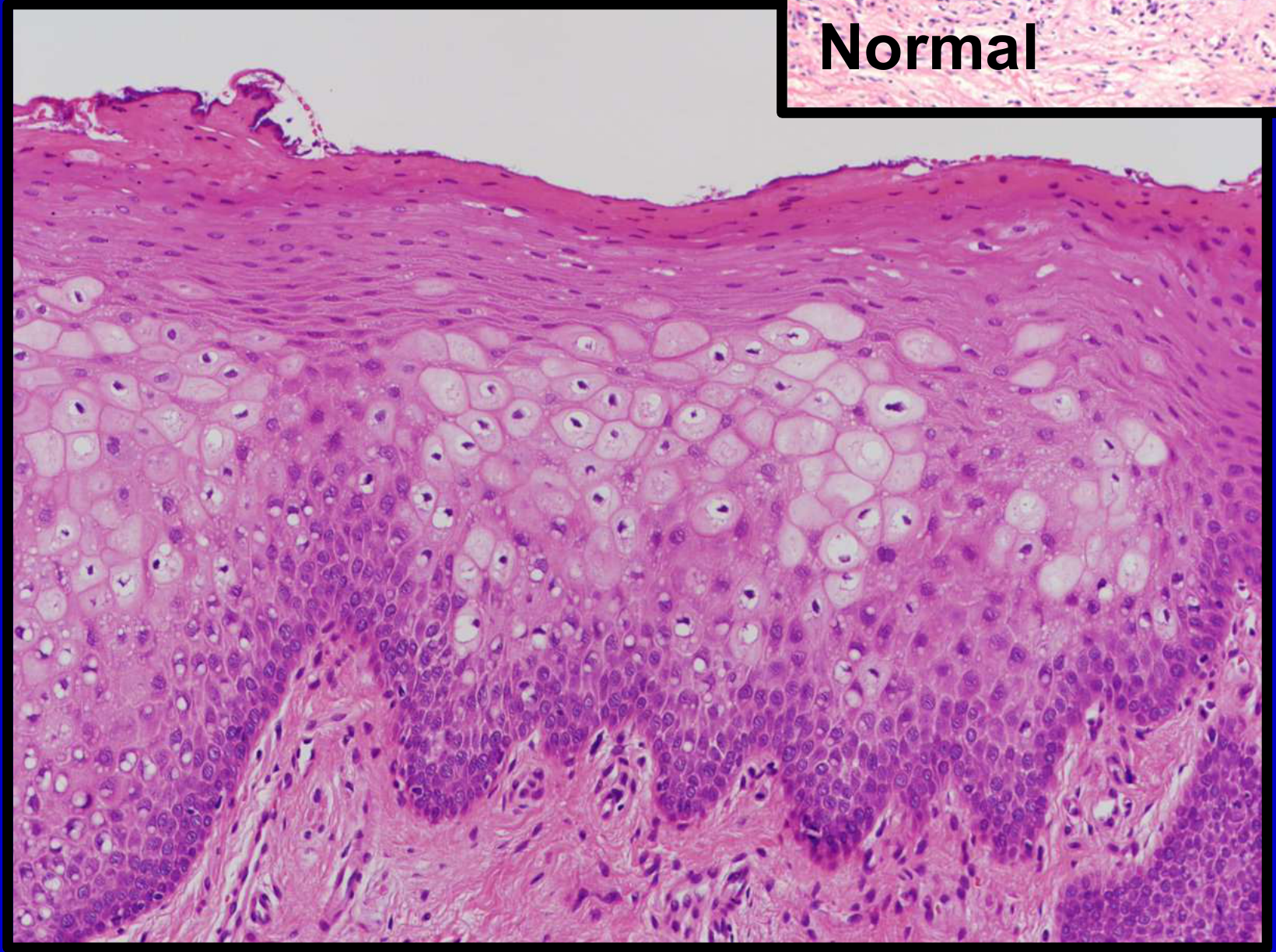
- Prominent hyperparakeratosis with marked acanthosis and a pale “spongy” epithelium. (W3 Slides; 1. Developmental disturbances of the oral cavity)
- Intracellular vacuolation and dyskeratosis sparing the basal cells; superficial layers show watery degeneration. (1. Developmental disturbances of the oral cavity; 1. DevelopmentalDisturbances Summary Table)
- Cytoplasmic clearing with perinuclear halos and perinuclear eosinophilic condensations (pathognomonic), representing aggregated abnormal keratin filaments; intracytoplasmic vacuolation is present (not spongiosis). (W3 Slides; 1. Developmental disturbances of the oral cavity; 1. DevelopmentalDisturbances Summary Table)
Clinical Features
Location
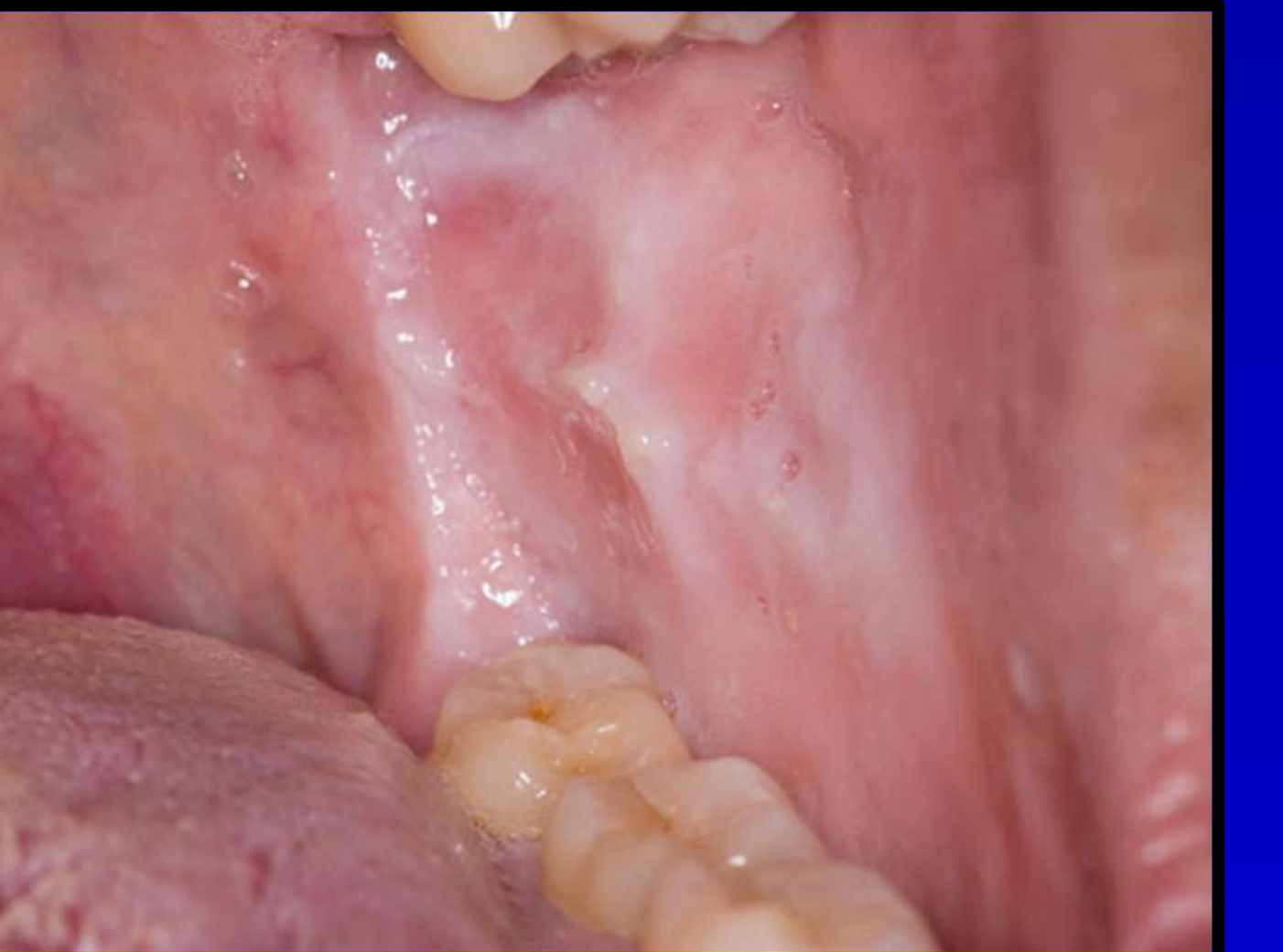
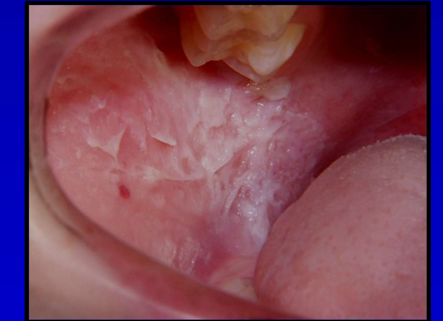
- Usually affects the buccal mucosa; may also involve ventral tongue, lip mucosa, floor of mouth, and soft palate. (Contemporary Oral Medicine white and red lesions of the oral Mucosa; 1. Developmental disturbances of the oral cavity)
- Extraoral mucosal involvement can include esophageal, upper airway, and genital sites; skin is not affected. (Contemporary Oral Medicine white and red lesions of the oral Mucosa; 1. Developmental disturbances of the oral cavity)
Appearance
- Bilateral, symmetric, asymptomatic, thick white spongy plaques; persistent and non-wipeable. (Contemporary Oral Medicine white and red lesions of the oral Mucosa; 1. Developmental disturbances of the oral cavity; 1. DevelopmentalDisturbances Summary Table)
- Diffusely white-to-grey, thickened, edematous, spongy mucosa. (1. Developmental disturbances of the oral cavity)
- Onset at birth/early childhood/adolescence with lifelong persistence. (Contemporary Oral Medicine white and red lesions of the oral Mucosa; 1. Developmental disturbances of the oral cavity)
Differential Diagnoses
- Chronic traumatic keratoses (clinically similar white plaques). (Contemporary Oral Medicine white and red lesions of the oral Mucosa)
- Leukoplakia (diagnosis of exclusion; white sponge nevus is excluded in the diagnostic pathway for persistent white patches). (9. OPMDs)
Relevant Clinical Investigations
- Biopsy is used to establish the diagnosis when clinically similar conditions (e.g., chronic traumatic keratoses) must be excluded. (Contemporary Oral Medicine white and red lesions of the oral Mucosa)
Patient Management
- No effective treatment; reported responses to antibiotics or antibacterial mouth rinses likely reflect reactive keratosis rather than true white sponge nevus. (Contemporary Oral Medicine white and red lesions of the oral Mucosa)